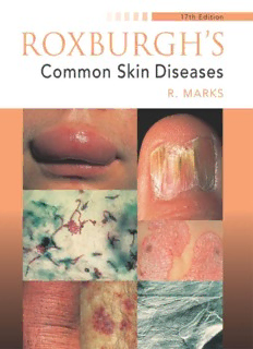
Preview Roxburgh's Common Skin Diseases
ROXBURGH’S Common Skin Diseases 17th Edition Ronald Marks Emeritus Professor of Dermatology and Former Head of Department of Dermatology University of Wales College of Medicine Cardiff,UK Clinical Professor Department of Dermatology and Skin Surgery University of Miami School of Medicine Miami,USA • • Hodder Arnold A member of the Hodder Headline Group London First published in Great Britain in 2003 by Arnold,a member of the Hodder Headline Group, 338 Euston Road,London NW1 3BH http://www.arnoldpublishers.com Distributed in the United States of America by Oxford University Press Inc., 198 Madison Avenue,New York,NY10016 Oxford is a registered trademark of Oxford University Press © 2003 Arnold All rights reserved. No part of this publication may be reproduced or transmitted in any form or by any means,electronically or mechanically,including photocopying,recording or any information storage or retrieval system,without either prior permission in writing from the publisher or a licence permitting restricted copying. In the United Kingdom such licences are issued by the Copyright Licensing Agency: 90 Tottenham Court Road,London W1T 4LP. Whilst the advice and information in this book are believed to be true and accurate at the date of going to press,neither the author nor the publisher can accept any legal responsibility or liability for any errors or omissions that may be made. In particular (but without limiting the generality of the preceding disclaimer) every effort has been made to check drug dosages; however it is still possible that errors have been missed. Furthermore,dosage schedules are constantly being revised and new side-effects recognized. For these reasons the reader is strongly urged to consult the drug companies’ printed instructions before administering any of the drugs recommended in this book. British Library Cataloguing in Publication Data A catalogue record for this book is available from the British Library Library of Congress Cataloging-in-Publication Data A catalog record for this book is available from the Library of Congress ISBN 0 340 76232 2 ISBN 0 340 76233 0 (International Students’ Edition – restricted territorial availability) 1 2 3 4 5 6 7 8 9 10 Commissioning Editor: Joanna Koster Project Editor: Anke Ueberberg Production Controller: Deborah Smith Cover Designer: Terry Griffiths Typeset in 10.5/12.5 Minion by Charon Tec Pvt. Ltd,Chennai,India Printed and bound in India What do you think about this book? Or any other Arnold title? Please send your comments to feedback.arnold@hodder.co.uk Contents Preface viii 1 An introduction to skin and skin disease 1 An overview 1 Skin structure and function 2 Summary 11 2 Signs and symptoms of skin disease 12 Alterations in skin colour 12 Alterations in the skin surface 14 The size,shape and thickness of skin lesions 15 Oedema,fluid-filled cavities and ulcers 17 Secondary changes 19 Symptoms of skin disorder 20 Summary 23 3 Skin damage from environmental hazards 25 Damage caused by toxic substances 26 Injury from solar ultraviolet irradiation 27 Chronic photodamage (photoageing) 29 Summary 35 4 Skin infections 37 Fungal disease of the skin/the superficial mycoses/infections with ringworm fungi (dermatophyte infections) 37 Bacterial infection of the skin 44 Viral infection of the skin 50 Summary 56 5 Infestations,insect bites and stings 58 Scabies 58 Pediculosis 63 Insect bites and stings 65 Helminthic infestations of the skin 69 Summary 70 iii Contents 6 Immunologically mediated skin disorders 71 Urticaria and angioedema 71 Erythema multiforme 75 Erythema nodosum 77 Annular erythemas 77 Autoimmune disorders 77 Systemic sclerosis 80 Morphoea 82 Dermatomyositis 83 The vasculitis group of diseases 84 Blistering diseases 87 Dermatitis herpetiformis 89 Epidermolysis bullosa 90 Pemphigus 91 Drug eruptions 92 Summary 95 7 Skin disorders in AIDS,immunodeficiency and venereal disease 97 Infections 98 Skin cancers 99 Other skin manifestations 99 Psoriasis 100 Treatment of skin manifestations of AIDS 100 Drug-induced immunodeficiency 100 Other causes of acquired immunodeficiency 101 Congenital immunodeficiencies 101 Dermatological aspects of venereal disease 102 Summary 104 8 Eczema (dermatitis) 105 Atopic dermatitis 105 Seborrhoeic dermatitis 114 Discoid eczema (nummular eczema) 117 Eczema craquelée (asteatotic eczema) 118 Lichen simplex chronicus (circumscribed neurodermatitis) 119 Contact dermatitis 121 Venous eczema (gravitational eczema; stasis dermatitis) 125 Summary 126 9 Psoriasis and lichen planus 128 Psoriasis 128 Pityriasis rubra pilaris 142 Lichen planus 144 Summary 147 iv Contents 10 Acne,rosacea and similar disorders 149 Acne 149 Rosacea 162 Perioral dermatitis 168 Summary 169 11 Wound healing and ulcers 171 Principles of wound healing 171 Venous hypertension,the gravitational syndrome and venous ulceration 173 Ischaemic ulceration 177 Decubitus ulceration 178 Neuropathic ulcers 179 Less common causes of ulceration 180 Diagnosis and assessment of ulcers 182 Summary 182 12 Benign tumours,moles,birthmarks and cysts 183 Introduction 183 Tumours of epidermal origin 184 Benign tumours of sweat gland origin 186 Benign tumours of hair follicle origin 188 Melanocytic naevi (moles) 188 Degenerative changes in naevi 192 Vascular malformations (angioma)/capillary naevi 194 Dermatofibroma (histiocytoma,sclerosing haemangioma) 197 Leiomyoma 198 Neural tumours 199 Lipoma 200 Collagen and elastic tissue naevi 200 Mast cell naevus and mastocytosis 201 Cysts 202 Treatment of benign tumours,moles and birthmarks 204 Summary 205 13 Malignant disease of the skin 207 Introduction 207 Non-melanoma skin cancer 207 Melanoma skin cancer 219 Lymphomas of skin (cutaneous T-cell lymphoma) 224 Summary 226 14 Skin problems in infancy and old age 227 Infancy 227 Old age 233 Summary 237 v Contents 15 Pregnancy and the skin 238 Physiological changes in the skin during pregnancy 238 Effects of pregnancy on intercurrent skin disease 240 Effects of intercurrent maternal disease on the fetus 240 Skin disorders occurring in pregnancy 241 Summary 242 16 Disorders of keratinization and other genodermatoses 243 Introduction 243 Xeroderma 245 Autosomal dominant ichthyosis 246 Sex-linked ichthyosis 247 Non-bullous ichthyosiform erythroderma 249 Bullous ichthyosiform erythroderma (epidermolytic hyperkeratosis) 251 Lamellar ichthyosis 252 Collodion baby 252 Other disorders of keratinization 254 Other genodermatoses 256 Summary 257 17 Metabolic disorders and reticulohistiocytic proliferative disorders 259 Porphyrias 259 Necrobiotic disorders 265 Reticulohistiocytic proliferative disorders 266 Summary 267 18 Disorders of hair and nails 268 Disorders of hair 268 Disorders of the nails 276 Summary 279 19 Systemic disease and the skin 281 Skin markers of malignant disease 281 Endocrine disease,diabetes and the skin 285 Skin infection and pruritus 288 Androgenization (virilization) 289 Nutrition and the skin 291 Skin and the gastrointestinal tract 292 Hepatic disease 292 Systemic causes of pruritus 293 Summary 293 20 Disorders of pigmentation 295 Generalized hypopigmentation 296 Localized hypopigmentation 297 vi Contents Hyperpigmentation 299 Summary 302 21 Management of skin disease 303 Psychological aspects of skin disorder 303 Skin disability 305 Topical treatments for skin disease 305 Surgical aspects of the management of skin disease 309 Systemic therapy 311 Phototherapy for skin disease 314 Summary 316 Bibliography 317 Index 319 vii Preface Recognition and treatment of skin disease is an important part of the practice of medicine. These skills should form an essential part of the undergraduate cur- riculum because skin disorders are common and often extremely disabling in one way or another.Apart from the fact that all physicians will inevitably have to cope with patients with rashes,itches,skin ulcerations,inflamed papules,nodules and tumours at some point in their careers,skin disorders themselves are intrinsically fascinating.The fact that their progress both in development and in relapse can be closely observed,and their clinical appearance easily correlated with their path- ology,should enable the student or young physician to obtain a better overall view ofthe way disease processes affect tissues. The division of the material in this book into chapters has been pragmatic, combining both traditional clinical and ‘disease process’categorization,and after much thought it seems to the author that no one classification is either universally applicable or completely acceptable. It is important that malfunction is seen as an extension of normal function rather than as an isolated and rather mysterious event.For this reason,basic struc- ture and function of the skin have been included,both in a separate chapter and where necessary in the descriptions ofthe various disorders. It is intended that the book fulfil both the educational needs of medical stu- dents and young doctors as well as being of assistance to general practitioners in their everyday professional lives.Hopefully it will also excite some who read it suf- ficiently to want to know more,so that they consult the appropriate monographs and larger,more specialized works. In this new edition ofRoxburgh’s Common Skin Diseasesaccount has been taken of recent advances both in the understanding of the pathogenesis of skin disease and in treatments for it.Please forgive any omissions as events move so fast it is really hard to catch up! viii C H A P T E R 1 An introduction to skin and skin disease An overview 1 Skin structure and function 2 Summary 11 An overview Skin is an extraordinary structure.We are absolutely dependent on this 1.7m2of barrier separating the potentially harmful environment from the body’s vulnerable interior.It is a composite of several types of tissue that have evolved to work in harmony one with the other,each ofwhich is modified regionally to serve a differ- entfunction (Fig.1.1).The large number of cell types (Fig.1.2) and functions of the skin and its proximity to the numerous potentially damaging stimuli in the environment result in two important considerations.The first is that the skin is frequently damaged because it is right in the ‘firing line’and the second is that Stratum corneum SC (15(cid:2)m) E (35–50(cid:2)m) Granular cell layer HF D (1–2mm) ESG SFL Malpighian layer Figure 1.1 Simple three-dimensional plan view of the Basal layer skin. HF (cid:2)hair follicle; ESG (cid:2)eccrine sweat gland; SC(cid:2)stratum corneum; E (cid:2)epidermis; D (cid:2)dermis; Figure 1.2 Diagram of the basic structure of the SFL(cid:2)subcutaneous fat layer. epidermis. 1
Description:The list of books you might like

The 5 Second Rule: Transform your Life, Work, and Confidence with Everyday Courage

The Spanish Love Deception

$100m Offers

The Sweetest Oblivion (Made Book 1)

Blumen für Algernon

Japp, Andrea H. - Sang Premier

Byron’s “Dithyramb on the Death of Napoleon and the

Svoboda-2006-49

Byron Alexis Mullo Naula
Afkondigingsblad van Aruba 2009 no. 1

bÈiþ
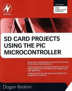
SD Card Projects Using the PIC Microcontroller

by Elmor van Staden submitted in accordance with the requirements
TS 129 199-5 - V6.4.0 - Universal Mobile Telecommunications System (UMTS); Open Service Access (OSA); Parlay X web services; Part 5: Multimedia messaging (3GPP TS 29.199-05 version 6.4.0 Release 6)

Henry is Twenty by Samuel Merwin

bölüm 1

The Japan Times 2006: Index

Bölüm 7 FET Karakteristikleri Deneyleri

Bölüm 2 sayfa 1.backup.fm

Bölüm 10 Kronik böbrek hastalığı

Centenary Magazine
Greek Government Gazette: Part 4, 2006 no. 74
