Polarized Delivery of Viral Glycoproteins to the Apical and Basolateral Plasma Membranes of ... PDF
Preview Polarized Delivery of Viral Glycoproteins to the Apical and Basolateral Plasma Membranes of ...
Polarized Delivery of Viral Glycoproteins ot the Apical dna laretalosaB amsalP Membranes of Madin-Darby Canine Kidney Cells Infected with Temperature-sensitive sesuriV MICHAEL .J RINDLER, IVAN EMANUILOV IVANOV, HEIDE PLESKEN, and DAVID D. SABATINI D o w Department of Cell Biology and ehT Kaplan Cancer Center, New York University Medical Center, New nlo a York 61001 de d fro m h TCARTSBA The intracellular route followed by viral envelope glycoproteins in polarized Madin- ttp Darby canine kidney cells was studied by using temperature-sensitive mutants of vesicular ://ru p re stomatitis virus (VSV) and influenza, in which, at the nonpermissive temperature (39.5°C), the ss.o newly synthesized glycoproteins (G proteins) and hemagglutinin (HA), respectively, are not rg transported out of the endoplasmic reticulum. /jcb/a After infection with VSV and incubation at 39.5°C for 4-5 h, synchronous transfer of G rticle protein to the plasma membrane was initiated by shifting to the permissive temperature -pd f/1 (32.5 °C). Immunoelectron microscopy showed that under these conditions the protein moved 00 /1 to the Golgi apparatus and from there directly to a region of the lateral plasma membrane /1 3 6 near this organelle. G protein then seemed to diffuse progressively to basal regions of the cell /1 0 5 surface and, only after it had accumulated in the basolateral domain, it began to appear on 11 1 6 the apical surface near the intercellular junctions. The results of these experiments indicate /1 3 6 that the VSV G protein must be sorted before sti arrival at the cell surface, and suggest that .p d passage to the apical domain occurs only late in infection when tight junctions are no longer f by g an effective barrier. ue nI complementary experiments, using the temperature-sensitive mutant of influenza, cul- st on 1 tures were first shifted from the nonpermissive temperature (39.5°C) to 18.5°C, to allow 9 F e entrance of the glycoprotein into the Golgi apparatus (see Matlin, .K ,.S and .K Simons, 1983, bru a Cell, 34:233-243). Under these conditions HA accumulated in Golgi stacks and vesicles but ry 2 0 did not reach the plasma membrane. When the temperature was subsequently shifted to 2 3 32.5°C, HA rapidly appeared in discrete regions of the apical surface near, and often directly above, the Golgi elements, and later diffused throughout this surface. To ensure that the anti- HA antibodies had access to lateral domains, monolayers were treated with a hypertonic medium to dilate the intercellular spaces. Some labeling was then observed in the lateral plasma membranes soon after the shift, but this never increased beyond t.0 gold particle/um, whereas characteristic densities of labeling in apical surfaces soon became much higher (~10 • particles/p,m). Our results suggest that the bulk of HA follows a direct pathway leading from the Golgi to regions of the apical surface close to trans-Golgi cisternae. Confluent monolayers of cultured Madin-Darby canine kid- larized (7, ,71 24) and provide a useful model to study the ney (MDCK) ~ cells are functionally and morphologically po- biogenesis of epithelial cell plasma membranes. After infec- tion with enveloped viruses, the polarized nature of the cells J Abbreviations used in this paper: ER, endoplasmic reticulum; G protein, glycoprotein; HA, hemagglutinin; MDCK, Madin-Darby is manifested in the assembly of specific virions on either one canine kidney; MEM, minimal essential medium; VSV, vesicular or the other plasma membrane domain (33). Influenza, sim- stomatitis virus. ian virus 5, or Sendal virions assemble exclusively on the EHT LANRUOJ FO CELL YGOLOIB • EMULOV 100 YRAUNAJ 1985 136 151 136 © The Rockefeller University Press 6116310t10- 158t5259-1200 $1.00 apical surface, whereas vesicular stomatitis virus (VSV) and replaced with MEM minus HCO3- (from a Gibco Laboratories Grand Island, NY kit), but containing 20 mM HEPES. CuRures were then transferred to an certain RNA tumor virus particles bud only from the baso- C"5.81 incubator without 2OC for 80-90 rain. When cultures were shifted to lateral regions of the plasma membrane (1, 28, 31, 32, 35, the permissive temperature, the medium was replaced with prewarmed new 37). medium at 32.5"C and incubation continued at this temperature for the The asymmetric budding of viruses from polarized epithe- indicated time intervals. For immunofluorescence studies, infection was carried lial cells is thought to be determined by a preferential accu- out on cells that were grown on 13-ram coverslips placed in 24-well dishes (Costar, Cambridge, MA). For preparation of frozen thin sections, cells that mulation of the envelope glycoproteins in the corresponding had been plated on collagen-coated 25-ram glass coverslips (-3 x l0 s cells/ membrane domain, which precedes viral assembly (32). It cm )2 were washed and then fixed with %2 glutaraldehyde (Polysciences lnc., appears that segregation of the glycoprotein does not require Wardngton, PA) in Dulbecco's phosphate-buffered saline (PBS) with 21CaC other viral components, in that it has been reported that 1.0( mM) for 60 rain at 4°C. Coverslips were washed in PBS and stored at C"4 accumulation of hemagglutinin (HA) in the apical surface until use. takes place in cells infected with recombinant SV40 viruses ecnecseroulfonummI :ypocsorciM Specimens fixed in %4 form- aldehyde in PBS for 30 min were treated with 0.2% Triton X-100 and incubated carrying the HA gene, but not encoding the other influenza with an IgG fraction (100 #g/ml) of rabbit anti-VSV glycoprotein (G protein) polypeptides (36). Thus, most likely, the mechanisms that antibody, or a mixture of two mouse monoclonal anti-HA antibodies (a gift of ensure the distribution of viral envelope glycoproteins in the Dr. .R G. Webster. .tS Jude Children's Hospital) applied at a 1:300 dilution. cell surface are the ones utilized for the segregation of cellular Two preparations of rabbit anti-G protein antibody were used (one of which was the gift of Dr. .J Lenard, Rutgers University). Both were reactive with only proteins to specific plasma membrane domains. the VSV G protein, as demonstrated by immunoprecipitation and Western Theoretically, plasma membrane proteins could be sorted immunoblots, and gave very low background labeling on uninfected cells. In a intracellularly by a mechanism that directs them to one or second step, samples were labeled with tetramethylrhodamine isothiocyanate- the other plasma membrane domain, or sorting would take conjugated goat anti-rabbit or anti-mouse IgG (Cappel Laboratories, Cochran- Do w place after arrival at the cell surfaces. The delivery of protein ville, PA) as previously described (28). Preparations were examined in a Leitz nlo Orthoplan microscope (E. Leitz, Inc., Rockleigh, N J) equipped with a Wild a molecules to the plasma membrane need not be restricted to camera (Wild Heerbrugg Instruments, Inc., Farmingdale,NY). ded the domain where the proteins eventually accumulate, be- nortceleonummI :ypocsorciM Monolayers were scraped, infused from cause a redistribution could be effected either through recy- with 0.6 M sucrose, and cryosectioned at -90°C using a Sorvall MT-2 ultra- h cling Using mechanisms MDCK cells or by doubly controlled infected lateral with difVfSusVi on. and influenza a microtome DuPont freezin(g DuPont attachment Instruments-Sorvall (FTS). Sections Biomedical 60-180 Div., nm Newtown, thick (estimated CT) with ttp://rup we have recently demonstrated (28) that glycoproteins of both ftiraolml y the by theinterference procedure color of Tokuyafter asu embedding and Singer and drying) (40, 41) were with prepared modifications essen- ress.o viruses can be found within the same Golgi apparatus. We, rg therefore, concluded that their sorting cannot take place be- described in a first step previously with either (15, 1 28). mg/ml Frozen of the sections rabbit attached anfi-G proteint o grids IgG were fractions incubated or /jcb/a fore arrival to this organelle. To determine whether the gly- a 1:15 dilution of mouse monoclonal ascites fluid. In a second step, affinity- rticle coproteins are sorted intracellulady or only after they have purified goat anti-rabbit or sheep anti-mouse IgG (Cappel Laboratories) con- -p d reached the cell surface, we have now employed temperature- jugated dures used to 18-wnemr e similar colloidal to those gold particles described was by applied. Geuze et The .la (11) conjugation with modifica- proce- f/100 sensitive mutants of VSV and influenza, in which, at the /1 tions detailed elsewhere (15, 28). Sectious were examined in a Philips 103 /1 nonpermissive temperature, the envelope glycoproteins ac- microscope (Philips Electronic Instruments, Inc., Mahwah, NJ) operated at 60 36/1 cumulate in the endoplasmic reticulum (ER). Their transfer kV. 05 to the cell surface was synchronized in one of two ways, by eninoihteM553 :gnilebaL Infected cells on 35-rnm dishes were 111 6 either directly shifting the temperature to the permissive one preincubated at 39.5"C for 30 rain with methionine-free MEM (from a GibeD /13 owrh ich by inletardso ducing to intracellular an intermediate accumulation cold tempeoraftu re the glycoprotein block, Laboratories ~SSmethionine MEM and incubated kit). (50 Labeling t~Ci/ml). for an was additional After carded 30 out 30 rain, rain in 5.0ceils at ml 39.5, were of fresh 32.5, washed mediumo r .C"5.81 three containing tiSmamepsle s in 6.pdf by g after its delivery to the Golgi apparatus (22). Our results were solubilized with 0.5% NP-40, 0.5% deoxycholate in a 01 mM Tris-HCl ue suggest that sorting takes place intracellularly, because the buffer at pH 7.6. After removing nuclei by sedimentation, samples were st o n bulk of the accumulated protein appeared to be transferred analyzed by electropboresis in a %01 polyacrylamide gel. Fluorography (5) was 19 performed using Enhance (New England Nuclear, Boston,MA) and autora- F directly to the domain from which viral budding takes place. diography with Kodak X-Omat film (Eastman Kodak Co., Rochester, NY). ebru A sensitive recent stmuudyt ant with of incfellluse nza, infected but with using the a dsiaffmeree nt temperature- immuno- STLUSER ary 202 cytochemical procedure, provided a similar conclusion for 3 the distribution of HA (3 l). Localization of the C protein in MDCK Cells Infected with the ts045 Mutant of VSV MATERIALS AND METHODS Cell Cultures and Viruses: The MDCK and Vero cell lines were IMMUNOFLUORESCENT LABELING: The pattern of cultured by standard procedures as previously described (7, 28). The ts045 immunofluorescence suggested that when MDCK cells in- mutant (16) of VSV (a gift of Dr. .J Lenard, Rutgers University, New Brunswick, fected with the ts045 mutant were maintained at 39.5°C, the N J) and the ts61 s mutant (42) of influenza (a gift of Dr. P. Palese, Mount Sinai newly synthesized G protein was retained in the ER. The School of Medicine, New York) were plaqued at 32.5"C on Vero and MDCK cells, respectively. For both mutants, viruses from different plaques were grown cytoplasm of permeabilized cells was diffusely labeled, and on Veto cells at 32.5"C and used to determine in microtiter plates the viral the periphery of the nucleus was well marked, but neither the cytopathic effects on %05 (CPE~o) of Veto and MDCK at the nonpermissive Golgi apparatus nor the plasma membrane were discernible (39.5'C) and permissive (32.5"C) temperatures. Only plaqued viruses that (Fig. I a). Electrophoretic analysis of cultures incubated with showed at least a l04 differential CPEsa were utilized. 35Smethionine (not shown) showed that the G protein syn- MDCK cells were infected with ts045 (15-20 plaque-forming units/cell) or ts6 Is (5-20 plaque-forming units/cell) in a small volume of minimal essential thesized at 39.5°C was of higher mobility than that found in medium (MEM), which in the case ofts045 contained 75 #g/ml DEAL Dextran infected MDCK cells incubated at the permissive tempera- (Pbarmacia Fine Chemicals, Piscataway, NJ). Cultures were kept for l-l.5 h ture, and corresponded in apparent molecular weight to the at 32.5"C in a %5 2OC incubator. The medium was then replaced with core glycosylated form of G protein (18). prewarmed MEM and incubation continued at 39.5"C for 4-5 h. To block exit from the Golgl apparatus by incubation at 18.5"C (22), the medium was As soon as 10 rain after cells incubated at the nonpermissive RIN DEER TEl AL. deziraloP yrevileD of lariV snietorpocyIG 1 3 7 D o w n lo a d e d fro m h ttp ://ru p re ss.o rg /jcb /a rticle -p d f/1 0 0 ERUGIF 1 Immunofluorescent localization of G protein in cells infected with the ts045 mutant of VSV. MDCK cells were infected /1 by incubation with the virus for 1 h at 32.5°C, maintained for 1 h at this temperature, and then incubated at 39.5°C for 4.5 h /13 6 before a temperature shift to 32.5°C (the permissive temperature). At different times, monolayers were fixed in formaldehyde, /10 5 permeabilized with 0.2% TX-t00, and treated with rabbit anti-G protein antibody followed by rhodamine-conjugated goat anti- 11 1 rabbit IgG. )a( In cells maintained at 39.5*C, fluorescence si observed around the nucleus and in a lacework of threadlike 6/1 cytoplasmic elements, sa expected from an accumulation of G protein in perinuclear and cytoplasmic RE cisternae. (b) After 51 36 .p rain at 32.5°C, the G protein si concentrated in the crescent shaped Golgi region surrounding the nucleus (arrows). )c( At 30 df b rain, significant label si also apparent in the lateral domain of the plasma membrane (d) (arrowheads). yB 021 rain, lateral domains y g are predominantly labeled. Photo exposure times were )a( rain, 1 (b and c) 30 ,s and (d) 20 .s x 2,900. ue st o n 1 9 F temperature were transferred to 32.5"C, a crescent-shaped surrounded the Golgi area. Labeling of the plasma membrane eb ru juoxretsacenntu,c lear as expected region of from the the cytoplasm transfer became to and concenitnrteantseiloyn flu- twiaasl ly minimal on lateral and membranes. when this occurred it took place preferen- ary 20 2 of the G protein in the Golgi apparatus. The intensity of The distribution of the G protein changed soon after shift- 3 labeling in the juxtanuclear region continued to increase for ing to the permissive temperature: by 51 rain, gold particles the next 5-10 rain (Fig. 1 b), when fluorescence in the lateral labeled the Golgi stacks (Fig. 2b). As was previously shown plasma membrane began to be detected. By 30 min after the in cells infected with the wild-type virus (28), the G protein temperature shift, the accumulation of G protein in lateral seemed to be randomly distributed over the Golgi cisternae. regions of the plasma membrane was very apparent (Fig. I c), Beginning at 51 rain and becoming more prominent at later and intense labeling of the lateral plasma membranes was the times, labeled small vesicular structures (60-120 rim) were most notable feature of cells that had been incubated for 2 h seen near the lateral plasma membrane (Fig. 3a). 30 rain after at 32.5°C before fixation (Fig. 1 d). shifting to the permissive temperature, focal labeling of the IMMUNOELECTRON MICROSCOPIC OBSERVATIONS: lateral membrane was conspicuous (Fig. 3, a and b), particu- Examination of ultrathin frozen sections immunolabeled with larly in regions where the two adjacent lateral membranes the gold-particle technique showed that in infected cells main- were well separated, as frequently happened just below the tained at 39.5°C the G protein was present in tubulovesicular tight junctions (Fig. 3a). However, the initial appearance of and cisternal elements, some of which could be identified as gold particles was not restricted to these regions and labeling belonging to the rough ER, as well as in the nuclear envelope seemed to take place at any point on the lateral plasma and in small round dense bodies resembling lysosomes (Fig. membrane (Fig. 3b). At these early times (up to 30 rain after 2a). Very few gold particles were found on Golgi cisternae, the shift), virtually no labeling of apical or basal surfaces was although they were frequently seen in vesicular elements that detected. During the first hour after transfer to 32.5"C, the G 138 ~HT LANRUOJ ~O LLEC YGOLOIB " EMULOV 100, 1985 D o w n lo a d e d fro m h ttp ://ru p re ss.o rg /jcb /a rticle -p d f/1 0 0 /1 /1 3 6 /1 0 5 1 1 1 6 /1 3 6 .p d f b y g u e st o n 1 9 F e b ru a ry 2 0 2 3 ERUGIF 2 lntracellular distribution of the G protein at the nonpermissive temperature and shortly after shift to the permissive one in MDCK cells infected with the ts045 mutant of VSV. Monolayers infected sa described in the legend to Fig. 1 were fixed in glutaraldehyde and cryosectioned according to the procedures detailed in Materials and Methods. Sections were treated with rabbit anti-G protein antibodies and goat anti-rabbit second antibodies complexed to colloidal gold particles 81( nm). )a( In monolayers kept at 39.5°C, the G protein si present primarily in RE cisternae (arrowheads) and multivesicular bodies (arrow). skcatS of Golgi cisternae (CA) were generfarleley of label, but frequently gold particles were found in vesicular structures at the Golgi periphery. Very few gold particles were associated with the apical (Ap) and basal )B( plasma membranes. (b) 51 rain after a temperature shift to 32.5°C, labeling for G protein si prominent in the Golgi apparatus (CA), but only very small amounts of G were detected in the lateral )L( plasma membrane. Apical (Ap) basal and )B( surfaces are virtually unlabeled. 0.2 Bar, #m. x 45,000. protein continued to accumulate over the lateral plasma G protein on the apical surface was first noted at this time. membrane, and after this period, heavy labeling of this area However, 2 h after the temperature shift--when the first was striking in many cells (Fig. 4a). The basal surface also budding of VSV particles was observed on lateral aspects of contained substantial amounts of G protein, and in a small the cells (Fig. 4 b)--gold particles could be found on all aspects fraction of the infected cells (not shown), the appearance of of the cell surface in many cells. In a number of instances it RELDNIR TE .LA deziraloP yrevileD of lariV snietorpocylC 1 39 D o w n lo a d e d fro m h ttp ://ru p re ss.o rg /jcb /a rticle -p d f/1 0 0 /1 /1 3 6 /1 0 5 1 1 1 6 /1 3 6 .p d f b y g u e st o n 1 9 F e b ru a ry 2 0 2 3 EIRUGIF 3 First appearance of the G protein on the lateral surface 30 rain after a temperature shift to 32.5°C. (a and b) By 30 rain after a shift to 32.5"C, the G protein began to accumulate in regions of the lateral plasma membrane (t) facing dilated intercellular spaces, either proximal to the tight junction (a) or closer to the base of the cell (b). A group of vesicular elements (a, arrow) near to the lateral surface is also labeled. The apical (Ap) and basal (B) surfaces remain very sparsely labeled, in contrast to stacks of the Golgi cisternae (GA) where gold particles are now abundant. Monolayers were infected and sections prepared sa described in Fig. 2. Bar, 0.5 pm. x 25,000. was apparent that initial labeling of the apical membrane G protein was not limited to basolateral domains, lateral domain was restricted to regions closest to the intercellular regions of the plasma membrane always remained the most junction (Fig. 5, a and b), as if G protein, which had accu- intensely labeled and, as was the case with the wild-type virus mulated in the lateral region, had now diffused through the (28), even at this and later times budding of VSV panicles junctions. Even though subsequently the distribution of the occurred only on lateral and basal surfaces. 041 EHT LANRUOJ FO CELL YGOLOIB • VOLUME 100, 1985 D o w n lo a d e d fro m h ttp ://ru p re ss.o rg /jcb /a rticle -p d f/1 0 0 /1 /1 3 6 /1 0 5 1 1 1 6 /1 3 6 .p d f b y g u e st o n 1 9 F e b ru a ry 2 0 2 3 ERUGIF 4 Accumulation of the G protein in the lateral and basal surfaces 1-2 h after the temperature shift. )a( yB 60 min after the temperature shift to 32.5"C, the lateral aspect of the plasma membrane )L( si heavily labeled. Gold particles are also found on the surface basal )B( of the cell. In contrast, the apical domain of the plasma membrane (Ap) si essentially free of label, while labeling of the Golgi complex (GA) continues to be prominent. Bar, 0.2/~m. x 42,000. (b) 2 h after the temperature shift, virions (arrow) are frequently seen budding into the lateral intercellular spaces. Bar, 0.2 ~m. x 38,000. Cells were infected and frozen thin sections were labeled sa outlined in Fig. 2. Localization of the HA Glycoprotein in MDCK migrating form of HA was synthesized at 39.5"C and that, Cells Infected with the ts61s Mutant of upon temperature shift to either 32.5 or 18.5"C, this was replaced by a slower-migrating form (not shown) of the pro- Influenza Virus tein shown. This result was essentially the same as that Electrophoretic analysis of products synthesized in infected reported for wild-type influenza HA as the protein matures cells incubated with [35S]methionine demonstrated that a fast- during its passage through the Golgi apparatus from an en- RELDNIR TE .LA Delivery Polarized of Viral snietorpocylG 141 D o w n lo a d e d fro m h ttp ://ru p re ss.o rg /jcb /a rticle -p d f/1 0 0 /1 /1 3 6 /1 0 5 1 1 1 6 /1 3 6 .p d f b y g u e st o n 1 9 F e b ru a ry 2 0 2 3 ERUGIF 5 Appearance of the G protein on the apical surface late after the temperature shift. (a and b) 180 min after the shift to 32.5°C, when labeling was prominent on the lateral and basal surfaces (L, B) and in the Golgi apparatus (CA), gold particles were also found on the apical surfaces, often concentrated near the junctional areas (arrows). Specimens were prepared as indicated in Fig. 2, Bar, 0.5 pm. × 30,000. doglycosidase H-sensitive form to a terminally glycosylated the Golgi apparatus was apparent (Fig. b). 6 In addition, after glycoprotein (1, 22, 31). 20-30 min, labeling of the plasma membrane began to be IMMUNOFLUORESCENT LABELING: No significant la- prominent (Fig. 6, c-f). An even better synchrony in transport beling of the apical plasma membrane was observed when of HA to the plasma membrane was obtained when, before anti-HA antibody was applied to MDCK cells that, after transfer to 32.50C, MDCK cells infected with the s ts61 mutant infection with the ts61s mutant, were kept at 39.5°C. When were maintained for 90 min at 18.5°C. It has previously been these cells were permeabilized with detergent, however, a weak shown, by using the wild-type virus (22), that incubation at and diffuse cytoplasmic labeling was detectable, consistent 18.5°C prevents release ofH A at the cell surface and leads to with the presence of the protein in the ER (Fig. 6a). 10-15 its accumulation in the Golgi region of the infected cells. A min after the shift to the permissive temperature (32.5°C), temperature shift to 18.5°C had the same effect in cells in- fluorescence in the juxtanuclear region presumed to contain fected with the s ts61 mutant: HA fluorescence became striking 241 EHT LANRUOJ FO CELL BIOLOGY • VOLUME 100, 1985 D o w n lo a d e d fro m h ttp ://ru p re ss.o rg /jcb /a rticle -p d f/1 0 0 /1 /1 3 6 /1 0 5 1 1 1 6 /1 3 6 .p d f b y g u e st o n 1 9 F e b ERUGIF 6 Immunofluorescent localization of HA in cells infected with the is61 mutant of influenza. MDCK cells were infected rua by incubation with the virus for 1.5 h at 32.5"C, and then incubated for 5 h at 39.5"C, before a temperature shift to 32.5"C (the ry 2 0 permissive temperature). At different times, formaldehyde-fixed monolayers were treated either directly with monoclonal anti- 23 HA, to label the apical plasma membrane (c, e), or after permeabilization with 0.2% TX-100, to label cytoplasmic structures now accessible to the antibodies (a, b, d, f). Rhodamine-conjugated goat anti-mouse IgG was applied in a second step. (a) In cells maintained at the nonpermissive temperature (39.5"C), a low level of cytoplasmic staining is detectable. The nucleus (N) is unlabeled. (b) 51 min after a temperature shift to 32.5°C, the HA si concentrated in the Golgi region (arrows). No labeling of the apical surface was detectable at this time. (c) 30 min after the shift the HA protein is found on the apical surface (arrowhead). (d) At this time the Golgi region remains heavily labeled but there is little detectable label in the lateral membrane. (e) After 60 min the apical plasma membrane is uniformly fluorescent. (f) At this time the Golgi si still a major site of accumulation but the lateral membrane contains little detectable HA. Photo exposure times were (a) 2 min, (c, e) 30 ,s and (b, d, f) 51 s.x 2,900. in a crescent-shaped region located towards one side of the membrane in an off-center position reminiscent of the asym- nucleus, thought to represent the Golgi apparatus (Fig. 7 b), metric location of the Golgi apparatus within the cell (Fig. but no label could be detected on the apical surface of 7d). By 30-40 min, however, the initially concentrated mol- nonpermeabilized cells (Fig. 7 a). After the temperature was ecules had diffused extensively over the entire apical surface, raised to 32.5"C, the HA that had accumulated intracellularly and by 45 min the brighter spot, which indicated the initial was rapidly transferred to the cell surface. Some cells mani- site of appearance of HA, was all but unrecognizable (Fig. fested apical fluorescence 5 min after the shift, but only after 7e). At no time during the first 45 min was a significant 10-20 min was surface staining readily observed on most cells amount of fluorescent label detectable on the lateral plasma (Fig. 7c). A single fluorescent spot was found on the apical membrane (Fig. 7, d and f). RELDNIR TE .LA Delivery Polarized of Glycoproteins Viral 341 D o w n lo a d e d fro m h ttp ://ru p re ss.o rg /jcb /a rticle -p d f/1 0 0 /1 /1 3 6 /1 0 5 1 1 1 6 /1 3 6 .p d f b y g u e st o n 1 9 F e b ru ERUGIbFlock to 7 synchronize Immunofluorescent transfer to the localization cell surface. of HA in MDCK cells infececllts ed were with infected the ts61 by mutant incubation of influenza with the and subjected virus for 1.5 h to at an 32.5°C, 18.5°C ary 20 2 maintained for 5 h at 39.5°C, and then for 1.5 h at 18.5°C before a temperature shift to 32.5°C. Individual coverslips of 3 formaldehyde-fixed cells were labeled sa described in the legend to Fig. I, either directly (a, c, e) or after permeabilization with detergent (b, d, f). (a and b) At 18.5°C, HA has not reached the plasma membrane (a) but accumulates in the Golgi apparatus (b) (arrow), which si heavily labeled. (c and d) 10 min after the temperature shift to 32.5°C, the HA is initially detected on the apical surface (c) (arrowheads), in off-center focal regions. Little or no fluorescence is found on the lateral surfaces at this time, but the Golgi region remains intensely labeled (d). (e and f) 45 min after the shift the HA is found over the entire apical surface (e) and in the Golgi apparatus (f). Labeling of the lateral surface is extremely low. Photo exposure times were (a, c, e) 30 s and (b, d, f) 15 .s x 2,900. IMMUNOELECTRON MICROSCOPIC OBSERVATIONS: mainly found on their dilated ends and over associated vesic- In ts6 Is-infected cells maintained at the nonpermissive tem- ular elements (Fig. 8 a). In agreement with the observations perature (39.5°C), the level of immunolabeling was quite low using fluorescence, virtually no label waso bserved at this time (not shown), but occasional grains that resembled ER cisternal on the plasma membrane. Only after the temperature was elements were seen on cytoplasmic vesicles and structures. raised did the HA begin to appear on the apical membrane. Upon temperature shift to 18.5°C, when fluorescence of the In some cells it was detected as soon as 5 min after the shift juxtanuclear region was striking, the gold particles were more (Fig. 8 b), but after 20 rain the surface localization was appar- frequently found in the Golgi region. However, they were not ent in most cells. Interestingly, HA first appeared in discrete particularly concentrated over the stacked cisternae but were regions, near and often directly above Golgi elements, sug- 144 EHT LANRUOJ FO - VOLUME BIOLOGY CELL 100, 1985 D o w n lo a d e d fro m h ttp ://ru p re ss.o rg /jcb /a rticle -p d f/1 0 0 /1 /1 3 6 /1 0 5 1 1 1 6 /1 3 6 .p d f b y g u e st o n 1 9 F e b ru a ry 2 0 2 3 ERUGIF 8 Intracellular distribution of HA during the cold-temperature block and shortly after shift to the permissive temperature in MDCK cells infected with the ts61 mutant of influenza. Cells were infected by incubation with the virus for 5.1 h at 32.5°C and then maintained for 5 h at °C 39.5 and 5.1 h at °C 18.5 before shift to 32.5°C. Cryosections of glutaraldehyde-fixed monolayers were treated with monoclonal anti-HA antibodies followed by colloidal gold-goat anti-mouse IgG complexes. (a) In cells maintained at 18.5 °C HA si found in Golgi apparatus (GA). Two sets of Golgi cisternae, possibly interconnected, are presented, each with a concentration of gold particles over vesicular elements toward one side of the stack. Apical plasma membranes (Ap) are unlabeled. Bar, 0.2 /zm. x 50,000. (b) 5 min after the shift to 32.5°C HA initially appears on the apical plasma membrane (Ap), in focal regions (arrowhead) that are not far from the Golgi apparatus (GA). Labeled vesicles are also found near the apical surfce (arrow). Very little labeling was seen on lateral or surfaces basal (not shown). Bar, 0.5 ~m. x 30,000. gesting that its transport from the Golgi apparatus to these often oblong shape with an average diameter varying between sites was direct (Figs. 8b and 9). At these and later times, 60 and 120 nm (Figs. 8b, 9, and 10a) were also visible labeled vesicles with an apparently smooth surface and an underlying the apical membrane. It may be presumed that RELDNIR TE .LA Polarized yrevileD of Glycoproteins Viral 145
Description:The list of books you might like

The Mountain Is You

Haunting Adeline

The Strength In Our Scars

A Thousand Boy Kisses

The Founding Fathers of American Intelligence
A NEW SPECIES OF CALLAEUM (MALPIGHIACEAE) FROM PUEBLA, MEXICO

IS 15644: Safety of Electric toys

ግእዝ አማርኛ መዝገበ ቃላት

Healthcare Financial Management 1993: Vol 47 Index

Bürofach-/Handelsdiplom VSH Med. Sachbearbeiterin+ Röntgen-Grundkurs

Bürgerliches Recht Band II Schuldrecht Allgemeiner Teil
Glossary

Polar Bear: 2006
Greek Government Gazette: Part 2, 2006 no. 1792
Greek Government Gazette: Part 2, 2006 no. 1757
Greek Government Gazette: Part 2, 2006 no. 1799

Ruth Fielding at Sunrise Farm

op1278241-1001

Achtung-Panzer!
Hydrodynamic source with continuous emission in Au+Au collisions at $\sqrt{s}=200$ GeV
Direct CP violation in B \to ρ^0(ω) PS

Semi-quantitative Approaches for Landslide Assessment and Prediction
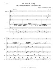
UN TEMA EN SWING
