Table Of Content11
Bull. not. Hist. Mus. Lond. (Zool.)64(2): 111-205 —Issued26November1998
1 TIICNATLffifrt
Partial revision of Paracyclops Claus, 1893
(Copepoda, Cyclopoida, Cyclopidae) with
PRESented
descriptions of four new species
xxTjqqs^
SUPHAN KARAYTUG_
/
DepartmentofZoology. The"NaturalHistoryMuseum, CromwellRoad. London SW75BD, UK& Schoolof
BiologicalSciences, Queen Maryand WestfieldCollege, MileEndRoad, London El 4NS, UK
GEOFFREY BOXSHALL
A.
DepartmentofZoology, TheNaturalHistoryMuseum, CromwellRoad, London SW75BD, UK
CONTENTS
Introduction 11
MaterialsandMethods 11
Paracyclopsaffinis(G.O.Sars, 1863) 113
Paracyclopspoppei(Rehberg, 1880) 120
Paracyclopsoligarthrus(G.O. Sars, 1909) 128
Paracyclopscanadensis(Willey. 1934) 136
ParacyclopsdilatatusLindberg. 1952 142
Paracyclopshardinginom. nov 145
ParacyclopsbaicalensisMazepova, 1961 151
ParacyclopsyeatmaniDaggett& Davies. 1974 151
ParacyclopswaiarikiLewis, 1974 162
ParacyclopspilosusDussart, 1984 169
Paracyclopscareclum Reid. 1987 172
ParacyclopsnovenariusReid, 1987 176
ParacyclopssmileyiStrayer, 1988 176
Paracyclopsreidaesp.nov 184
Paracyclopsbromeliacolasp.nov 189
Paracyclopspunctatussp.nov 195
Paracyclopsrothatsp.nov 200
Acknowledgements 200
References 204
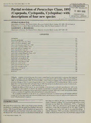
SYNOPSIS. A partial revision ofthe genus Paracyclops is made based on type material and on collections from numerous
localitiesaroundtheworld.ThefollowingParacyclopsspeciesareredescribed: P.affinis(G.O.Sars, 1863),P.poppei(Rehberg,
1880),P.oligarthrus(G.O.Sars, 1909),P.canadensis(Willey, 1934),P.dilatatusLindberg, 1952,P.hardinginomennovum,P.
baicalensisMazepova. 1961,P.yeatmaniDaggett&Davis, 1974,P.waiarikiLewis,1974,P.pilosusDussart, 1984,P.carectum
Reid, 1987.P.novenariusReid, 1987andP.smileyiStrayer. 1988.Fourspeciesaredescribedasnewtoscience:P.reidaesp.nov.,
P. rochaisp. nov.,P.punctatussp. nov.,andP. bromeliacolasp.nov.
Detailed descriptions of these species are given including several previously overlooked microcharacters, such as the
ornamentation of the coxobasis of antenna, the cuticular ornamentation of urosomal somites and the posterior spinular
ornamentationoftheswimminglegs,thatareshowntohavesignificanttaxonomicvalueatspecieslevel.Thedetaileddescription
ofmalesisrevealedtobe importantindifferentiatingbetweencloselyrelatedspecies.
The geographical distributions ofthe species are re-evaluated on the basis ofexamined material and verifiable published
records.ItisrevealedthatP.affinisdoesnotoccurinNorthAmericaandallpreviousrecordsofP.affinisinNorthAmericarefer
tothenewlydiscoveredP. canadensis.
INTRODUCTION have been recorded in all types offreshwater habitats (Karaytug,
1998):RdilatatusLindberg, 1952wasfoundintheDniesterestuary
(Ukraine) on the Black Sea (Monchenko, 1977), P. baicalensis
Thegenus ParacyclopsClaus, 1893 isoneofnine generacurrently Mazepova, 1961 was collected from great depths in Lake Baikal
recognisedasconstitutingthe sub-family Eucyclopinae (Dussart& (Mazepova, 1978), and P. bromeliacola sp. nov. and P. reidae sp.
Defaye, 1985; Pospisil & Stoch, 1997).All speciesareknowntobe nov. inhabit pools in the leaf axils of terrestrial Bromeliads. P.
benthic although they can sometimes occurin the watercolumn in chiltoni (Thomson, 1882) was recently collected from freshwater
the littoralzone. Paracyclopsspeciesaredistributedworldwideand bodies on EasterIsland and is theonly freshwatercopepodon this
©TheNaturalHistoryMuseum, 1998
112 S. KARAYTUG ANDG.A. BOXSHALL
Fig.1 P.affinis.Adultfemale.A.maxillule;B,maxilliped;C,body,dorsal;D,maxilla;E,labrum;EG,mandible;H.detailofcaudalseta.Scalebarsin|im.
1
PARACYCLOPS REVISION 13
remote island (Dumont & Martens, 1996). P. oligarthrus (G. O. SPECIES DESCRIPTIONS
Sars, 1909) occurs only in LakeTanganyika.
The lack of sufficient detail in the original description of the
Paracyclops affinis (G. O. Sars, 1863)
type-species P.fimbriates (Fischer, 1853) and the publication of
various incompletely described species or forms that are closely (Figures 1-7)
related to the type-species has created considerable taxonomic
CyclopsaffinisSars, 1863: Brady (1878),Vosseler(1886), Schmeil
confusion.This has beenexacerbatedby the use ofalimited setof (1892). Brady (1892),Van Douwe (1909), Lilljeborg (1901).
traditional characters for differentiating between species within
Cyclopspygmaeus Rehberg. 880
the genus, such as the morphology of the caudal rami and leg 5. 1
Cyclops (Heterocyclops)affinis Sars, 1863: Claus (1893a)
The P. fimbriatus complex is a particular problem and has been Platycyclops affinis (Sars, 1913-18): Lowndes (1930, 1932)
addressed in aseparate paper in which a neotype is designated for
Paracyclopssitiseiensis Harada. 1931: Kiefer (1938)
P1.9f2i9mbarrieataullsraenddesPc.rfiibmebdri(aKtuasr,ayPt.ucghi<oBnoixasnhadlPl.,iimnmiprneustsusa).KiMefoesrt. Cyclops (Paracyclops) affinis Sars, 1863: Gurney (1933)
early records of Paracyclops species are unreliable (Karaytug, OriginalDESCRIPTION. CyclopsaffinisSars, 1863:Fork.VidensL-
1998). Selskab. Christiana (Jahr 1862); p. 256.
Thegenusnowcontains26speciesand2subspecies.P.fimbriatus Typelocality. Norway
is the type species ofthe genus. The redescription of P.fimbriatus
(Karaytug&Boxshall,inpressa)fromaneotypecollectedfromone TYPE MATERIAL. Three specimens of P. affinis collected by Sars
ofthetypelocalitieshasstabilisedthetaxonomyofP.fimbriatusand including 1 slide (1 female. Reg. No: F7380Zool. Mus. Oslo);one
its closely related species P. chiltoni (Thomson, 1882) and P. tube with 1cfand 1 cop. V9( Reg. No: F 20480) examined. Since
imminutus Kiefer 1929. Two new species, P. longispina and P. the locality data of Sars' material are not known precisely, the
altissimus, fromAfricaaredescribedelsewhere (Karaytugetal., in redescription ofP. affinis is basedon all material examined.
press). No material of P. aioiensis Ito, 1957. P. itenoi Ito, 1962, P. Other material examined
timmsi Kiefer, 1969, P.fimbriatuspawpamisi Lindberg, 1960, P. - TheNaturalHistoryMuseum.London:229 9 lcffromRingmere,
eucyclopoides Kiefer, 1929, P.fimbriatus euchaetus Kiefer, 1939 .
England, Reg. No: 1950. 9. 20. 194.Coll: R. Gurney:Calthorpe,
cinoeudldinbtehiosbptaaipneerdi.nTdehteairleimnacilnuidnigngsnpeucmieersooufsPparreavciyoculsolypsovaererleoxoakme-d E1n6g9lan9d..23c9fc9f.,1BcfM,NBHMN1H9371.9501.1.9.1260..611993;;NDoervfoolnk,,EEnnggllaanndd,,
microcharactersthathavesignificanttaxonomicvalueatthespecies 29 9. Normancoll., BMNH 1911. 11.8.40555-556.
dleivlealt.atOusnlLyindpabretriga,l 1r9ed5e2scarnidptPi.opnisloosfusPD.usssmairlte,yi19S8tr4ayweerr.ep1o9s8s8i,blPe. - Germany, Karlsruhe, 1 9dissected on 2 slides, coll: Kiefer in
1935.
duetothepoorconditionoftheoriginalslides. Fournewspeciesare
nroevc.o,gnainzdedP;. bPr.ormeeildiaaecoslp.ansop.v.n,ovP.. rochai sp. nov., P. punctatus sp. -- TTShhweeedeNNanat,tuurNraaollrmHHiaisnsttocororylly.M.uMBsueMsueNmuH,m,L1o9L1nod1n.odn1o:1n.:81.9l.4c0f5lf5cr0fo-fm5r5o4mPa.lUepsstailnae,,
BMNH1938. 3. 9. 83-89 (1030).
- Japan. 39 9. Hokkaido, coll: T. Ishida on 4 Nov 1987; 299.
MATERIALS AND METHODS Ryuky; Lake Biwa, 59 9dissected on 5 slides; Desaru Beach,
Malaya(0o21'N. 104°4'E), 29 9undissectedandmountedon 1
Specimens were dissected and mounted in lactophenol. Broken slide, 1 9dissectedon 1 slide;Abiro,Hokkaido, 1 9dissectedon
glass-fibres were added to prevent the appendages from being I slide(42°48'N, 141°50'E); R. Hichi, 29 9. lcfdissectedon 3
slides.
ctioomnpwrheiscshedalblyotwheedcvoiveerwsilnigpfarnodmtaollfasciidleista.tAelrlotdartaiwoinnagnsdwmearneipmualdae- - Ethiopia. 1 slide (1 &), Lac Haik. Coll: C. H. Fernando on 1
withtheaidofacameraIucidausinganOlympus BH-2 microscope Aug. 1984. Dissected on 1 slide: Urosome (dorsally), leg 4
with Nomarski differential interference contrast and all measure- (anteriorly) and antennule could be examined but all other ap-
mentsmadewithanocularmicrometer.Bodylengthsweremeasured pendages were in poorcondition.
fromthebaseoftherostrumtotheposterioredgeofthecaudalrami. Redescription ofadultfemale
Body width is given as the widestpart ofthe cephalothorax. In the Body length and width not including caudal setae given in Table
spine and setaformulaofthe swimming legs Roman numerals and 1. Genital double-somite, second and third abdominal somites
Arabic numerals are used for spines and setae, respectively. The withdorsal surfaceridgesextendinground sides to ventral surface
terminology proposedby Huys & Boxshall (1991) is adopted. The as figured (Figure 2A.B). Seminal receptacle divided into broad
new nomenclature systemforthe setation elementsofcaudal rami butterfly-shaped anterior and posterior lobes (Figure 2A). Anal
was established by Huys (1988) who identified 7 setae (Figure cleft with irregularly arranged spinules (Figure 2B.D). Caudal
2B): anterolateral accessory seta (I) is usually missing in mem- rami short, about twice as long as broad (Figure 2A,B); outer
bers ofthe family Cyclopidae but is present in some, forexample terminal seta (IV) and inner terminal seta (V) with complex
Metacyclops pseudoanceps (Boxshall & Braide, 1991), II - the spinular ornamentation (Figure 1C); spinular row at base ofante-
anterolateral seta, III - the posterolateral seta, IV - the outer rolateral seta (II) extending proximally near inner margin, almost
terminal seta, V - the inner terminal seta, VI - the terminal halfway along ramus; terminal accessory seta (VI) shorter than
accessory seta, VII - the dorsal seta. The terminology proposed posterolateral seta (III).
by Karaytug & Boxshall (in press b) to identify the individual Antennule 11-segmented(Figure3C). Segment6withspiniform
setae on the first segment of male antennule is used. The terms seta.Segment9withaesthetasc(Figure3C).Setalformula8,4,2,6,
'frontal' and 'caudal' introducedbyVan deVelde (1984) todenote 4,2,2,3,4+aesthetasc,2+aesthetasc,7+aesthetasc.Coxobasisof
the anterior and posterior surfaces ofthe antennary coxobasis are antennawithcomplexornamentationoncaudalandfrontalsurfaces
adopted here. asfigured(Figure3A.B);withspinularrownearinnersetae(arrowed
114 S. KARAYTUGANDG.A. BOXSHALL
inFigure3B). Firstendopodal segmentwithspinularrownearbase setae, plus 1 modified element (Figure 7F), main part of element
ofinnerdistal setacaudally (arrowedinFigure 3B). lying along surface of segment and ornamented with longitudinal
Labrum with 3 spinules at either side of free posterior margin ridgesandsmallcentralpore.SegmentalfusionpatternasfollowsI-
(arrowed in Figure IE). Mandible with spinular row near base of V,VI-VII,VIII, IX, X, XI, XII, XIII, XIV,XV, XVI, XVII, XVIII,
gnathobasic blades (arrowed in Figure IF). Maxillule with XIX-XX, XXI-XXIII, XXIV-XXVIII.
proximalmost spine ornamented with spinules (arrowed in Figure Coxobasis ofantenna with spinules nearbase ofinner setae but
1A).Maxilla(Figure 1D)withpraecoxabearingspinularrowdorsally spinulessmallerthanthoseoffemale(Figure6E). Sixthleg(Figure
andwithspinularrowonoutermargin.Coxawithscatteredspinules 6C)armedwith 1 innerspinesurroundedbyspinulesatbase;middle
alongouteredge. Syncoxaofmaxillipedwithoutspinulesnearbase setaplumoseandaslongasinnerspine;outersetanakedandabout
of3setae(arrowedinFigure IB);basiswithspinularrowonanterior halfas longas innerspine.
surface and 2 diffuse groups ofspinules on posterior surface. First
endopodal segmentwith2tiny spinules on anteriorsurface. Strong Variability, females. Arrangements of spinules on anal cleft
may vary (Figure 2D). Coxobasis ofantenna sometimes withextra
setafusedtosecondendopodal segment,claw-likeandornamented
spinularrow on caudal surface (Figure 3D).
with spinules (arrowed inFigure IB).
Legs 1 to3withoutmid-distalspinularrowonposteriorsurfaceof Differentialdiagnosis,female. P.affinisisdistinguishedfrom
coxa(arrowedinFigures4B,C;5C).Coxaeoflegs2-4withspinular other Paracyclops species by the combination ofits 11-segmented
row on anteriorsurface andwith innerspine bearing largepostero- antennule; the surface ridges on the urosomal somites, the spinular
lateral spinule (arrowed in Figures 4A; 5A,B); basis with spinular ornamentation ofthe anal cleft, and the presence of 1 seta on the
row on anterior surface nearinnermargin (arrowed in Figures4A; secondendopodal segmentofleg4.
5A,B). Innercoxal seta ofleg 1 semispinulose (arrowed in Figure P. affinisandP. canadensisareverycloselyrelated,butP. affinis
4D).Terminalendopodal segmentofleg 3 with spine abouthalfas caneasilybedifferentiatedfromP. canadensisbythepossessionof
longassegment(Figure5B).Coxaofleg4withcomplexornamen- threespinesontheterminalexopodal segmentofleg3 (Figure5B),
tationonposteriorsurface;intercoxalscleritewithspinularrowson by the presence of spinules at the base of the outer seta of leg 5
anterior and posterior surfaces, and along distal margin (Figure (arrowed in Figure 2C); by having fewer surface ridges on the
5A,D). genital, secondandthirdfreeabdominal somites (Figure2A,B);by
Spine and setaformulaas follows: the spinular row not extending either side of anal cleft (Figure
2B,D); bythe structureofthe innercoxal spinesoflegs 2to4; and
Coxa Basis Exopod Endopod bythepresenceofaspinularrowontheanteriorsurfaceofthebasis
oflegs 2 to4neartothe innermargin (Figures4A; 5A,B).
Legl 0-1 1-1 I-1;I-1;III,5 0-1;0-l;1,1,4
Leg2 0-1 1-0 I-1;I-1;III,I,5 0-1:0-1;1,1,4 Remarks and comparisons
Leg3 0-1 1-0 I—1;I—1;III,5 0-1:0-2:1,1,4 Historicallytherehasbeensomedisagreementaboutthetaxonomic
Leg4 0-1 1-0 I-1;I-1;III,5 0-l;0-l;1,11,2 position ofP. affinis.This species wasoriginally publishedby Sars
(1863) under the name Cyclops affinis and this name was used by
Leg5 (Figure2C)withlonginnerspine,about4timesaslongas
several subsequent authors (Brady, 1878, 1892; Vosseler, 1886;
segment;outersetasimple,justlessthanhalfaslongasinnerspine
Schmeil, 1892;VanDouwe, 1909)eventhoughSars(1863)didnot
and with spinules atbase (arrowed in Figure 2C).
mention the ornamentation ofthe caudal rami and did not include
Description ofadultmale anydrawings intheoriginalpublication. Rehberg (1880)described
BodylengthofspecimenfromEngland(Norfolk):619urnandbody Cyclopspygmaeusasanewspeciesonthebasisofthelengthofthe
width: 213 urn. Differing from adult female as follows: Genital caudalsetaeandtheornamentationofthecaudalramiwhichheused
somite separate, ornamented with 3 complete, 1 incomplete dorsal todistinguish itfrom C. affinis. Cpygmaeus wasregardedby Sars
surfaceridgesand4incompleteventralsurfaceridges;first, second (1913-18) asasynonymofC. affinisandisherealsoconsideredto
and third free abdominal somites each with 2 complete dorsal and beasynonymofP. affinis. Claus (1893a)placed C. affinisinanew
ventral surface ridges (Figure 6A.B). subgenus,Heterocyclopsonthebasisofthepatternofdevelopment
Antennuledigeniculate(Figure7A,B),indistinctly 16-segmented. oftheantennule. LaterSars (1913-18) included C. affinisin anew
Segment 1 armedwith8setae;setaAsimple(arrowedinFigure7E). genus, Platycyclops, but ignored or overlooked earlier work by
Segment 10 (= ancestral segment XV) produced on one side into Claus (1893). Platycyclops is a synonym of Paracyclops Claus,
sheath enclosing segment 11 ventrally: armed with 2 setae, one of 1893.TheinadequacyofSars'sdescriptionofC.affinis(Sars, 1913-
which pear-shaped and constricted apically, constricted part bent 18) prompted Lowndes (1932) to redescribe C. affinis, correcting
slightlyinwardsandwithsmallterminalseta-likeprocess,otherseta some errors in Sars's descriptions. Harada (1931) distinguished P.
long and naked. Segment 11 bearing curved seta ornamented with sitiseiensisfromP. affinisonthebasisoftheproportional lengthof
doublerow ofstrong denticles but notas strong as in P.fimbhatus the spines ofleg 4 and the stronger inner spine ofleg 5, however,
group; plus 1 naked seta (Figure 7E,F). Segment 12 armed with these characters are not significantly different from P. affinis des-
curved seta similar to that ofeleventh segment, plus short highly cribedherein. Therefore P. sitiseiensisis regardedas asynonymof
chitinized seta. Segment 13 armed with 2 short naked setae. Seg- P. affinis,asalready indicatedbyMonchenko(1974).Thelengthof
ment 14armedwith 1 shortspinulatesetaeproximally,2shortnaked the innerspines offifth and sixth legsofmale P. affinis from Lake
Table1 Bodylength(BL)andwidth(BW)measurements(inurn)ofP.affinisfromvariouslocalities(N=numberofspecimensmeasured)
Locality Sex BL(mean±SD) Range BW(mean±SD) Range N
England(Ringmere) 9 709± 12 684-723 267±5.5 254-272 10
England(Norfolk) 9 692±20.2 657-731 261 ±11.7 244-281 13
Sweden(Upsala) 9 827±60 753-877 269±7.8 262-277 4
115
PARACYCLOPS REVISION
Fig.2 P. affinis.Adultfemale.A,urosome,ventral;B,urosome,dorsal;C,leg5,ventral;D,analsomite,dorsal.Scalebarsin\im.
116 S. KARAYTUGANDG.A. BOXSHALL
Fig.3 P. affinis.Adultfemale.A.antenna,coxobasis,frontal;B,antenna,caudalshowingtypicalspinulation;C,antennule;D,antenna,coxobasis,caudal
showingvariantpatternofspinulation.Scalebarsinu.m.
PARACYCLOPS REVISION
117
50
Fig.4 P.affinis.Adultfemale.A,leg2,anterior;B,intercoxalscleriteandcoxaofleg2,posterior;C,intercoxalscleriteandcoxaofleg 1,posterior;D,
leg 1,anterior.Scalebarin\xm.
118 S. KARAYTUGANDG.A. BOXSHALL
Fig.5 P. affinis.Adultfemale.A,leg4.anterior;B,leg3,anterior;C,intercoxalscleriteandcoxaofleg3,posterior;D. intercoxalscleriteandcoxaofleg
4,posterior. Scalebarin|im.
PARACYCLOPS REVISION
119
Fig.6 P. affinis.Adultmale.A,urosome,ventral;B,urosome,dorsal;C,detailofleg6,ventral;D,detailofleg5,ventral;E,antenna,coxobasis,caudal.
Scalebarsinp.m.
120 S. KARAYTUGANDG.A. BOXSHALL
Fig.7 P.ajfinis.Adultmale.A,antennule,ventralshowingsegmentation;B,dorsalshowingsegmentation;C,body,dorsal;D,detailofsetationelements
ofcaudalrami;E,anteroventralshowingsetation;F,detailofsegments 12to 15.Scalebarsinurn.

