Table Of ContentFUNDAMENTAL PRINCIPLES OF DESCRIPTIVE
ANATOMIC PATHOLOGY
(How to Describe and Interpret What You See)
Paul C. Stromberg DVM, PhD
Diplomate, American College of Veterinary Pathologists
Professor of Veterinary Pathology
Ohio State University
[email protected]
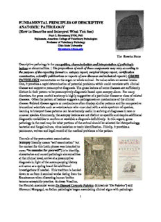
The Rosetta Stone
Descriptive pathology is the recognition, characterization and interpretation of pathologic
lesions or abnormalities. [ The proportions of each of these components may vary according to
the purpose of the reporting format i.e. autopsy report, surgical biopsy report, certification
examination, scientific publications or reports of new diseases and technical reports]. GROSS
PATHOLOGY concentrates on the organ or whole animal. Its value exists on several levels.
One, it provides a rapid determination of potential problems which could correlate with clinical
disease and support a presumptive diagnosis. The gross lesions of some diseases are sufficiently
distinct in their pattern to be presumptively diagnostic based upon autopsy alone. For many
disorders, the gross morbid anatomy is highly suggestive of a particular disease or class of related
diseases. Often the pattern of lesions suggests a pathogenesis or mechanisms of the clinical
disease. Related disease agents or mechanisms often display similar patterns and for comparative
biomedical scientists such as veterinarians who must deal with a wide spectrum of species,
learning to interpret these patterns can be extremely useful in arriving at diagnoses in rare or
unusual species. Commonly, the autopsy lesions are not distinct or specific and require additional
diagnostic modalities to confirm or establish a diagnosis definitively. In this regard, gross
pathology is the road map for what portions of the animal should be selected for histopathology,
bacterial and fungal culture, virus isolation or toxin identification. Thirdly, it provides a
permanent, written and legal record of the medical problems of the patient.
The role of the postmortem examination
(autopsy literally means “self examination” but
the context for this Latin phrase was intended to
mean “to examine for yourself”) is to identify,
characterize and record pathologic abnormalities
at the clinical level, arrive at a presumptive
diagnosis in light of the accompanying history
and serve as a spring board for additional
investigations if needed. This tradition is handed
down to us from 2 seminal works dating from the
Renaissance when dissecting human bodies
became acceptable practice. Andreas Vesalius,
the Flemish anatomist wrote De Humani Corporis Fabrica (known as “De Fabrica”) and
Giovanni Morgagni, an Italian pathologist began associating clinical signs with pathologic
1
changes in De Sedibus et Causis Morborum ( called “De Sedibus”). Performance of the autopsy
in veterinary medicine, (frequently called necropsy which means “death examination”) is
unique in its execution when compared to human medicine. Except for clinicians practicing in
training institutions where a resident staff of pathologists performs the autopsies, most private
veterinary practitioners must do the autopsy themselves. Thus, it is important to understand the
fundamentals of the autopsy to get the most out of it. The autopsy is an ephemeral event and
when it’s over, it’s over! This makes the accurate description and interpretation of the autopsy
findings critical because that is what remains in the permanent record as the basis for later
historical, medical and legal review and interpretation. A principal limitation of retrospective
clinical studies is the lack of or a poorly conducted autopsy with inconsistent or incomplete
characterization of the lesions. Although our human counterparts occasionally have the
opportunity to exhume a body for re-examination at a later date that option is rarely open to
veterinary pathologists.
Contrary to popular belief, the definitive diagnosis of disease is NOT always made at the
microscopic level. The microscopic examination evaluates a tiny fraction of the organs and
tissues. These histopathologic findings are integrated with the results of the autopsy and
interpreted in light of the signalment, history, clinical appearance and laboratory tests to arrive at
a diagnosis. What every histopathologist wants to know above all else is “What was the
gross appearance?” This is easy when the pathologist performs both the autopsy and
histopathologic examinations. However, the culture of veterinary medicine is such that most of
the time the autopsy is performed by a clinician and the tissues are sent to a pathologist (often far
away) for histopathology. To acquire precision and efficiency in the pathologic diagnosis of
disease and maximize the results of the autopsy, the clinician must accurately observe, describe,
record, interpret and communicate the results of the autopsy to the pathologist. When this is
done properly, pathologists can maximize their contribution to the process often by providing not
just a summary of the lesions they observe (called a “morphologic diagnosis”) but making a
specific clinical disease diagnosis. All too often veterinary students do not understand this.
Although trained to perform the autopsy, clinicians do not assign sufficient importance to it or
perform it often enough to reduce it to reliable practice and get the most out of it when needed.
The things the pathologist most wants to know, indeed frequently needs to know, for
definitive interpretation, are often out of his/her control. As a result communication between
clinician and pathologist breaks down leaving all parties dissatisfied, including the pet owners
who depend upon correct diagnosis for prognosis, decision making, grief counseling, absolution
of guilt and closure. Clinicians are frustrated because they are getting only morphologic
diagnoses which they cannot relate to clinical diseases instead of definitive clinical disease
diagnoses which they understand and know how to treat or explain to their clients. Pathologists
become frustrated because they know they can provide more but lack the information essential to
make a definitive diagnosis.
A post graduate continuing education effort for clinicians to rediscover the autopsy skills learned
in professional school would help considerably in this matter. An organized, systematic
approach to observation, description and interpretation of the lesions and changes found at
autopsy would provide clinicians with the means to fill in the gaps, deliver the information
2
pathologists most want to know, improve communication with the pathologist, assist in their own
knowledge of the case, provide immediate feedback to interested animal owners and maximize
the yield from the autopsy. In addition, the rules of gross pathology are easy to learn and apply
and with minimal guidance, the practitioner can improve their interpretation skills with feedback
from the pathologist. In short, continued learning and the satisfaction of professional growth at
minimal expense could result in the increased information flow to clients and better management
of clinical disease problems.
Although this discussion focuses upon interpreting autopsy lesions, the principals are exactly
the same for surgical pathology specimens. The skills learned here should be applied to the
submission of surgical biopsies with markedly improved results in a clinical setting where the
patient is still alive and treatment or therapeutic options may be significantly impacted by poor
communication. Often the difference between my making a specific disease diagnosis and a
generic pathologic process is determined by what the gross lesion looked like, where the lesion
occurred, how the lesions were distributed, the age, breed, sex of the patient and what the
clinician’s working diagnosis was. As incredible as it may seem, I frequently get surgical
biopsies in which I am not even told the species or the location of the sample much less the other
mentioned information. Some clinicians mistakenly believe you should not “bias” pathologists
by sharing information with them. Nothing could be further from the truth. I have often said that
if the object of the autopsy/biopsy is to see if you can fool me, I will tell you right now, You can
fool me. Easily! If, on the other hand, the object is to acquire a rapid, accurate, specific
diagnosis, share what you have observed and what you think with the pathologist. Do you think
physicians withhold information from human pathologists? Imagine what the malpractice
attorneys would do with THAT bit of information if they did? The most common reason biopsies
are returned to clinicians in some human medical centers is insufficient information. With the
heightened interest in animal rights and the uncontrolled proliferation of attorneys, it’s only a
matter of time before medical liability issues with pets are going to be tested in court. Delayed
treatment or misdiagnosis due to poor communication resulting in an “untoward outcome” may
elicit legal action in the near future.
Description versus Interpretation
Gross observations are objective and should never change. Interpretations are subjective, open to
discussion and can be altered retrospectively. Interpretation is always a guess but with proper
training and experience it can be a very, very good guess. Interpretation is what pathologists get
paid to do but they should always justify their interpretation by accurate descriptions.
Interpretation:
Description:
Describe first, then
interpret Diffuse pulmonary
The lung was diffuse dark
congestion and
red to plum colored,
edema
heavy, wet and
foamy fluid freely ran
Diffuse acute
from the cut surface.
interstitial
It felt firmer tha n 3
pneumonia
normal and not
crepitant
ELEMENTS OF THE GROSS DESCRIPTION
The interpretation of gross lesions begins with the observation and
characterization of the abnormal findings. To do this one must know what
attributes are important and what they mean when observed. The following is a
list of those attributes which I believe form the elements of a good gross
description of abnormalities seen at autopsy, or surgical biopsy for that matter:
1. DISTRIBUTION – What is the spatial arrangement of lesions?
2. DEMARCATION – How clearly set off from the adjacent normal
tissue is it?
3. CONTOUR - Are the lesions raised, flat or depressed
4. SHAPE – Do the lesions have a geometric shape?
5. COLOR - “Well, what color is it”? Pick one.
6. SIZE – absolute vs. relative; lesion, whole organ, paired organs
7. TEXTURE - What does the cut surface look like? Amorphous or solid
8. CONSISTENCY - How does the lesion feel? Fluid, soft, firm, hard
9. SPECIAL FEATURES - Odor, sound
10. EXTENT – How much of the organ or tissue is affected?
11. CHRONICITY
A subjective assessment; usually difficult to be precise.
What is the” real time” definition of chronic?
Often the following terms are used:
Acute – a change that can be produced in seconds to hours? Days?
Subacute – what are the gross criteria?
Acute and chronic – a mixture of acute and ongoing changes
Chronic-active – same as acute and chronic
Chronic - at least days.
4
GROSS HALLMARKS OF CHRONICITY
a. Proliferation of cells takes time. Thus evidence of cellular proliferation makes it
likely the lesion is chronic
b. Deposition of stroma or extra cellular matrix – Fibrosis, hyperostosis or Periosteal
new bone (PNB)
c. Size- large changes in organs either way (increased or decreased) imply a passage of
time. Hypertrophy, atrophy.
You can see fibrovascular proliferation (granulation tissue) microscopically as early as 3-
5 days after the damage. So if you can see it grossly, the lesion must be at least 1-2 wks
old. Is that chronic?
THE LOGIC TEST: Ask yourself the question…
“Is the lesion…seconds, minutes, hours, days, weeks, months or years old?”
Arrive at an estimated range of the logical time you think it took to make the
lesion you observed.
12. SEVERITY
Also a subjective assessment; a sliding scale that
is often relative and variable among pathologists.
Often a 5 point scale used:
Minimal
Mild
Moderate
Marked
Severe
A. DISTRIBUTION = the spatial arrangement of the lesions in the organ or tissue.
Lesions or abnormalities may occur with distinct
distribution patterns which when recognized are
clues to the disease process and assist in
estimating severity or significance of the observed
findings. Among the attributes of gross lesions,
distribution is one of the most important and
should be included in nearly every morphologic
diagnosis. In many dermatopathies, the lesion
distribution is the key to diagnosis. Distribution
may reflect pathogenesis.
5
1. Random - the lesion occurs without reference to architecture or relationship to
particular organ or tissue structures. The scattering of abscesses or tumors
through out a lung or liver may be random.
2. Symmetrical - a pattern with some degree of organization is apparent in the
abnormality.
Linear pattern
Suggests some
organization
Bilateral lesions may imply a metabolic or systemic disorder affecting a certain group of
related cells present in distinctly separated areas of the organ or tissue or the same
location in paired organs. Bilateral symmetrical flank alopecia in dogs;
polioencephalomalacia in ruminant brains. A symmetrical or organized appearance
occurs when a pathologic process highlights or outlines a certain anatomic or
physiological subunit; often it outlines a vascular unit or circulatory bed; airways in
the lung, portal tracts in the liver, glomeruli or tubules in kidney.
3. Focal - a single defined lesion on a
background which is either normal or itself
abnormal but containing or reflecting a different
process than present in the focal lesion. i.e. a
6
solitary tumor in the liver; an abscess in a congested lung. One of the easiest
distributions to see but with little discriminative power as to pathogenesis.
4. Multifocal - more than a single discrete lesion on a background.
Highly variable; several to many lesions. This may require a further
characterization such multifocal widespread to emphasize that the
abnormality was not just 2 or 3 foci. “Multifocality often suggests an
embolic shower” Also easy to appreciate.
5. Multifocal to Coalescing - when there are
many lesions present that may appear to be
growing together or fusing. This reflects an
active process which is expanding or not
otherwise contained or limited by the host
defense mechanisms.
6. Miliary - a special case of”multifocal” in which there are numerous tiny foci present that
are too numerous to count The miliary pattern stems from miliarius which is the
7
Pale = necrosis or Red = blood.
exudate Platelets, DIC
Vascular damage
Latin word for millet seed. Miliary distributions may reflect a recent embolic shower to the
organ or tissue. Because the lesions are small, the implication is that the event is recent.
7. Segmental - a well defined portion or segment of the tissue is abnormal; sometimes
a distinct geometric shape. This distribution implies that the pathologic process is
restricted by anatomic or physiologic factors and so occupies a discrete portion.
*Segmental lesions often define a vascular bed.
Distal “segment” of the tail
8. Diffuse - Everything in the frame of reference is abnormal or affected. This
generally implies greater severity and therefore significance than focal or multifocal
lesions. Also because it takes time for a process to affect the entire tissue, diffuse
lesions MAY (but not necessarily) be more chronic or older. **Often difficult to
appreciate because there is no contrast with normal.
8
Everything in the field of view looks pretty much the same
THE PARADOX OF DESCRIPTIVE PATHOLOGY
“The most severe lesion may be the easiest to overlook
because there is no normal for contrast”
Is this lung normal or no t? You have to
remember what the normal post mortem variation
in color of the lung is to properly interpret your
observation
B. DEMARCATION =The degree to which the lesion is set off or defined from the
adjacent tissue.
Iridium-rich
Cretaceous-Tertiary
boundary ~ extinction
of the dinosaurs
9
1. Well demarcated – The boundary between normal and abnormal is
abrupt, discrete and easily seen.
Implication = The lesion
represents a different tissue
or is well contained or
separated from the adjacent
normal tissue. Tumors,
abscesses with capsules,
pus, a rim of necrosis often
produce a well demarcated
lesion.
2. Poorly Demarcated – The
boundary between normal and
abnormal is blurred or not
easily seen.
Implication = the lesion
and adjacent tissue may be
similar; the process
gradually infiltrates into
normal or may be poorly
contained.
C. CONTOUR = the degree to which the lesion is elevated or depressed with respect to
the adjacent tissue
10
Description:The Rosetta Stone training institutions where a resident staff of pathologists performs the autopsies, most private .. Foreign Material – plant material, parasites. 2. toxin, genetic defect (deletion, recessive gene, mutation etc) or metabolic . even if you're a positive genius humiliation and r

