Table Of ContentAdvanced Techniques in
Biological Electron Microscopy II
Specific Ultrastructural Probes
Edited by
J.
K. Koehler
With Contributions by
S. S. Brown R. W. Davis P. Echlin J. Ferguson
S. W. Hui J. K. Koehler J. A. Lake
G. 1. Nicolson D. F. Parsons W. D. Perkins
J.-P. Revel
With 105 Figures
Springer-Verlag Berlin Heidelberg New York 1978
JAMES K. KOEHLER, Ph. D.
University of Washington
Department of Biological Structures
School of Medicine
Seattle, W A 98195/USA
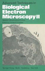
The cover illustration shows a rat lymphocyte from bone marrow sequentially labeled
with tritiated uridine (nuclear silver grains) and hemocyanin conjugated to anti Ig
(hemocyanin on cell surface, arrows).
ISBN-13: 978-3-642-66811-1 e-ISBN-13: 978-3-642-66809-8
DOl: 10.1007/978-3-642-66809-8
This work IS subJect to copynght All nghts are reserved, whether the whole or part of the material IS concern
ed, specIfically those of translanon, reprinting, re-use of illustrations, broadcastIng, reproduction by photoco
pymg machIne or similar means, and storage in data banks. Under § 54 of the German Copyright Law, where
copIes are made for other than pnvate use, a fee is payable to the publisher, the amount of the fee to be deter-
mined by agreement with the publisher.
© by Springer-Verlag BerlIn Heidelberg 1978
Softcover reprint of the hardcover 1st edition 1978
The use of registered names, trademarks, etc in this publication does not imply, even in the absence of a spe
CIfic statement, that such names are exempt from the relevant protectIve laws and regulations and therefore
free for general use
TypesettIng, offsetprinting and bookbinding: Konrad Triltsch, Graphischer Betneb, Wiuzburg
213113130-543210
Preface
The use of the term "advanced" in the title of this book is somewhat ar
bitrary and very much relative with respect to time. Many techniques which
were considered at the "cutting edge" of ultrastructural methodology just a
few years ago are now rou tin ely used in numerous laboratories. One could
cite freeze-fracture, cryothin sectioning, or indeed most of the field of scan
ning electron microscopy as concrete examples. Thus the use of the term "ad
vanced techniques" must be interpreted with regard to the present state of
the art, and is useful only in informing the potential reader that this volume
is not a primer to be used as an initial introduction into basic biological elec
tron microscopy. Many excellent volumes have filled that niche in the past
few years, and it is not intended that this modest book be a complete com
pendium of the field. Furthermore, any limited selection of papers on advanc
ed techniques necessarily reflects the preferences and arbitrary whims of the
editor, thereby excluding many equally important procedures which the
knowledgeable reader will readily identify.
The first volume of this series appeared approximately five years ago and
illustrated techniques which were thought to represent advanced and yet ba
sically morphological methods for gaining increased ultrastructural informa
tion from biological specimens. The present volume, on the other hand,
stresses techniques which provide specific physicochemical data on the speci
mens in addition to the structural information. The future importance of
fine structural investigations would seem to be strongly dependent on our
abiliry to adopt such methods to help answer some of the outstanding ques
tions in cell biology.
Three of the contributions of this volume deal with the use of various
surface probes having specific affinities for cell surface molecules. The utiliza
tion of labeled lectins to explore cell surfaces has grown explosively during
the past few years, and is discussed in the chapter by G. L. NICOLSON. Anti
body labels have also become a very powerful specific probe of surface activi
J.
ties and are treated in the chapter by W. D. PERKINS and K. KOEHLER.
J.
S. S. BROWN and P. REVEL deal with the use of these and other rypes of la
beled probes in the scanning electron microscope. The use of these methods has
already contributed some very important new information to improve our
concepts of antigen processing, cell fusion, and exocytosis, to mention just a
few examples. The area of scanning electron microscopy is further represent
ed by the chapter on low temperature preparations contributed by P. ECH-
IV Preface
LIN. The localization of highly labile substances in biological materials con
tinues to be a nagging problem and considerable promise for a solution seems
to lie in such cryo techniques. Another procedure that will be useful for
such investigations involves the use of hydrated specimens which is discussed
from the point of view of electron diffraction as well as electron microscopic
studies by S. W. HUI and D. PARSONS. Finally, in the area of nucleoprotein
fine structure research, two of the most elegant procedures are presented in
J. J.
chapters by LAKE, FERGUSON and R. W. DAVIS. The first of these is an
account of the detailed structure of the ribosome as determined from nascent
antibody. labeling studies, and the second describes the use of heteroduplex
analysis in genetic mapping research.
My sincere appreciation is extended to the authors of these chapters for
their time-consuming efforts and patience, to the staff of the Springer-Verlag
for their dedication to the production of the highest quality scientific publi
cations, and to Ms. DoRIS RINGER for help in the editorial processing of
the manuscripts.
Seattle, February 1978 JAMES K. KOEHLER
Contents
Ultrastructural Localization of Lectin Receptors
G. L. NICOLSON (With 7 Figures)
A. Introduction. . . . 1
B. Purification of Lectins 2
C. Purification of Markers 2
I. Hemocyanin. . 7
II. Ferri tin. . . . 7
1. Cadmium Sulfate Crystallization 7
2. Ammonium Sulfate Precipitation 8
3. Ultracentrifugation . 8
III. Peroxidase . . . . . . . . . 8
IV. Mannan-Iron Complex. . . . . 9
D. Synthesis of Probes and Labeling Techniques 9
I. Lectin-Hemocyanin . . . . 9
1. Labeling Procedures. . . 10
2. Platinum-Carbon Replicas 10
II. Lectin-Ferritin Conjugates 12
1. One-Step Glutaraldehyde Coupling 12
2. Two-Step Glutaraldehyde Method 15
3. Labeling Procedures. . . . . . 15
III. Lectin-Peroxidase Techniques . . . 20
1. Two-Step Lectin-Peroxidase Labeling 20
2. Single-Step Lectin-Peroxidase Labeling 24
IV. Lectin-Polysaccharide-Iron Complexes 26
1. Lectin-Dextran-Iron Complexes 26
2. Lectin-Mannan-Iron Complexes 26
References . . . . . . . . . . . . . 27
Antibody-labeling Techniques
J.
W. D. PERKINS and K. KOEHLER (With 8 Figures)
A. Rationale . . . . . . . . . . . . 39
B. Antibody Labels . . . . . . . . . 40
C. Methods for Coupling Label to Antibody 41
VI Contents
I. One-Step Method 41
II. Two-Step Method 41
D. Iodination of Antibody 42
I. Iodination of Antibody with Chloramine T 43
II. 125I-Labeled Antibody for Transmission Electron Microscopy 43
III. Lactoperoxidase Labeling of Antibody 45
IV. Antibody Labeling with an Acylating Agent 45
E. Hemocyanin Label of Antibody ....... 45
I. Purification of Hemocyanin ...... 46
II. Conjugation of Hemocyanin with Antibody 47
F. Reaction of Antibody with Cells . . . . . . . 47
G. Clotting Procedure for Handling Single Cell Suspensions 49
H. Radioautography . . . . . . 51
I. Replica Techniques . . . . . 53
I. Surface Replica Technique 54
II. Freeze-etching Technique . 58
J. Conclusions 58
References . . . . . . . . . . 60
Cell Surface Labeling for the Scanning Electron Microscope
S. S. BROWN andJ.-P. REVEL (With 3 Figures)
A. Introduction. 65
B. Labeling Techniques for the SEM 66
I. The Label 66
II. The Marker. 67
1. Electron-Dense Markers 68
2. Markers Recognizable by Their Shapes 71
3. Cathodoluminescent and Other Markers 72
III. Coupling Label to Marker 73
1. Direct Coupling 73
2. Indirect Coupling 74
3. Purification and Analysis of Conjugates 74
C. Interpretation of Cell Surface Labeling in the SEM 75
I. Quantitation 76
1. Influence of Valence of the Label 76
2. Stoichiometry of the Binding of Label to Marker 76
3. Influence of the Size of the Marker 77
II. Resolution 77
1. Size of the Marker 77
2. Size of the Label-Marker Complex 78
Contents VII
III. The Sample. . . . . . . . 78
1. Label-Induced Rearrangements. 78
2. Sources of 'False' Labeling 79
3. Types of Samples 80
4. Subsequent Sample Preparation for the SEM 81
D. Summary 81
References . . . . . . . . . . . . . . . . . . 82
Low-Temperature Biological Scanning Electron Microscopy
P. ECHLIN (With 18 Figures)
A. Introduction. . . . . . . . . . . . . . . . . 89
R Low-Temperature Solidification of Cell and Tissue Fluids 90
C. Pre-treatment Before the Cooling Process 91
I. Chemical Fixation 91
II. Artificial Nucleators 92
III. Cryoprotection. . . 92
IV. Embedding Agents . 96
V. Non-chemical Pre-treatment. 99
D. Specimen Cooling. . . . . . . 101
E. Post-freezing Preparative Procedures 105
I. Frozen-dried or Frozen-hydrated 106
II. External Surfaces of Internal Details 109
F. Specimen Transfer. . . . . . 115
G. Low-temperature Specimen Stages 116
H. Specimen Examination 116
I. Conclusions 117
References . . . . . . 118
Quantitative Electron Microscopy of Nucleic Acids
J.
FERGUSON and R. W. DAVIS (With 26 Figures)
A. Introduction. 123
B. Basic Protein Film Method 123
I. Aqueous Technique. 124
II. Formamide Technique 126
III. Reagents. 127
IV. Problems Related to Contrast 128
V. Double-Strand/Single-Strand Distinction and Length Ratios 129
C. Heteroduplex Molecules 130
I. Experimental Procedure 130
VIII Contents
II. Examples. . . . . . . . . . . . . . . . . 132
III. Complications Which May Arise in Constructing and
Examining Heteroduplex Molecules ...... 134
IV. Branch Migration . . . . . . . . . . . . . 135
V. Terminology, Topology, and Stability of Branch Points 137
VI. Diheteroduplexes. . . . . 138
VII. Partial Sequence Homology . 140
VIII. Partial Denaturation Mapping 143
D. Measuring and Error Analysis 143
I. Measurement Procedures . . 143
II. Reference Markers and Orientation 144
III. Error Analysis . . . . . . . . 144
IV. Determination of Number Average Molecular Weight 145
1. DNA Standard . . . . . . . . . . 146
2. Unbiased Sampling of Molecules 146
3. Background Subtraction of Contaminating
DNA Molecules . . . . . . . . . . 146
V. Determination of DNA Concentration by Electron
Microscopy . . . . . . . . 147
E. Artifacts and Topology Considerations 147
I. Flowers . . . . . . 147
II. Lateral Aggregation. . . . . 148
III. Intermolecular Overlap 149
IV. Branch Peelback in Heteroduplex Molecules 150
V. 2: 2 Branch Point Configuration .... 151
VI. Renaturation of Single-Stranded Circular Molecules 151
VII. Topologic Restriction to Renaturation in Linear Molecules
-Renaturation of 'Knotted' DNA 152
F. RNA and Transcription Complexes . 154
I. Techniques for Preparing RNA 154
II. Secondary Structure Maps 156
III. Transcription Complexes. . . 157
IV. Mapping of Complementary RNA Sequences in DNA 158
1. R-Loop Method . . . . . . . . 158
2. Single-Strand Binding Protein Method 160
G. Tagging Methods. . . . 161
I. RNA-Ferritin Tags . . . . . . 161
II. Protein-Ferritin Tags . . . . . 163
III. General Comments and Problems 164
H. Protein-free Spreading . . . . 164
I. Direct Visualization. . . 164
II. Intercalating Dye Method 166
Contents IX
III. Benzyldimethylalkylammonium Chloride Method 166
IV. Other Methods 167
References . . . . . . . . . . . . . . . . . . . 167
Electron Microscopy of Specific Proteins: Three-Dimensional
Mapping of Ribosomal Proteins Using Antibody Labels
J. A. LAKE (With 32 Figures)
A. Introduction. 173
B. Techniques 173
C. Interpretation 179
References 209
Electron Microscopy and Electron Diffraction Studies
on Hydrated Membranes
S. W. HUI and D. F. PARSONS (With 11 Figures)
A. Introduction. . . . . . . . . . . . . 213
B. Operation of Hydration Chamber in an Electron Microscope 216
I. Hydration Chamber. . . . . . . . . . . . 216
1. Principles. . . . . . . . . . . . . . . 216
2. Chambers for Fixed-beam Transmission Electron
Microscope . . . . . . . . . . . . . 217
3. Chambers for Scanning Electron Microscope 218
II. Preparation of Wet Membrane Specimens 218
C. Electron Microscopy 220
I. Dark Field . . . 220
II. Energy Filters . . 223
III. Image Intensifiers 224
D. Electron Diffraction (ED) 224
I. Selective Area Electron Diffraction 225
II. Small-angle Electron Diffraction . 227
III. Phase Transition and Phase Separation in Membranes 229
E. Conclusions and Future Development 231
References . . . . . . . . . . . . . . . . . . . . 232
Subject Index 237
Con tribu tors
BROWN, SUSAN S., Department of Structural Biology, Stanford University
School of Medicine, Stanford CA 94305, USA
DAVIS, RONALD W., Department of Biochemistry, Stanford Universiry
School of Medicine, Stanford CA 94305, USA
ECHLIN, PATRICK, The Botany School, Downing Street, Cambridge,
CB2 3EA, Great Britain
FERGUSON, JILL, Department of Biochemistry, University of Washington,
Seattle, W A 98195, USA
HUI, SEK-WEN, Electron Optics Laboratory, Biophysics Department, Ros
well Park Memorial Institute, Elm Street, Buffalo, NY 14263, USA
KOEHLER, JAMES K., Universiry of Washington, Department of Biological
Structures, Seattle, W A 98195, USA
LAKE, JAMES A., Molecular Biology Institute and Department of Biology,
Universiry of California, Los Angeles, CA 90024, USA
NICOLSON, GARTH 1., Department of Developmental and Cell Biology, Uni
versity of California, Irvine, CA 92717, USA
PARSONS, DoNALD F., Electron Optics Lab., Division of Laboratories and
Research, New York State Department of Health, Empire State Plaza,
Albany, NY 12201, USA
PERKINS, WILLIAM D., Department of Biological Structure, University of
Washington, School of Medicine, Seattle, W A 98195, USA
REVEL, JEAN-PAUL, Division of Biology 156-29, California Institute of Tech
nology, Pasadena, CA 91125, USA

