Table Of ContentX-ray pushing of a mechanical microswing
A. Siria1,2, M. S. Rodrigues3, O. Dhez3, W. Schwartz1,2, G. Torricelli4, S. LeDenmat3, N. Rochat2,
G. Auvert2,5, O. Bikondoa3, T. H. Metzger3, D. Wermeille3, R. Felici3, F. Comin3 and J. Chevrier1
1 Institut N´eel, CNRS-Universit´e Joseph Fourier Grenoble, BP 166 38042 Grenoble Cedex 9, France
2 CEA-LETI, 17 Avenue des Martyrs 38054 Grenoble Cedex 9, France
3 ESRF, 6 rue Jules Horowitz 38043 Grenoble Cedex 9, France
4 Department of Physics and Astronomy, University of Leicester, University Road Leicester LE1 7RH, England
5 STMicroelectronics, 850 rue Jean Monnet, 38926 Crolles, France
(Dated: February 3, 2008)
Nanoelectromechanical Systems (NEMS) are
8 among the best candidates to measure interac-
0
tionsatnanoscale[1,2,3,4,5,6],especiallywhen
0
resonating oscillators are used with high quality
2
factor [7, 8]. Despite many efforts [9, 10], ef-
n
ficient and easy actuation in NEMS remains an
a
issue [11]. The mechanism that we propose, ther-
J
mally mediated Center Of Mass (COM) displace-
2
ments, represents a new actuation scheme for
2
NEMS and MEMS. To demonstrate this scheme
] efficiency we show how mechanical nanodisplace-
t
e ments of a MEMS is triggered using modulated
d X-ray microbeams. The MEMS is a microswing
- constituted by a Ge microcrystal attached to a
s
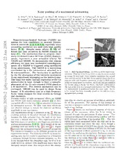
FIG.1: Set-Updescription. (a)Schemaoftheexperimen-
n Si microcantilever. The interaction is mediated
talsetup. BluerayistheX-raybeamontheGemicro-crystal
i by the Ge absorption of the intensity modulated
. at orange Si lever end. Grey cylinder represents the optical
s X-ray microbeam impinging on the microcrystal.
c fiberandtheredrayisthelaserbeamusedtodetectthelever
The small but finite thermal expansion of the Ge
i position with sub-Angstrom precision. (b and c) SEM image
ys microcrystal is large enough to force a nanodis- oftheGecubesgluedonSilevers. In(b)thecutandsoldered
h placement of the Ge microcrystal COM glued on Ge crystal using a Focus Ion Beam, has been positioned at
p a Si microlever. The inverse mechanism can be theendoftheleverinasymmetricalposition(i.e. TheCOM
[ envisaged: MEMS can be used to shape X-ray Ge microcrystal is positioned below the lever end). In (c)
beams. A Si microlever can be a high frequency a Ge crystal has been manually glued on the side in a very
2 X-ray beam chopper for time studies in biology asymmetrical position.
v
and chemistry.
0
5 Previous studies of light mechanical effects on MEMS
0 and NEMS have shown radiation pressure [12] or ther- tion edge we observe an increase of oscillation amplitude
2 malswitcheffectinthelever[13]asactuationmechanism for all the geometries. The amount of this increase as
1. for mechanical systems. We show that these effects are functionofgeometryandmicroswingcharacteristicisthe
0 not effective enough to induce the observed oscillation basis of our findings.
8 amplitude in our experiments. Figure 3 reports the mechanical response of the can-
0 The experimental set-up is presented in fig. 1. The mi- tilever at the resonance, when the X-ray energy is
v: croswing position is measured through the interference scanned through the germanium K-edge energy. The
i between the light reflected from the back of the lever mechanical response of the microswing matches well the
X and from a cleaved fiber end. This experimental set-up XAS reference spectrum of germanium [14] . The two
r has been shown to produce a sub-Angstrom precision in curves have been normalised below the edge and in the
a
positionmeasurements[5, 6, 13]. SEMimagesofthemi- continuum atomic part above the edge. Even though a
croswings used are shown in fig. 1(b) and 1(c). Figure mechanical detection of EXAFS has already been shown
2presentsthemechanicalresponsemeasuredaroundthe [15],thisisthefirsttimeutilisingaMEMS.Onthebasis
first resonance frequency ω for different geometries and of the experimental evidence presented in fig. 2, we can
0
experimentalsetups. TheintensityoftheX-raybeamim- identify the oscillation driving force. Radiation pressure
pacting onto the Ge crystal is modulated at a frequency can be ruled out as the oscillation is the same whatever
ω sweepingthroughtheleverresonantfrequencyω . For the direction of the beam (figure 2(c) and 2(d)) with re-
0
X-rayenergiesbelowtheabsorptionedge, theleverisal- spect to the oscillation direction.
ready forced to oscillate with amplitudes larger than the From fig. 2 and 3 it is evident that the oscillation am-
thermally induced noise. For energies above the absorp- plitude is a function of the absorption cross section. In-
deed, its spectrum follows well the absorption coefficient
2
reabsorbed. The overall number of photons I that in-
h
duce the temperature increase is then:
I =I (1−TE )(1−wE TEf ) (1)
h 0 Ge Ge wGe
where I is the incoming intensity, TE the Ge trans-
0 Ge
mission coefficient, function of the photon energy and
sample thickness, and wE the fluorescence yield. TEf
Ge wGe
is the rate of fluorence at energy E which escape from
f
the sample. This last coefficient is dependent on sample
thickness.
AtenergiesbelowtheGe-Kedge,themainprocessisthe
Auger electron production [16]. Most of the absorbed
photons contribute then to the heating because of short
mean free path (few nanometer) of the Auger electrons
FIG. 2: Measured resonance curve of the first oscil-
and their cascades. At energies higher than the Ge K
lating mode for all levers. In red the X-ray beam energy
edgetheabsorbedphotonsgeneratefluorescence,Coster-
issetbelowtheK1sedge(E =11.07keV),inblackitisset
ph
at the K1s edge (E =11.103keV). Kronig and Auger electrons.
ph
(a) Uncoated cantilever (k = 0.025 N/m, Q = 86, I = The decreased amplitude of the XAFS peak and oscilla-
0
7.41010ph/s)withGeblockgluedonthesideandX-raybeam tionsaftertheK-edgewithrespecttothereferencespec-
parallel to the oscillation direction. tra are due to this intrinsic self-absorption effect. In ta-
(b) Coated cantilever (k = 0.027 N/m, Q = 60, I0 = ble I the absorbed photon flux Ih is calculated for two
3.51010ph/s)withGeblockgluedonthesideandX-raybeam lever/crystal configurations, for two X-ray beam direc-
parallel to the oscillation direction..
tions, and for coated and uncoated levers. The ratio of
(c) Uncoated cantilever (k = 0.135 N/m, Q = 75, I =
0 themeasuredoscillationamplitudesx(ω )aboveandbe-
2.41012 ph/s) with Ge block glued below and X-ray beam 0
lowK-edgeenergyisconsistentwiththeratioofabsorbed
parallel to the oscillation direction.
photons.
(d) Same than (c) with X-ray beam perpendicular to the os-
The temperature increase ∆T can be calculated taking
cillation direction.
into account the overall energy deposited in the crystal
and the heat flow throughthe lever (cooling by radiation
for germanium bulk. We explored then the hypothesis and convection is here negligible). The absorbed power
that the absorbed energy is promptly turned into heat W is then:
leading to a temperature increase dependent on how the
heat is evacuated. W =CT˙(t)+G(T(t)−T0) (2)
As a first approximation, the number of photons that W (cid:16) (cid:17)
contributes to a temperature increase, is the difference T(t)=T0+ G 1−e−CGt (3)
between the number of absorbed photons and fluores-
cence photons that escape from the sample considering
that the fluorescence emission can be photoelectrically l =23µm Uncoated Coated
0
E T w T I x(ω ) [nm] x(ω ) [nm]
ph Ge Ge wGe h 0 0
11.07 0.72 0 - 0.28 I 1.053 0.113
0
11.103 0.083 0.535 0.83 0.51 I 1.902 0.199
0
Ratio 1.82 1.81 1.76
l =43µm Paral. Perp.
0
E T w T I x(ω ) x(ω )
ph Ge Ge wGe h 0 0
11.07 0.54 0 - 0.47 I 4.066 4.713
0
11.103 0.009 0.535 0.33 0.63 I 5.898 6.959
0
Ratio 1.34 1.47 1.48
TABLE I: Correspondance between absorbed photon
and oscillation amplitude for different levers and ge-
ometries. The top part presents the comparison , for a
coatedandanuncoatedleverwithanasymmetricalgeometry
likeinFig. 1(c). TheX-raybeamishereparalleltothedirec-
FIG. 3: Cantilever oscillation amplitude in function tion of oscillation. The second part presents the comparison
of beam energy. We show in black, our experimental data foranuncoatedleverwithasymmetricgeometry. TheX-ray
and in red, the handbook reference EXAFS spectrum at Ge beam is here either parallel or perpendicular to the direction
K edge. of oscillation
3
where T is the ambient temperature and T(t) the block by the thermal fluctuations of the lever position and is
0
temperature as function of time. ∆T(ω) is then x (k T)=1.6pm.
i B
The system in fig. 1(b) presents a much more symmetri-
W 1
cal geometry. l value in this case must be smaller than
∆T(ω) = (4) 0
G (1+ωτ) the one in the case of fig. 1(c), but it is not easily mea-
C surable. A rough estimate of the residual misalignment
τ = (5)
G between the COM of Ge microcrystal and the Si lever
axis is the incertitude in the FIB positioning device that
ω is the beam chopper frequency, τ is the ratio between is about 1 µm.
thethermalcapacityoftheGeblockandthethermalcon- Thedistancel thatbestfitsthedatawhileallotherpa-
0
ductivityoftheSilever. Fortheuncoatedandthecoated rametersareknownis1.5µmwhichisindeedclosetothe
lever of (fig. 2(a) and 2(b)) the experimental conditions precisionoftheFIBmotor. Thecomparisonbetweenthe
are nearly identical whereas the oscillation amplitude is model (equation 7) and the measured oscillation is pre-
10 times larger in 2(a) than in 2(b). This difference can sented in figure 4 as the excitation frequency is swept
be described using those last equations. The presence of from 100 Hz to 2500 Hz. The agreement further estab-
the metallic coating increases the thermal conductivity lishesthatthethermallyforceddisplacementoftheCOM
G of the system and therefore induces a consequential is at the origin of the observed lever oscillation equiped
decrease of ∆T compared to the uncoated lever. with the Ge crystal. Results for all configurations are
Howeverthisdescriptioncannotexplainthedifferenceof then consistently explained using this single actuation
the amplitude of oscillation between the (fig. 2(a) and mechanism.
2(c)). The oscillation amplitude in fig. 2(c) is 3 times The MEMS actuation mechanism shown here can be ex-
larger than in 2(c) against a photon flux 40 times bigger tended to NEMS actuation. Considering a Si lever of
and an absorption rate 25% higher because of the differ- 1×0.1×0.1 µm and a Ge block of 100×100×100 nm
ence in Ge-crystal dimensions. The difference in the me- with thermal conductivity of G = 3.7·10−8 W/K and
chanicalpropertiesofthecantilever(2(a)k =0.025N/m, thermal capacity of C = 1.7·10−15 J/K [17] leads, ac-
2(c)k =0.135N/m)cannotexplainsuchalargedescrep- cording to Eq. 4, to a substantial temperature increase
ancy. However, the position of the Ge crystal and this at a frequency in the MHz regime, typical for the reso-
symmetry with respect to the lever has not been consid- nanceofsuchaNEMS.Ifa1µW laserbeamisabsorbed
ered. This remark is essential to the conclusion of this in this Ge block, the induced thermal expansion will be
paper. We show that the thermally induced change in several pm. As NEMS with lateral size close to 100nm
the distance between the Ge crystal COM and the lever canexhibitqualityfactorsof1000,aforcedCOMoscilla-
axis controls the system dynamics
The thermally induced change in the COM position is
determined by :
∆l(ω) = l α∆T(ω) (6)
0
l is the distance between the block COM and the lever
0
axis and α the linear thermal expansion coefficient.
Forasimple1Dmechanicaloscillatortheoscillationam-
plitude is given by:
(cid:112)
x(ω) = x (ω) |ψ(ω)|2
i
(cid:118)
(cid:117) ω2Q2
= xi(ω)(cid:117)(cid:116)Q2(ω2−0ω2)2+ω2 (7)
ω2 0
0
ψ(ω) is the oscillator transfer function. Here, x (ω) cor-
i
responds to ∆l(ω).
FIG. 4: Response function of the lever shown in fig-
For the system in fig. 1(c), l =13µm close to half the
0 ure1(b). Blackcurveisthemeasuredamplitudeofthelever
Ge crystal thickness. For an intensity I =7.4 1010ph/s
0 oscillation as the beam intensity is modulated from 100 Hz
thetemperatureincreaseisfoundtobe∆T(ω )=0.24K.
0 to2500Hz. Redcurveisthecalculatedexpressionusingex-
Using α = 5.910−6K−1, according to equation 6, the
Ge perimental parameters characteristics of the X-ray beam, of
induced COM displacement is ∆l(ω0) = 19pm. Using the X-ray absorption around the Ge K edge and of the lever
equation 7, with the measured quality factor of 86 and described as a single mode oscillator. The error bar in red
the amplitude at the resonance of 1.9nm, the COM dis- curve has been determined using the Brownian motion. Red
placement is found to be ∆l(ω ) = 22pm which is con- curvecalculationinvolvesthemisalignementoftheGemicro-
0
sistent with the value calculated from equation 6. The crystal on the Si lever as the single adjustable parameter. In
error bar on the measured lever position is determined the inset a zoom on the resonant peak is presented.
4
tionwithamplitudeofseveralpmcanresultatresonance position of metal. The cubic Ge crystal is 43 µm thick.
in a nanometric NEMS oscillation amplitude. This is far It is soldered to the Si lever in a symmetrical position.
above the thermally induced fluctuations of NEMS posi- The lever is a standard Silicon AFM cantilever whose
tion. ThisstrategyofNEMSexcitationcanbecompared dimensions are 350x35x2 µm3. This lever has no metal-
to photothermal actuation based on thermally induced lic coating. The second Ge microcrystal is about 23 µm
strain [18]. The essential difference is in the origin of the thick (fig. 1(c)). It has been manually glued on the
NEMS displacement. This origin is, in the mechanism side of the cantilever in a very asymmetrical position.
that we propose, a strain-free thermally induced change Forasymmetricallymountedcrystals,twotypesoflevers
in mass spatial distribution in asymmetric structure. have been used: one bare and another with a metallic
Due to limited performances of current X-ray choppers, coating.
MEMS is here operated close to 1 kHz i.e. at very low
frequencies. TheuseofMEMSasSisinglecrystalmicro-
oscillators can provide X-ray choppers at much higher
frequencies. We have already produced experimental ev- B. BEAMLINE SET-UP
idence of such an effect at 13 kHz. Using diffraction, Si
single crystal MEMS appear as a good candidate for the
TheexperienceswereperformedattheEuropeanSyn-
high frequency manipulation of X-ray microbeam. This
chrotron Radiation Facility (ESRF). The beamlines in-
could offer new tools either to change phase X-ray wave-
volved were the Anomalous Scattering Beamline (ID01)
front,ortoproducerapidlymodulatedintensityofX-ray
andSurfaceScienceX-RayDiffraction(SXRD)Beamline
beams that are so important in real time studies of fast
(ID03). The Anomalous Scattering Beamline ID01 has
dynamical processes in chemistry and in biology [19].
been designed to combine small and wide angle X-ray
scattering techniques with anomalous diffraction. The
radiation from the undulators can be tuned from 2.5 to
I. METHODS
40keVwithaSi(111)doublecrystalmonochromator(en-
ergy resolution ∆E/E ≈ 10−4). Focusing is achieved
A. MICROSWING REALISATION byusingberylliumCompoundRefractiveLenses(CRLs)
[20]. The effective focus size is ≈4×6µm2 with ≈1010
ThefirstGemicrocrystalinfig. 1(b)hasbeendirectly photons/second on the focal spot. At the SXRD beam-
cut to Ge wafer by a Focus Ion Beam (FIB). In order line the photons were tuned at the Ge K edge using a
to fabricate the micro-oscillator, a cubic like germanium liquid nitrogen cooled monolithic double crystal Si (111)
crystal has been etched from a (1 0 0) oriented germa- monochromator. The beam was focused at the sample
niumwaferusingtheFIBStrata400fromFEI.Then,the by a Kirkpatrick-Baez (KB) mirror system located 43 m
cube has been extracted using a motorized tungsten tip from the photon source. The beam size at the sample,
andplacedclosetothesiliconcantileverend. Finally,the 1 m from the KB system, is ≈ 3×5µm2 with ≈ 1012
cubewasweldedtothecantileverusinglocalizedFIBde- photons/second on the focal spot.
[1] M. Li, H. X. Tang and M. L. Roukes, Ultra-sensitive in fluid, Nature 446, 1066-1069 (2007)
NEMS-based cantilevers for sensing, scanned probe and [8] S. S. Verbridge, J. M. Parpia, R. B. Reichenbach, L. M.
veryhigh-frequencyapplications,NatureNanotechnology Bellan and H. G. Craighead, High Quality Factor reso-
2, 114-120 (2007) nanceatroomtemperaturewithnanostringsunderhigh
[2] K. L. Ekinci, X. M. H. Huang and M. L. Roukes, Ul- tensile stress, J. Appl. Phys 99, 124304 (2006)
trasensitivenanoelectromechanicalmassdetection,Appl. [9] I.Bargatin,I.KozinskiandM.L.Roukes,Efficientelec-
Phys. Lett. 84, 4469-4471 (2004) trothermalactuationofmultiplemodesofhighfrequency
[3] Y. T. Yang, C. Callegari, X. L. Feng, K. L. Ekinci and nanoelectromechanical resonator, Appl. Phys. Lett. 90,
M. L. Roukes, Zeptogram-Scale Nanomechanical Mass 0931161-0931163 (2007)
Sensing, Nano Lett. 6, 583-586 (2006) [10] S. Srinivasan, J. Hiller, B. Kabius and O. Au-
[4] H.J.MaminandD.Rugar,Sub-attonewtonforcedetec- ciello, Piezoelectric/ultrananocrystalline diamond het-
tion at millikelvin temperatures, Appl. Phys. Lett. 79, erostructures for high-performance multifunctional mi-
3358-3360 (2001) cro/nanoelectromechanical systems, Appl. Phys. Lett.
[5] A.N.ClelandandM.L.Roukes,Ananometre-scaleme- 90, 134101-134103 (2007)
chanical electrometer, Nature 392, 160-162 (1998) [11] A.N.ClelandandM.L.Roukes,Nanoelectromechanical
[6] D.Rugar,R.Budakian,H.J.MaminandW.Chui,Single systems face the future, Phys. World 14, 25-31 (2001)
spin detection by magnetic resonance force microscopy, [12] O. Arcizet, P. F. Cohadon, T. Briant, M. Pinard and
Nature 430, 329-332 (2004) A. Heidmann, Radiation-pressure cooling and optome-
[7] T.P.Burg,M.Godin,S.M.Knudsen,W.Shen,G.Carl- chanical instability of a micromirror, Nature 444, 71-74
son, J. S. Foster, K. Babcock and S. R. Manalis, Weigh- (2006)
ing of biomolecules, single cells and single nanoparticul [13] C. H. Metzger and K. Karrai, Cavity cooling of a mi-
5
crolever, Nature 432, 1002-1005 (2004)
[14] EXAFS-Data-Base-(Standards) URL http://www.
nsls.bnl. gov/beamlines/x18b/data.htm
[15] T. Masujima, H. Kawata, M. Kataka, H. Shiwaku, H.
Yoshida, H. Imai, T. Toyoda, T. Sano, M. Nomura, A.
Iida, K. Kobayashi and M. Ando, Instrumentation for
thephotoacusticEXAFSmethod,Rev. Sci. Instrum 60,
2522-2524 (1989)
[16] M. O. Krause, Atomic radiative and radiationless yields
forKandLshells,J. Phys. Chem. Ref. Data 8,307-327
(1979)
[17] D. Li, Y. Wu, P. Kim, L. Shi, P. Yang and A. Majum-
dar,Thermalconductivityofindividualsiliconnanowires,
Appl. Phys. Lett. 83, 2934-2936 (2003)
[18] D. R Koenig, C. Metzger, S. Camerer and J. P Kot-
thaus, Non-linear operation of nanomechanical systems
combining photothermal excitation and magneto-motive
detection, Nanotechnology 17, 5260-5263 (2006)
[19] M. Wulff, F. Schotte, G. Naylor, D. Bourgeois, K. Mof-
fat and G. Mourou, Time-resolved structures of macro-
molecules at the ESRF: single-pulse Laue diffraction,
stroboscopic data collection and femtosecond flash pho-
tolysis,NuclearInstrumentsandMethodsinPhysicsRe-
search A 398, 69-84 (1997)
[20] A. Snigirev, V. Kohn, I. Snigireva and B. Lengerer, A
compoudrefractivelensforfocusinghigh-energyX-rays,
Nature 384, 49-51 (1996)

