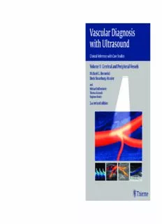
Vascular diagnosis with ultrasound/ 1, Cerebral and peripheral vessels: 49 tables. / Michael G. Hennerici; Doris Neuerburg-Heusler PDF
Preview Vascular diagnosis with ultrasound/ 1, Cerebral and peripheral vessels: 49 tables. / Michael G. Hennerici; Doris Neuerburg-Heusler
Hennerici_Vasc_103832 11.10.2005 11:48 Uhr Seite 1 N H Vascular Diagnosis e e u n e n r e b r u i with Ultrasound c r g i - H e u s l Vascular Diagnosis with Ultrasound e Clinical Reference with Case Studies r One of the most powerful non-invasive diagnostic tools available to clinicians, vascular V Volume 1: Cerebral and Peripheral Vessels a ultrasound technology has undergone dramatic changes in recent years, creating exciting s new diagnostic and treatment options for a growing array of disorders. The completely c u Michael G. Hennerici revised new edition of Vascular Diagnosis of Ultrasound offers the most comprehensive l a and up-to-date information on the broad spectrum of vascular ultrasound applications. r Doris Neuerburg-Heusler D Now in two volumes—1: Cerebral and Peripheral Vessels and 2: Abdominal and Renal ia Vessels—this edition retains the accessible design and logical structure of the first edition g and n and adds a new team of expert contributors and more than 500 illustrations and images. o Michael Daffertshofer s i Volume 1 features: s Thomas Karasch w Stephen Meairs i • Comprehensive coverage of vascular ultrasonography in the arteries and veins t h of the cerebral circulation and the peripheral upper and lower limb circulation. U 2nd revised edition • Systematic presentation of all available ultrasound technologies—including l t continuous and pulsed-wave Doppler mode, B-mode, conventional and color- ra coded duplex analysis in frequency and amplitude power modes. s o u • In-depth chapters on anatomy and physiology, normal and abnormal findings, n test accuracy and sensitivity. d , • Helpful comparison with data from other diagnostic methods (e.g., conventional V and noninvasive MR angiography) used in each region, as well as authoritative o l assessments of recent developments in ultrasound technology, such as tissue u m perfusion studies, 3D and 4D imaging, contrast enhancement, and microbubble applications, and their diagnostic and therapeutic implications (sonothrombolysis e 1 and thrombus micro-fragmentation). • A revised section with many new, challenging case studies for review both for the novice and the expert in the field. Praise for the first edition of Vascular Diagnosis with Ultrasound: “A huge asset to the library of the ultrasound department…will be the first source of information for any vascular ultrasound questions. Highly recommended.”—RAD Magazine “An exhaustive review of vascular diagnosis… I recommend it to any physician in training or practice… vivid illustrations and high-quality color images complement the excellent 2 n discussions.”—Journal of Vascular Surgery d e d i t i o n The Americas ISBN 3-13-103832-2 (GTV) Rest of World ISBN 1-58890-144-0 (TNY) ISBN 1-58890-144-0 ISBN 3-13-103832-2 ,!7IB5I8-jabeeg! ,!7ID1D1-adidcg! www.thieme.com I II III Vascular Diagnosis with Ultrasound Clinical Reference with Case Studies Volume 1: Cerebral and Peripheral Vessels Michael G. Hennerici, M.D., Ph.D. Doris Neuerburg-Heusler, M.D. Professor and Chairman Former Director of the Department Department of Neurology of Noninvasive Diagnostics University of Heidelberg Aggertalklinik, Engelskirchen Klinikum Mannheim Cologne, Germany Mannheim, Germany With contributions by Michael Daffertshofer, M.D, Ph.D. Thomas Karasch, M.D. Stephen Meairs, M.D., Ph.D. Associate Professor Department of Associate Professor Department of Neurology Cardiology/Angiology Department of Neurology University of Heidelberg University of Cologne University of Heidelberg Klinikum Mannheim Cologne, Germany Klinikum Mannheim Mannheim, Germany Mannheim, Germany 2nd revised edition 532 illustrations 49 tables Thieme Stuttgart · New York IV LibraryofCongressCataloging-in-PublicationData Important note: Medicine is an ever-changing science Hennerici,M.(Michael)G. undergoing continual development. Research and clini- [GefässdiagnostikmitUltraschall.English] calexperiencearecontinuallyexpandingourknowledge, Vasculardiagnosiswithultrasound:clinicalreferences in particular our knowledge of proper treatment and with case studies / Michael G. Hennerici, Doris Neuer- drugtherapy.Insofarasthisbookmentionsanydosageor burg-Heusler ; with contributions by Michael Dafferts- application, readers may rest assured that the authors, hofer,ThomasKarasch,StephenMeairs. editors,andpublishershavemadeeveryefforttoensure p.cm. thatsuchreferencesareinaccordancewiththestateof Rev.translationof:GefässdiagnostikmitUltraschall.2. knowledgeatthetimeofproductionofthebook. Aufl.1995. Nevertheless,thisdoesnotinvolve,imply,orexpressany Includesbibliographicalreferencesandindex. guaranteeorresponsibilityonthepartofthepublishers ISBN 3−13-103832−2 (alk. paper) −- ISBN 1−58890- inrespecttoanydosageinstructionsandformsofappli- 144−0(alk.paper) cationsstatedinthebook.Everyuserisrequestedtoex- 1.Blood-vessels-−Ultrasonic imaging. 2.Blood-vessels-− aminecarefullythemanufacturers’leafletsaccompany- Ultrasonicimaging-−Casestudies. ingeachdrugandtocheck,ifnecessaryinconsultation [DNLM: 1.Vascular Diseases-−ultrasonography.WG withaphysicianorspecialist,whetherthedosagesched- 500 H515g 2005a]I. Neuerburg-Heusler, Doris. II. Hen- ules mentioned therein or the contraindications stated nerici, M. (Michael) Gefässdiagnostik mit Ultraschall. bythemanufacturersdifferfromthestatementsmadein EnglishIII.Title. the present book. Such examination is particularly im- RC691.6.U47H46132005 portant with drugs that are either rarely used or have 616.1’307543-−dc22 been newly released on the market. Every dosage 2005010530 scheduleoreveryformofapplicationusedisentirelyat the user’s own risk and responsibility. The authors and publishersrequesteveryusertoreporttothepublishers 1stEnglishedition1998 anydiscrepanciesorinaccuraciesnoticed.Iferrorsinthis workarefoundafterpublication,erratawillbepostedat www.thieme.comontheproductdescriptionpage. Someoftheproductnames,patents,andregisteredde- signsreferredtointhisbookareinfactregisteredtrade- marksorproprietarynameseventhoughspecificrefer- encetothisfactisnotalwaysmadeinthetext.Therefore, the appearance of a name without designation as pro- prietaryisnottobeconstruedasarepresentationbythe publisherthatitisinthepublicdomain. ©1998,2006GeorgThiemeVerlag, Thisbook,includingallpartsthereof,islegallyprotected Rüdigerstrasse14,70469Stuttgart,Germany bycopyright.Anyuse,exploitation,orcommercialization http://www.thieme.de outside the narrow limits set by copyright legislation, ThiemeNewYork,333SeventhAvenue, without the publisher’s consent, is illegal and liable to NewYork,NY10001USA prosecution.Thisappliesinparticulartophotostatrepro- http://www.thieme.com duction,copying,mimeographing,preparationofmicro- films,andelectronicdataprocessingandstorage. Coverdesign:MartinaBerge,Erbach TypesettingbyprimustypeHurlerGmbH,Notzingen PrintedinGermanybyGrammlich,Pliezhausen ISBN3-13-103832-2(GTV) ISBN1-58890-144-0(TNY) 12345 V Preface Diagnosisandtreatmentofvasculardiseaseshavemade largestudytrialsandbasicresearch.Italsointroduced tremendousprogresssincetheintroductionofnon-in- experimental techniques just investigated in research vasiveultrasoundtechnologiesinclinicalpracticeinthe laboratories at that time (harmonic imaging, 3-D and early1970s.Todayultrasoundiscapableofmonitoring 4-Dimaging,flowvolumemeasurements,intra-arterial the early silent stages of atherogenesis as well as the and interventional applications, functional and moni- morphological features of advanced atherosclerosis toringstudies,etc.).AthirdGermaneditionwaspub- duringitsprogressionandregressioninallmajorarter- lished very soon thereafter in 1999; due to the fast ies of the body. The ability of ultrasound to visualize developmentofultrasound,thiseditionincludednew botharterialandvenousthrombusformationhasbeen dataonimagingofperfusionindifferentorgansandin- expanded to include detection and quantification of vestigationsofsmallvesselnetworksbymeansofnew circulating microemboli. Vascular ultrasound studies echocontrastmedia. areimportanttoolsinindividualpatientdiagnosisand Sincetheamountofmaterialtobeincludedinthe duringfollow-up,andnotablyinrandomized,prospec- newsecondEnglisheditionof"VascularDiagnosiswith tive clinical trials that serve to strengthen evidence- Ultrasound"hasbeengrowingsorapidlyandbecause basedmedicineforclinicalpractice.Therecentuseof wedidnotwanttomissthebroadspectrumofvascular state-of-the-artultrasoundmonitoringinsuchstudies ultrasoundapplicationsaddressingalargecommunity hashelpedtointroducenewpathwaysforthemanage- ofbothinvestigatorsandclinicians(whoarenowcon- ment of our patients, both in modern industrialized fronted with results of ultrasound studies from societies and in developing countries. Increasing age throughout the world using different instruments, combinedwiththeunfortunatelyremaininghighprev- differenttechnologies,anddifferenteconomicrestric- alencesandincidencesofmyocardialinfarction,stroke, tions),wedecidedtocompletelyrevisethetextandsplit andperipheralvasculardiseaseunderlinetheneedfor thebookintotwovolumes.Thefirstvolumedealswith earlyidentificationofsubjectsathighriskfortreatment cerebral and peripheral vessels. The second volume inayetasymptomaticperiodwithpotentialtherapeutic addresses the abdominal vessels, small parts vessels, impactinprimaryprevention,andformeanstoimprove andtopicssuchastumorvascularization,whichhasbe- monitoring for secondary prevention. Ultrasound will come a fascinating area of vascular ultrasound. The continuetoplayamajorroleinrealizingtheseimpor- completerevisionincludesbothdiagnosticandthera- tantgoalsofpreventivemedicine;asitisnon-invasive, peuticaspectsofultrasoundthataddress,particularlyin alwaysavailable,andeconomicallyviable,ithasdistinct theatlas,theadvantagesofrecentneurosonologictech- advantagesoverallothervasculardiagnostictools. nologies and applications. This edition also provides The first German edition of this book (1988) was newinformationonultrasoundproceduresinperiph- welcomed by both novice and experienced sono- eral angiology, reflecting the results of recent ultra- graphers due to its strict illustrative composition. A sound studies that provide normative parameters for majorrevisionpublishedin1994wasnecessaryforin- therapeuticdecisions.Newerhorizonsinvascularsono- clusionofrapidlydevelopingtechnologies.Thisedition graphy,suchassonothrombolysisinstroke,peripheral firstintroducedacollectionofindividualcasehistories artery thrombolysis using intra-arterial ultrasound to andvascularfindings,whichbecameofmajorinterestto causemicro-fragmentationofthrombi,andmolecular manyreadersofthebook.Theatlasillustratedfromthe imaging for non-invasive detection of diseases using very beginning the combined use of ultrasound tech- microbubblestargetedtodisease-associatedmolecular nologieswithclinicaldataandothermethodsappliedin structures,areonlyafewofthefascinatingperspectives clinicalpracticeandwasparticularlyusefulandwellac- addressedinthisnewvolume. cepted both by the specialized collaborations in the Thefirstvolumeofthisnewsecondeditionhascon- vascularlaboratory,aswellasbycliniciansunfamiliar tinuedtobewrittenbyM.G.HennericiandD.Neuer- withspecificultrasoundtestsbutusingtheirreports.In burg-Heusler. However, this would not have been 1997whenthefirstEnglisheditionwaspublished,rare possiblewithoutthehelpandcooperationofnewco- findingsandspecificproblemswereonlyoccasionally workers, such as Michael Daffertshofer and Stephen includedandselectedrepetitionswereintentionalfor MeairsfromtheDepartmentofNeurology,Universityof educationalpurposes.Thiseditionwasalsoacomplete Heidelberg,UniversitätsklinikumMannheimandongo- revisionofthesecondGermanmonographandincluded ingcooperationwithThomasKarasch,Departmentof advancedultrasoundapplicationsaswellasdatafrom InternalMedicine(Cardiology/Angiology),Universityof VI Preface Cologne,whoseexpertisewassowellacknowledgedal- volume,withthesupportofourteamfromThiemepub- ready for previous editions. We are grateful to our lishers headed by Cliff Bergman. We are especially vasculartechniciansandsecretarieswhohelpeduswith grateful for the continuous support of our families, the enormous work on the manuscript, including the Marion Hennerici, and the late Helmut Neuerburg; continuous preparation of the abundant literature in withouttheirloveandsupportwewouldhavefailed. thisexcitinganddevelopingfield.Ittookuslongerthan expected but we finally managed to finish the first MichaelG.HennericiandDorisNeuerburg-Heusler VII Table of Contents 1 Physics and Technology of Ultrasound 1 BasicUltrasoundPhysics ..................... 1 Time-IntervalHistograms ................... 13 PropertiesofWaves ........................... 1 SpectralAnalysis ........................... 13 Reflection ..................................... 1 Three-DimensionalUltrasound ................. 14 Refraction..................................... 2 ImageAcquisition .......................... 15 ScatteringandDiffraction ...................... 2 MonitoringSpatialPosition............... 15 Attenuation ................................... 2 ComputerizedMotor-DrivenSystems ..... 15 IntensityandPower ........................... 2 SemiregistrationTechniques ............. 15 TheDopplerEffect............................. 2 POMScanheadTracking.................. 16 ReconstructionTechniques .................. 16 UltrasoundTechnology ....................... 3 VisualizationTechniques .................... 16 DopplerSystems .............................. 3 Four-DimensionalApplications ................. 17 Continuous-WaveDopplerSonography ...... 3 Pulsed-WaveDopplerSonography ........... 3 ContrastImaging ............................. 18 B-ModeImaging............................... 6 NonlinearCharacteristicsofMicrobubbles ...... 18 ColorDopplerFlowImaging ................... 7 MicrobubbleDestruction....................... 18 PowerDopplerImaging ........................ 9 LowMechanicalIndexImaging................. 19 DuplexSystems ............................... 9 StimulatedAcousticEmission .................. 19 CompoundImaging............................ 10 HarmonicImaging............................. 19 B-FlowImaging ............................... 10 PulseInversionHarmonicImaging ............. 20 Transducers ................................... 11 PowerPulseInversionImaging ................. 20 SignalAnalysis ................................ 12 MicrovascularImaging......................... 20 AudioSignalAnalysis ....................... 12 MicrobubbleRefillKinetics..................... 21 Zero-CrossingCounter ...................... 12 References .................................... 22 2 Extracranial Cerebral Arteries 27 Examination.................................. 27 AnatomyandFindings ...................... 36 SpecialEquipmentandDocumentation ......... 27 Anatomy ................................ 36 ExaminationConditions ....................... 27 Findings................................. 37 PatientandExaminer ....................... 27 Evaluation .................................... 37 ConductingtheExamination ................ 27 SourcesofError ............................... 37 ExaminationSequence ...................... 29 NeckArteries ................................. 38 OrbitalArteries................................ 29 Principle ................................... 38 NeckArteries ................................. 29 AnatomyandFindings ...................... 39 CommonCarotidArtery .................. 29 CarotidSystem .......................... 39 InternalandExternalCarotidArteries..... 31 Anatomy ............................. 39 Vertebral−SubclavianSystem ............. 33 CommonCarotidArtery ............... 39 VertebralArtery ...................... 33 InternalCarotidArtery ................ 41 SubclavianArtery ..................... 36 ExternalCarotidArtery................ 43 InnominateArtery(Brachiocephalic Vertebral−SubclavianSystem ............. 44 Trunk) .................................. 36 Anatomy ............................. 44 ThoracicAorta ........................... 36 VertebralArtery ...................... 44 SubclavianArtery ..................... 45 NormalFindings.............................. 36 InnominateArtery−AorticArch ........... 46 OrbitalArteries................................ 36 Evaluation ................................. 46 Principle ................................... 36 VIII TableofContents SourcesofError ............................ 47 SequentialIpsilateralStenosesinthe UltrasoundIncidentAngle................ 47 CarotidSystem ....................... 72 CardiacArrhythmia ...................... 47 MultivesselDisease ................... 73 VascularWidth .......................... 47 CarotidArteryDissection .............. 73 AnomaliesintheVascularCourse......... 47 PostoperativeFindings ................ 73 VenousSuperimposition ................. 47 PostinterventionalFindings(Stenting) . 73 Echogenicity............................. 48 SourcesofError ............................ 73 Postintervention ......................... 48 DiagnosticEffectiveness..................... 74 DiagnosticEffectiveness..................... 50 VertebralArterySystem ....................... 75 Principle ................................... 75 PathologicalFindings ......................... 52 Findings ................................... 75 OrbitalArteries................................ 52 IncreasedFlowVelocities.............. 76 Principle ................................... 52 DecreasedFlowVelocities ............. 76 Findings ................................... 52 SloshPhenomenon.................... 77 Evaluation ................................. 53 IntermediateBloodFlow .............. 77 SourcesofError ............................ 53 AbsentSignal ......................... 77 DiagnosticEffectiveness..................... 54 RetrogradeBloodFlow ................ 78 CarotidSystem ................................ 55 Evaluation ................................. 78 Principle ................................... 55 SourcesofError ............................ 78 Findings ................................... 62 DiagnosticEffectiveness..................... 79 CommonCarotidArtery ............... 62 Supra-AorticSystem ........................... 80 InternalCarotidArtery ................ 65 Principle ................................... 80 ExternalCarotidArtery................ 69 Findings ................................... 80 Evaluation ................................. 71 SubclavianArtery ..................... 80 VascularDegenerationandAthero- ObstructionProximaltotheOrigin genesis ............................... 71 oftheVertebralArtery ................ 81 InternalCarotidArteryStenosis........ 71 ObstructionDistaltotheOriginofthe InternalCarotidArteryOcclusion ...... 71 VertebralArtery ...................... 81 CarotidBifurcationStenosis ........... 72 InnominateArtery .................... 81 InternalCarotidArteryOcclusionand AorticArch ........................... 81 IpsilateralExternalCarotidArtery Evaluation ................................. 82 Stenosis .............................. 72 SourcesofError ............................ 82 ExternalCarotidArteryOcclusionand DiagnosticEffectiveness..................... 82 InternalCarotidArteryStenosis........ 72 References .................................... 82 3 Intracranial Cerebral Arteries 89 Examination.................................. 89 CO ReactivityTest.................... 97 2 SpecialEquipmentandDocumentation ......... 89 AutoregulationTests .................. 98 ExaminationConditions ....................... 90 VasoneuralCoupling .................. 101 PatientandExaminer ....................... 90 PosteriorCerebralArteryStimulation .. 101 ConductingtheExamination ................ 91 MiddleCerebralArteryStimulation .... 102 UltrasoundApplicationandProbePosition ... 91 Techniques ........................... 103 TranstemporalUltrasound............. 91 TranscranialMonitoring(TCM) ........ 103 TransorbitalUltrasound ............... 91 UltrasoundContrastAdministration.... 104 TransnuchalUltrasound ............... 92 NormalFindings.............................. 105 OphthalmicArteryandInternalCarotid Principle ...................................... 105 Artery(Siphon) .......................... 92 AnatomyandFindings ......................... 106 MiddleCerebralArtery(MCA) ............ 92 CarotidSiphon—OphthalmicArtery ....... 106 AnteriorCerebralArtery(ACA) ........... 94 Anatomy ............................. 106 IntracranialInternalCarotidArtery Findings .............................. 106 andT-Junction........................... 94 MiddleCerebralArtery................... 107 PosteriorCerebralArtery(PCA) ........... 94 Anatomy ............................. 107 VertebralArtery(VA) .................... 94 Findings .............................. 107 BasilarArtery(BA) ....................... 95 AnteriorCerebralArteryandAnterior FunctionalTests ............................ 96 CommunicatingArtery ................... 108 CompressionTests .................... 96 Anatomy ............................. 108 VasomotorReactivityTests ............ 97 TableofContents IX Findings .............................. 108 Evaluation .................................... 121 NormalValues ........................ 109 DilativeArteriopathy ....................... 121 PosteriorCerebralArteryandPosterior CarotidCavernousFistula ................... 121 CommunicatingArtery ................... 109 ArteriovenousMalformation ................ 121 Anatomy ............................. 109 SpasmsandAneurysms ..................... 122 Findings .............................. 109 Extracranial−IntracranialBypass ............. 123 VertebralArteryandBasilarArtery ....... 109 IncreasedIntracranialPressureandCerebral Anatomy ............................. 109 CirculatoryArrest........................... 124 Findings .............................. 109 IntracranialFindingsinStealPhenomena .... 125 FunctionalTests ......................... 110 FunctionalDisturbances .................... 125 CompressionTests .................... 110 Monitoring ................................. 126 VasomotorReactivityTest ............. 110 AcuteStroke.......................... 126 CO ReactivityTests ................... 111 SpontaneousMicroemboli(MES) 2 AutoregulationTests .................. 112 andHigh-IntensitySignals(HITS) ...... 128 VasoneuralCoupling .................. 112 MonitoringDuringInterventional Monitoring ........................... 113 Procedures ........................... 128 Evaluation .................................... 113 Right-to-LeftShuntDetection.......... 128 SourcesofError ............................... 114 TCDandMigraine..................... 129 WeakorAbsentSignals ............... 114 TCDandEpilepsy ..................... 129 AnatomicalReasonsforError .......... 114 SourcesofError ............................... 129 TechnicalArtifacts .................... 114 IncorrectAnatomicIdentification HemodynamicCauses ................. 114 ofFlowAccelerations ................. 129 PathophysiologicalCauses ............. 114 AbsentSignal ......................... 129 IdentificationProblems................ 115 InadequateControl ................... 129 DiagnosticEffectiveness ....................... 115 NormalVariants ...................... 129 SpecialApplications ........................... 129 PathologicalFindings ......................... 115 BrainStructureImaging ............... 129 Principle ...................................... 115 Hemorrhage,MidlineShift ............ 130 Findings ...................................... 117 TissuePerfusion ...................... 130 Stenoses ................................... 117 DiagnosticEffectiveness ....................... 131 Occlusions ................................. 118 References .................................... 132 CollateralizationofExtracranialCarotid ArteryObstructions......................... 120 4 Cerebral Veins 139 Examination.................................. 139 Findings ...................................... 143 SpecialEquipmentandDocumentation ......... 139 ExtracranialVeins .......................... 143 ExaminationConditions ....................... 139 JugularVeinThrombosis ................. 143 PatientandExaminer ....................... 139 EctasiasandAneurysms.................. 143 ConductingtheExamination ................ 139 VenousCongestion ...................... 143 FunctionalTests(ProvokedFlowSignals) .... 139 ArteriovenousFistulas ................... 143 IntracranialVeins........................... 143 NormalFindings.............................. 139 SinusThrombosis ........................ 143 AnatomyandFindings ......................... 139 CavernousSinusFistula .................. 145 ExtracranialVeins—InternalJugularVein .. 141 ArteriovenousMalformations............. 145 IntracranialVeins ........................ 142 VenousMalformations ................... 146 Evaluation .................................... 143 Evaluation .................................... 146 PathologicalFindings ......................... 143 SourcesofError ............................... 146 Principle ...................................... 143 DiagnosticEffectiveness ....................... 146 References .................................... 147
Description: