Table Of ContentUnbiased Stereology
Three-Dimensional Measurement in Microscopy
Second Edition
Unbiased Stereology
Three-Dimensional Measurement in Microscopy
Second Edition
C.V.Howard & M.Reed
Developmental Toxico-Pathology Research Group,
Department of Human Anatomy and Cell Biology,
University of Liverpool, Liverpool, UK
This edition published in the Taylor & Francis e-Library, 2008.
“To purchase your own copy of this or any of Taylor & Francis or Routledge’s
collection of thousands of eBooks please go to www.eBookstore.tandf.co.uk.”
© Garland Science/BIOS Scientific Publishers, 2005
First edition published 1998
Second edition published 2005
All rights reserved. No part of this book may be reprinted or reproduced or utilized in any
form or by any electronic, mechanical, or other means, now known or hereafter invented,
including photocopying and recording, or in any information storage or retrieval system,
without permission in writing from the publishers.
A CIP catalogue record for this book is available from the British Library.
ISBN 0-203-00639-9 Master e-book ISBN
ISBN 1 85996 0898 (Print Edition)
Garland Science/BIOS Scientific Publishers
4 Park Square, Milton Park, Abingdon, Oxon, OX14 4RN, UK and
270 Madison Avenue, New York, NY 10016, USA
World Wide Web home page: www.garlandscience.com
Garland Science/BIOS Scientific Publishers is a member of the Taylor & Francis Group.
Distributed in the USA by
Fulfilment Center
Taylor & Francis
10650 Toebben Drive
Independence, KY 41051, USA
Toll Free Tel: +1 800 634 7064; E-mail: [email protected]
Distributed in Canada by
Taylor & Francis
74 Rolark Drive
Scarborough, Ontario MIR 4G2, Canada
Toll Free Tel.: +1 877 226 2237; E-mail: [email protected]
Distributed in the rest of the world by
Thomson Publishing Services
Cheriton House
North Way
Andover, Hampshire SP10 5BE, UK
Tel.: +44 (0)1264 332424; E-mail: [email protected]
Library of Congress Cataloging-in-Publication Data
Howard, Vyvyan.
Unbiased stereology/C.V.Howard, M.Reed.—[2nd ed.].
p. cm.—(Advanced methods)
Includes bibliographical references and index.
ISBN 1-85996-089-8
1. Stereology. 2. Microstructure-Measurement. I. Reed, M.G.(Matt G.) II. Title
III. Series.
Q175.H86 2004
5022.822–dc22 2004015841
Production Editor: Catherine Jones
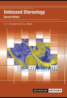
Pseudocolored image of neurons from Layer II of rat neocortex.
Image provided by Dr Gesa Staats de Yanes.
Contents
Abbreviations ix
Preface to the first edition xi
Preface to the second edition xiii
Acknowledgments xiv
Dedication xv
Safety xvi
Foreword—L.Wolpert xvii
1. Concepts 1
1.1 Sampling and bias 2
1.2 Why unbiasedness is desirable 3
1.3 Sources of bias in microscopy 4
Sampling bias 5
Systematic bias 5
1.4 The hierarchical nature of microscopical investigations 6
1.5 Stereology—geometrical quantification in 3D 7
1.6 Think in three dimensions—a macroscopic analogy by
thought experiment 7
Points probe volume 8
Lines probe surface 8
Planes probe length 8
Volumes probe number 9
1.7 Dimensions and sectioning 9
1.8 Geometrical probes 11
1.9 Ratios and densities 12
1.10 The reference space 12
2. Random sampling and random geometry 17
2.1 Stereology and randomness 17
2.2 Two simple experiments 18
2.3 Take a sweet, any sweet 19
2.4 A uniform random (UR) sample 20
2.5 A single UR point in 1D, 2D and 3D 21
2.6 Repeat the process… 23
2.7 A systematic random sample in 2D and 3D 26
2.8 Random geometry 26
2.9 Randomizing directions—isotropy in the plane 30
vi Contents
3. Estimation of reference volume using the Cavalieri method 35
Exercise 3.1 43
Exercise 3.2 44
Exercise 3.3 45
Exercise 3.4 47
Exercise 3.5 50
4. Estimation of component volume and volume fraction 53
4.1 Estimation of volume fraction 53
4.2 Estimation of total volume of a defined component 55
Exercise 4.1 62
Exercise 4.2 63
Exercise 4.3 64
5. Number estimation 65
5.1 Some useful ‘definitions’ 66
5.2 Some ‘non-definitions’ 66
5.3 A cautionary tale 66
5.4 On the right road 68
5.5 Continuous scanning of a plane 68
5.6 The disector principle 69
5.7 From theory to practise 71
5.8 Implementation of the physical disector 74
5.9 Typical sampling regime for the physical disector 77
5.10 Optical section ‘scanning’ methods—the unbiased
brick and optical disector 78
5.11 The unbiased brick-counting rule 82
5.12 Application of the optical disector counting rule 82
5.13 Typical sampling regime for the optical disector 83
5.14 Direct estimation of number—the fractionator 85
5.15 A 2D example of the f ractionator principle 85
5.16 The multi-stage fractionator 87
5.17 The optical fractionator 87
5.18 Some special designs for counting in 3D 89
The single section or ‘cheating’ disector! 89
The double disector 90
The ‘molecular’ or ‘golden’ disector 91
Counting closed space curves—Terminal bronchial
ducts in lung 92
Counting complex shaped objects in 3D and
connectivity estimation 93
Exercise 5.1 94
Exercise 5.2 96
Exercise 5.3 98
Exercise 5.4 99
6. Estimation of total surface area and surface density 103
6.1 Estimation of surface density 104
6.2 Random directions and orientations in 3D space 105
6.3 Generating isotropic line probes—1 Vertical sections 108
Contents vii
6.4 Generating isotropic line probes—2 Isotropic sections 110
6.5 Estimation procedure 110
6.6 Examples of vertical sectioning protocols 111
Exercise 6.1 113
Exercise 6.2 117
7. Length estimation 119
7.1 Generation of IUR sections—the orientator and isector 122
Exercise 7.1 125
8. Stereological analysis of layered structures 127
8.1 Stereological ratios 127
8.2 Vertical sections of layered structures 127
8.3 Practical example 129
8.4 Application areas 130
8.5 Worked example of application 130
9. Particle sizing 133
9.1 Step 1—selecting particles 133
9.2 Step 2—measuring the size of the selected particles 134
9.3 The difference between the number-and volume-weighted
distributions of size 134
9.4 Direct estimators of mean particle volume 136
The ‘point-sampled intercept’ (PSI) method 136
The ‘selector’ 137
The ‘nucleator’ 138
9.5 Indirect estimation of mean particle size from Stereological
ratios 141
9.6 Distributions of particle volume 141
Indirect estimation of the second moment of the
number-weighted particle volume distribution 141
Direct estimation of particle volume distributions 142
10. Statistics for stereologists 143
10.1 ‘Quantifying is a committing task’ (Cruz-Orive, 1994) 143
10.2 Preliminary concepts 144
Population 144
Parameter 144
Sampling unit 144
Sample 144
Estimate 145
Estimator 145
Uniform random sample 145
10.3 Unbiased estimates 145
10.4 Elements of good statistical practice 146
10.5 Quantities of interest 148
10.6 Application to a single object 152
10.7 Addition of variances 152
10.8 Two-stage estimation 154
10.9 Calculation of CE for the Cavalieri method 154
10.10 Calculation of the CE of a ratio estimator 157
10.11 Prediction of CE for two-stage estimation 159
viii Contents
10.12 Precision of Cavalieri estimation for single objects 160
Exercise 10.1 162
Exercise 10.2 163
11. Single-object stereology 165
11.1 Introduction 165
11.2 Volume of single objects—arbitrary orientation designs 166
11.3 Surface of single objects—isotropic or vertical orientation
designs 167
The isotropic ‘spatial grid’ 167
The vertical spatial grid 175
11.4 Length estimation in vertical orientation designs 175
Length density 175
Total feature length 177
Exercise 11.1 180
12. ‘Petri-metrics’ 183
12.1 Introduction 183
12.2 Counting methods 183
The 2D fractionator 183
Practical application at the microscope 185
12.3 Length estimation in 2D 187
12.4 Combined length and number estimation 189
Exercise 12.1 191
Exercise 12.2 193
13. Second-order stereology 195
13.1 Introduction 195
13.2 Second-order methods for point patterns 195
13.3 Second-order methods for volumetric features 198
13.4 The covariance estimator 199
13.5 Linear dipole probes 203
13.6 Making sense of covariance 205
13.7 Example of the application of linear dipole probes 207
13.8 Using isotropic rulers to get ‘one-stop stereology’ 208
Appendix A:Practical gadgets for stereology 211
Appendix B: Set of stereological grids 217
Appendix C:Worked answers to exercises 239
Appendix D:Useful addresses 259
References 261
Index 271
Abbreviations
0D zero-dimensional MRI magnetic resonance imaging
1D one-dimensional mean star volume
2D two-dimensional mean number-weighted
3D three-dimensional particle volume
A total area mean surface-weighted
a/f area of frame particle volume
a/p area per point mean volume-weighted
asf area sampling fraction particle volume
AUR arbitrary uniform random N total number
CCD charge-coupled device N numerical density
v
CD-ROM compact disk read-only memory NA numerical aperture
CE coefficient of error NDT non-destructive testing
CV coefficient of variation P point count
(cid:1)x inter-point spacing in horizontal PSI point-sampled intercept
direction Q– disector count
(cid:1)y inter-point spacing in vertical (cid:2) summation
direction S total surface
FSU fundamental sampling unit S surface density
v
h disector height SD standard deviation
hsf height sampling fraction ssf slice or section sampling fraction
I intersection count T inter-plane spacing
IUR isotropic uniform random TVP total vertical projection
L total length UR uniform random
l/p length per point V total volume
L length density V volume density
v v
M linear magnification VUR vertical uniform random
ix

