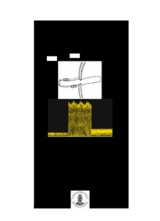
Umbilical vein constriction at the abdominal wall PDF
Preview Umbilical vein constriction at the abdominal wall
Umbilical vein constriction at the abdominal wall An ultrasound study in low risk pregnancies Svein Magne Skulstad Institute of Clinical Medicine Division of Obstetrics and Gynecology University of Bergen and Department of Obstetrics and Gynecology Haukeland University Hospital Bergen, Norway 2005 Umbilical vein constriction at the abdominal wall An ultrasound study in low risk pregnancies Table of contents II Abbreviations IV List of original papers VI Acknowledgements VII Summary IX 1 Introduction 1 1.1 History 1 1.2 Developmental anatomy and physiology 3 1.2.1 Developmental anatomy 3 1.2.2 Developmental physiology 7 1.2.3 Umbilical cord growth 8 1.3 Some aspects of the fetal circulation 10 1.3.1 Cardiac function, output and blood pressure 10 1.3.2 Umbilical venous blood flow 10 1.3.3 Umbilical venous blood flow in fetal disease 12 1.3.4 Umbilical vein pulsation 14 1.3.5 Umbilical vein pulsations in fetal disease 16 1.4 Umbilical cord complications 17 1.5 The ultrasound examination 23 1.5.1 Physics 23 The transabdominal transducer 23 Resolution of the ultrasound image 25 Doppler investigations 26 Continous wave Doppler 28 Pulsed wave Doppler 28 Colour Doppler 28 1.5.2 Safety 29 II 2 Hypothesis, aims and objectives 34 2.1 Hypothesis 34 2.2 Aims and objectives 34 3 Subjects and methods 34 3.1 Selection of subjects 34 3.2 Methods 36 3.2.1 Ultrasound equipment 36 3.2.2 2D–imaging 36 3.2.3 Colour Doppler 36 3.2.4 Doppler velocimetry 37 3.2.5 Data quality assurance 38 3.2.6 Statistical analysis 38 4 Results 39 5 Discussion 42 5.1 Methodological considerations 42 5.1.1 Subjects studied 42 5.1.2 Reproducibility of measurements 42 Ultrasound measurements 42 Weighing of the infant and the placenta 46 5.2 Discussion of results 47 6 Conclusions 51 7 Perspectives 52 8 References 53 9 Research papers I – IV 66 III Abbreviatons 2D–imaging Two-dimensional ultrasound, gray scale ultrasound, AD Anno Domini, after Christ BC Before Christ BW Birthweight BW/PW Birthweight/placental weight ratio CI Confidence intervals DV Ductus venosus ECMUS European Committee for Medical Ultrasound Safety EFSUMB European Federation of Societies for Ultrasound in Medicine and Biology EPOR Erythropoietin receptor gene ET Endothelin FDA Food and Drug Administration (United States government agency) fs Sampling frequency in Doppler IP Index of pulsation of the pressure in the umbilical vein in a mathematical model I Spatial Peak Temporal Average Intensity (mW/cm2); commonly spta used measure of the acoustic energy that the tissues are exposed to IVC Inferior vena cava kHz Kilohertz λ Wavelength LHV Left hepatic vein MHz Megahertz MI Mechanical index; empirical factor correlated to the formation of bubbles in living tissue (cavitation) mm Hg Pressure expressed in terms of the weight of a column of mercury of unit cross section MPa Megapascal; million Newton per square metre (pressure) MRG Multi range gated mW/cm2 Milliwatt per square centimetre (energy disposal in the tissue) IV NO Nitric oxide pCO Partial pressure of carbon dioxide in arterial blood 2 pH Quantitative measure of the acidity or basicity of blood PI Pulsatility index: (systolic velocity – diastolic velocity)/mean velocity pO Partial pressure of oxygen in arterial blood 2 PRF Pulse repetition frequency in Doppler PW Placental weight at birth PW Pulsed wave Doppler RC Reflection coefficient Re Critical Reynolds number when a transition from laminar flow to d turbulence occurs Reynolds number In fluid mechanics: a number that expresses the risk of laminar flow developing into turbulence; it depends on vessel dimension, density, velocity and viscosity of the fluid SD Standard deviation UV Umbilical vein V Maximum time averaged blood velocity in a vessel measured by max pulsed Doppler technique V Maximum time averaged blood velocity in the umbilical vein at max.abd the abdominal wall V Maximum time averaged blood velocity in the umbilical vein in max.cord the cord V Mean time averaged blood velocity in a vessel mean Z Impedance, resistance to pulsatile flow Z Impedance in the ductus venosus DV z–score The distance in standard deviations between the observation and the mean: (observed value–mean)/SD Z Impedance in the umbilical vein UV V List of original papers 1. Skulstad SM, Rasmussen S, Iversen OE, Kiserud T. The development of high venous velocity at the fetal umbilical ring during gestational weeks 11–19. Br J Obstet Gynaecol 2001; 108: 248–253. 2. Skulstad SM, Kiserud T, Rasmussen S. Degree of fetal umbilical venous constriction at the abdominal wall in a low–risk population at 20–40 weeks of gestation. Prenat Diagn 2002; 22: 1022–1027. 3. Skulstad SM, Kiserud T, Rasmussen, S. The effect of vascular constriction on umbilical venous pulsation. Ultrasound Obstet Gynecol 2004; 23: 126–130. 4. Skulstad SM, Rasmussen S, Seglem S, Svanaes RH, Aareskjold HM. The effect of umbilical venous constriction on placental development, cord length and perinatal outcome. Early Human Dev 2004; In press VI Acknowledgments The work presented in this thesis was performed at the Fetal Medicine Unit, Department of Obstetrics and Gynecology, Haukeland University Hospital, University of Bergen during the years 1997 – 2004. Many have supported my efforts, and I extend my sincere gratitude and appreciation to them all. My warmest thoughts go to the late Professor Sverre Stray Pedersen, head of Department of Obstetrics and Gynecology at the time when my study started, for letting me have a part–time position to do research. I will always remember his encouragement and constructive criticism in the initial phase of the study, coloured by his never–ending enthusiasm regarding science and towards younger clinicians and investigators in general. Another important person in the early phases of the study was professor Ole–Erik Iversen, who supported me with his rich experience in the art of publication, and provided a sound scepticism in the discussion of the preliminary results of the study. Most of all my gratitude goes to my tutor, co–writer and friend professor Torvid Kiserud. He introduced me into scientific work and the art of the fetal ultrasound examination, and supervised me throughout the project. His encouragement, uncompromising demands for quality, constructive criticism, time and patience have been invaluable for this work. In addition, his sense for proportions, order and perfection and his extensive knowledge improved my papers and this book. I am greatly indebted to him for his teaching and his never–ending optimistic creativity and readiness for help and support, often far beyond the level expected from a supervisor. Professor Svein Rasmussen, my friend and co–supervisor in this project, generously shared both his devotion to statistics and his ability to encourage in the most humoristic way. He showed the rare ability of promoting both improvement and development in my work, in addition to providing emotional support when needed. This thesis and three of the papers in it were written during my period of employment at The Blood Bank, Haukeland University Hospital. I am deeply obliged to Tor Herwig, the head of the department, for kindly providing all the practical means to accomplish this work. Associate professor Einar Kristoffersen shared his devotion to and experience with Macintosh computers and software. He also generously shared his cognition of the medical presentation. I also thank my office–mate through these years, Anne Bakken, for interesting discussions and unselfish support. The Centre for clinical research, Haukeland University Hospital provided a scholarship for three months, during which I enjoyed a most relaxed and creative scientific atmosphere under the leadership of professor Ernst Omenaas. My gratitude goes to the statisticians, associate professor Geir Egil Eide, and Tore Wentzel–Larsen, for their kind help with part of the statistics in paper IV. I further thank each and all of the staff members at the Unit for Fetal Medicine at the Department of Obstetrics and Gynecology, in particular the midwives for recruiting participators to the study, especially Helga Bognø and Margot Waardal. I also thank my fellow colleagues in the unit for all support, and among them particularly Knut VII Gjelland, for introducing me to the fascinating world of obstetrical ultrasound, and Synnøve Lian Johnsen, for providing valuable comments on this manuscript. Silje Seglem, Ragnhild H. Svanaes and Hanne May Aareskjold are thanked for their contributions to the collection of perinatal data and the writing of paper IV. Kjersti Boge provided excellent secretarial assistance. I owe special thanks to professor emeritus Per Bergsjø for his revision of the language in this book. His knowledge of the art of medical writing has improved this manuscript significantly. I acknowledge the grants from the University of Bergen and the Norwegian Society of Ultrasound in Medicine. A contribution has also been provided by Vingmed Sound, Horten, Norway. Last but not least, my warm thanks goes to my beloved family for tolerating the long working hours necessary. A huge hug belongs to my wife Britt for patiently standing beside me during all these years, and our children for not giving up on their stressed and absent–minded father. I thank Eivor for always allowing me to listen when she was playing the piano and singing, Vegard for dragging me out fishing, Tarjei for always sharing his latest inventions with me, and Runar for wanting me to tell him the fairy tales of H.C. Andersen. Bergen, September 2004 Svein Magne Skulstad VIII Summary The umbilical vein is the only vessel supplying the fetus with blood supplying oxygen and nutrients from the placenta. Case reports indicate that the fetal end of the umbilical cord is susceptible to mechanical complications. Hypothesis: We assume that umbilical ring constriction may affect the umbilical vein and have haemodynamic effects on fetal development and birth. Aims: To describe the occurrence and degree of umbilical venous constriction in low risk–pregnancies. To establish reference ranges. To determine whether such a constriction has a haemodynamic effect, or any effect on fetal development or perinatal outcome. Material and methods: 384 low–risk singleton pregnancies were included in the cross–sectional studies after written consent and ethical approval. 2D–imaging, colour Doppler and pulsed Doppler were used to measure diameters and blood velocity in the umbilical vein before, at or beyond the abdominal wall. All blood flow velocimetry was performed during fetal quiescence. Perinatal outcome was noted. Results: From 13 weeks onwards, after the period of physiological umbilical herniation, umbilical venous constriction was noted in increasing numbers and severity until 19 weeks of gestation (paper I). For the latter half of the pregnancy, during gestational weeks 20–40, the pattern remained constant; 41/191 (21%) had a venous constriction corresponding to a diameter reduction to the half, while the corresponding venous blood velocity increment was ≥300%, and 5% of the fetuses had velocities ≥107 cm/s, which is exceptionally high compared with other blood velocities in the body, whether arterial or venous (paper II). The reproducibility study showed that the measurements of venous blood velocity in the cord and at the umbilical ring had SD of 0.58 and 1.83 cm/s respectively and that the diameter measurements both at the umbilical ring and at the cord had a mean SD of 0.07 mm (paper II). The incidence of umbilical venous pulsation was higher at the umbilical ring in the abdominal wall, 242/279 (87%) than in the cord, 43/198 (22%) or intra–abdominally, 84/277 (30%). When pulsation was observed intra–abdominally, the pulsatility was not different from that at the umbilical ring. The lowest pulsatility was found in the cord vein, where the largest vein diameter was found (paper III). Umbilical venous constriction had a significant negative correlation to the birthweight/placental weight ratio in male but not in female fetuses. Umbilical venous constriction was also associated with and increased length of the cord, but only in female fetuses. Constriction was also associated with Apgar score ≤7 at one minute after birth but not after five, and was not associated with emergency delivery (paper IV). Conclusions: The umbilical ring seams to tighten in the following weeks after the period of physiological herniation, causing an increasing number and degree of umbilical vein constriction. After 19 weeks of gestation, the occurrence of umbilical vein constriction is constant until term. The degree of constriction can be considerable, and in 20% of the fetuses the umbilical vein diameter is ≤50% of that in the cord. We have established reference ranges. Within physiological ranges, i.e. in a low–risk population, such constrictions have a haemodynamic effect leading to increased incidence of pulsations and gender specific effects on fetal, cordal and placental growth. The significant effect on Apgar score at one minute warrants further studies of the effect of extreme constriction on perinatal morbidity. IX 1 Introduction 1.1 History The umbilical cord is the fetal lifeline, which supplies the oxygen and nutrition needed, and transports the waste products from the fetus to the mother. In Japanese folklore the umbilical cord has been called the “The flower stalk of life”, and it has been surrounded by an aura of magic and the supernatural in different cultures all over the world. The legitimacy of the child was determined by the specific gravity of the umbilical cord, the child’s future virility by the size of its stump. Sterility or diminished fecundity was forestalled by tasting its blood or by eating its substance. The umbilical cord was worn as a talisman to protect its bearer from various sicknesses and misfortunes (Spivack 1946). In the Kingdom of Buganda (the largest of the four kingdoms in the western region of Uganda, East Africa), from the dawn of legend up to the middle of the nineteenth century, the jawbone and umbilical cord of the kabaka (king or clan leader) were preserved in a special shrine after his death, and became the means through which his successor could consult him in affairs of state (Welbourn 1964). Since long, the existence and function of the cord has been related to fetal survival. In the Brahmanas, a constituent of the Vedas (ancient Indian literature, compiled around 3500 BC), this description is to find: "The dhamanis (ducts with thick walls equivalent to arteries) in the fetus take their rise from the umbilical cord, thus bringing nourishment from the mother. The embryo is held at the navel. It grows without taking food, that is, there is no effort made on the part of the embryo to take food and no food is specially served to it. The food in its final form is assimilated automatically and directly into the system of the embryo. The child is nourished of its own accord as it were. The mother is not conscious of the nourishment given to the young one below her heart" (Bhargava and Chakrabarti 1995). In Hippocrates’ (460 – 377 BC) treatise on “The Nature of the Child” it was stated that the embryo is nourished by maternal blood, which flows to the fetus and coagulates, forming the embryonic flesh (Hippocrates et al. 1978). According to Hippocrates the first nutrient for the fetus is supplied through the umbilical cord. The umbilical cord is also considered the 1
Description: