The skull of Mesenosaurus romeri, a small varanopseid (Synapsida: Eupelycosauria) from the Upper Permian of the Mezen River basin, northern Russia PDF
Preview The skull of Mesenosaurus romeri, a small varanopseid (Synapsida: Eupelycosauria) from the Upper Permian of the Mezen River basin, northern Russia
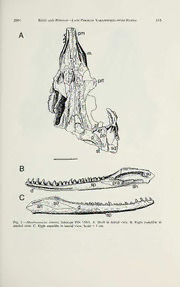
ANNALS OF CARNEGIE MUSEUM VoL. 70, Number 2, Pp. 113-132 24 May 2001 THE SKULL OF MESENOSAURUS ROMERI, A SMALL VARANOPSEID (SYNAPSIDA: EUPELYCOSAURIA) FROM THE UPPER PERMIAN OF THE MEZEN RIVER BASIN, NORTHERN RUSSIA Robert R. Reisz^ Research Associate, Section ofVertebrate Fossils David S Berman Curator, Section ofVertebrate Paleontology Abstract Restudy ofMesenosaurus romeri, based on new and previously described cranial materials from the Upper Permian of the Mezen River basin of northern Russia, confirms its assignment to the synapsid eupelycosaurian family Varanopseidae. Comparisons with othermembers ofthefamily supportapattern of relationship that recognizes two clades: one is composed of Mesenosaurus and Mycterosaurus for which the subfamily designation Myctersaurinae is proposed, and the otherincludes the remainingwell- known varanopseids Elliotsmithia, Varanops, Varanodon, andAerosaurus for which the subfamily des- ignationVaranodontinaeis proposed. AmongthelatePaleozoic synapsids, Varanopseidaehasthelongest fossil record, extending from the end ofthe Carboniferous to well into the Late Permian, andthe widest geographical distribution, including North America, South Africa, and Russia. Key Words: Varanopseidae {Mesenosaurus), Synapsida, Upper Permian, Mezen River Basin, Russia Introduction The Mezen River basin of northern Russia has extensive exposures of Upper Permian sediments along the edges of several rivers, especially the Peza and Kimja rivers, both affluents ofthe Mezen River. These sediments, although visited only sporadically by paleontologists and geologists, have produced the skeletal remains of a diverse assemblage of amniotes, including numerous enigmatic par- areptiles, at least two therapsids, and most interestingly a small synapsid of var- anopseid affinities, Mesenosaurus romeri Efremov (1938). Mesenosaurus romeri was originally described on the basis of a partial skull, but its assignment by Romer and Price (1940) to the synapsid family Varanopseidae was only tentative, owing to the incompleteness of the holotype and only known specimen. A few additional specimens of M. romeri were recovered in the 1950s, which led to its more recent restudy by Ivachnenko (1978). These new specimens, including a poorly preserved articulated skeleton, were the basis of Ivachnenko’s argument that Mesenosaurus was the oldest known archosaur (Evans, 1988; Carroll, 1988). As part of a systematic program of collecting in this area, several new specimens have been recovered and prepared, allowing a reevaluation of the anatomy and phylogenetic relationships of this interesting Paleozoic amniote. This study reaf- firms the assignment of Mesenosaurus to the synapsid family Varanopseidae. Anatomical structures are identified by the following abbreviations: an, angular; bo, basioccipital; co, posterior coronoid; d, dentary; ec, ectopterygoid; ex, exoc- ' Department of Biology, Erindale Campus, University of Toronto, Mississauga, Ontario L5L 1C6, Canada. Submitted 15 December 1999. 113 14 Annals of Carnegie Museum VOL. 70 cipital; f, frontal;j,jugal; 1, lacrimal; m, maxilla; n, nasal; o, opisthotic; p, parietal; pal, palatine; pf, postfrontal; pm, premaxilla; po, postorbital; pp, postparietal; pra, prearticular; prf, prefrontal; ps, parasphenoid; q, quadrate; qj, quadratojugal; sc, sclerotic element; so, supraoccipital; sp, splenial; sq, squamosal; st, supratemporal; su, surangular; t, tabular; v, vomer. Systematic Paleontology Class Amniota Subclass Synapsida Eupelycosauria Kemp, 1982 Family Varanopseidae Romer and Price, 1940 Mycterosaurinae, new subfamily — Definition. Varanopseid synapsids more closely related to Mycterosaurus than to Varanops.— Diagnosis. Small varanopseid synapsids characterized by greatly expandeddor- sal lamina ofmaxilla that contacts the prefrontal, resulting in the anterior shortening of the lacrimal to a level less than half the distance from the orbit to the naris and the loss of a nasal-lacrimal contact; prefrontal with well-developed ventral orbital process that contacts the palatine; paroccipital process ofopisthotic anteroposterally, ratherthan dorsoventrally, expanded oval in cross section; caniniformregionlocated far forward and at a level immediately behind the external naris. Mesenosaurus Efremov, 1938 Type species Mesenosaurus romeri Efremov, 1938 — Revised Diagnosis. Myterosaurine eupelycosaur characterized by the follow- ing cranial features: 1) premaxilla slender and with mate forms a narrowly rect- angular snout in dorsal and ventral views; 2) dorsal process of premaxilla long and forms anterior half of dorsal margin of external naris; 3) deep excavation of the lateral surface of the body of the premaxilla narrows the base of the dorsal process to produce an expanded narial shelf that extends nearly to the snout tip; 4) palatal process of premaxilla with unusually long median suture; 5) well-de- veloped depression on the lateral surface of the nasal that extends posteriorly from the narial border to nearly the anterior end of the prefrontal; 6) slight lateral swelling of the maxilla at the level of the caniniform tooth; 7) short posterior process of the maxilla fails to reach the level of the postorbital bar; 8) first pre- maxillary tooth smaller than the second and third teeth; 9) single, median vo- merine tooth row; 10) postorbital cheek region of skull unusually broad and low, with nearly vertical posterior margin; 11) posterior edge of transverse flange of the ptyergoid is angled slightly anterolaterally from basal articulation; 12) stapes slender, short, and rodlike, with modestly developed footplate and distally ex- panded quadrate process. Mesenosaurus romeri Efremov, 1938 — Holotype. PIN (Paleontological Institute, Russian Academy ofSciences, Mos- cow) 158/1, partial skull and nearly complete right mandible (Fig. 1). — Referred Specimens. PIN 3586/8a, partial skull of large individual (Fig. 2); PIN 3706/11, 3706/ 15, partial skulls ofjuvenile individuals (Fig. 3, 4), SGU (Saratov Geological Institute, Russia) 104 V/1558, partial skull, similar in size to the holotype (Fig. 5). — 2001 Reisz and Berman Late Permian Varanopseid from Russia 1 — Fig. 1. Mesenosaurus romeri, holotype PIN 158/1. A. Skull in dorsal view. B. Right mandible medial view. C. Right mandibe in lateral view. Scale = 1 cm. 116 Annals of Carnegie Museum VOL. 70 — Fig. 2. Mesenosaurus romeri, PIN 3586/8a, anteriorpartoflargestknown skull in ventral anddorsal views. Scale = 1 cm. — Horizon and Locality. Mezen River Basin, northern Russia, Lower Tatarian, Upper Permian. — Diagnosis. Same as for genus. Description and Comparisons — Skull. General. The reconstruction of the skull shown in Figure 6 is a com- posite based primarily on the holotype, relying on the referred specimens only when necessary. Details of the snout region were available only in the larger specimens, whereas the posterior portions of the palate and braincase were well preserved only in the smaller, juvenile specimens. The pattern of the dentition was based largely on more mature specimens. As discussed below in the Discus- sion section, the Varanopseidae is recognized as being divisible into two subfam- ilies: the stem-based Mycterosaurinae is proposed for Mycterosaurus and Mes- enosaurus, whereas all other known genera, Elliotsmithia, Aerosaurus, Varanops, and Varanodon, are included in the proposed stem-based Varanodontinae. With reference to this subdivision, the following description not only compares Mes- enosaurus with Mycterosaurus, but also emphasizes features defining Varanodon- tinae as including taxa which are more closely related to Varanodon than to Mycterosaurus. — 2001 Reisz and Berman Late Permian Varanopseid from Russia 117 — Fig. 3. Mesenosaurus romeri, PIN 3706/11, nearly complete skull ofjuvenile in dorsal, and ventral views. Scale = 1cm. In profile the skull has a low, subrectangular outline, with the occipital margin being normal to the jaw line. The skull outline is very distinctive in dorsal view, with the cheek regions diverging widely to about the level of the postorbital bar, where a sharply defined angulation then orients the temporal margins parallel to the midline. The snout of Mesenosaurus is unique among Paleozoic amniotes in being formed essentially by only the premaxilla and having a narrowly rectangular outline with a truncated tip in dorsal view. The orbit is unusually large and appears anteroposteriorly elongate because of the reduced height of the skull. The dorsal rim ofthe orbit is expanded slightly above the skull table as a rounded ridge. The parapineal foramen is large and located close to the posterior border of the skull roof. The lateral temporal fenestra is tall, occupying nearly the entire height of the skull. A pronounced sculpturing, consisting of a distinct pattern of grooves, covers most of the skull roof. In addition, a welLdeveloped tubercular or nodu- larlike ornamentation extends along the orbital margins of the prefrontal, post- orbital, and jugal. The internal nares are greatly elongated, equaling one-half the length between the snout tip and the anterior margin of the subtemporal fossa, and the palatal surface bears a complex pattern of tooth-bearing ridges. The un- usually slende—r proportions of the lower jaw match those in other varanopseids. Skull Roof. The premaxilla, present in the holotype (Fig. 1) and PIN 4586/8 (Fig. 2), is large and possesses a minimum of five marginal teeth. In both dorsal or ventral views the paired premaxillae form a narrow, abruptly truncated snout tip with nearly parallel lateral margins, giving it a rectangular outline. Just above 118 Annals of Carnegie Museum VOL. 70 — Fig. 4. Mesenosaurus romeri, PIN 3706/15, partial skull in dorsal and occipital views. Scale = 1cm. the marginal tooth row the lateral surface of the body of the premaxilla is deeply excavated so as to produce an expanded narial shelf that extends nearly to the tip of the snout. The excavation results in a narrowing of the base of the dorsal process, which is otherwise well developed in both length and width. The pro- cesses contact one another throughout their length, expanding slightly as they form the anterior halfofthe dorsal margin ofthe greatly elongated external nares, — 2001 Reisz and Berman Late Permian Varanopseid from Russia 19 — Fig. 5. Mesenosaurus romeri, SGU 104V/1558, nearly complete skull with right mandible in dorsal and lateral views. Scale — 1 cm. then narrowing as they extend between the nasals to a level well beyond the posterior borders of the external nares. The premaxilla forms nearly the entire ventral margin of the external naris. In contrast to all other late Paleozoic amni- otes, there is no sharp, angular union between the dorsal and lateral surfaces of its subnarial bar, but rather, as seen only in varanopseids, the external surface of the subnarial bar is broadly rounded in transverse section. As on the skull roof, the extraordinarily narrow snout appears to be responsible for the extensive palatal contact between the premaxillae. Here their midline union extends over half the anterior, palatal length of the bone. Their remaining, posterior portions form a short palatal process which are narrowly separated along the midline by anterior 120 Annals of Carnegie Museum VOL. 70 — 2001 Reisz and Berman Late Permian Varanopseid from Russia 121 processes of the vomers before extending onto the ventral surface of the vomers. The premaxillary contribution to the internal naris is restricted to a very small portion of the anterior lateral border. A maximum of five teeth is borne by the premaxilla: the first is smaller than the second and third, but larger than the last two. The teeth are similar to those in other varanopseids in being closely spaced, strongly recurved, sharply pointed, and flattened from side to side with the larger teeth possessing a well-developed cutting edge along the distal half of the pos- terior edge. The nasal exhibits a unique, well-developed depression that extends posteriorly from its anterolateral margin bordering the external naris to nearly the anterior end of the prefrontal (Fig. 2B). The width of the nasal in this region is narrowed A greatly by the dorsal process of the premaxilla. wide abutment, rather than the typical overlapping suture, marks the contact between the nasal and the dorsal lamina of the maxilla. The lateral margin of the broader, posterior half of the nasal is gently bowed ventrally to its contact with the maxilla and prefrontal. The frontal, preserved in all the specimens, exceeds slightly the nasal as the longest bone of the midline series, and its anterior process exceeds greatly the posterior process in width. As in other varanopseids, the contribution of the frontal to the orbital margin is extensive and is achieved by a medial emargination at the orbits, rather than by a lateral extension or lappet of the frontal as in sphenacodontids. The posterior process of the frontal is like that in Mycterosaurus in forming a narrow, triangular extension that diverges from the midline as it contacts the medial margin of the postfrontal. The broad parietals not only occupy most of the postorbital skull table (Fig. 3, 5), but also form a broadly triangular, anterior, midline process that extends well into the supraorbital region. The occipital mar- gin of the parietal is broadly concave, with the posterolateral corner being drawn A out into a winglike process. well-developed occipital flange of the parietal is overlapped externally by the postparietal and tabular and therefore is not visible in the articulated skull. A deep, narrow groove on the posterolateral wing of the parietal received the anterior portion of the supratemporal; a distal portion of the supratemporal, not represented in any of the specimens, is presumed to have overlapped the squamosal, A short, narrow strip ofthe parietal posterolateral wing is exposed dorsally between the supratemporal and postorbital and is bordered distally by the squamosal. The unusually large, transversely oval parapineal fo- ramen lies close to the posterior margin of the skull table. The large, roughly rectangular postparietals are restricted entirely to the occiput, have a deeply con- cave occipital surface, and unite in a median occipital ridge (Fig. 5). The median ridge ends just short of the ventral margin of the postparietals, below which each possesses a small, distinct, ventral medial process. Reexamination ofthe holotypic skull of Mycterosaurus reveals clearly the presence of paired postparietals and the same small process that defines the ventral limit of the median ridge. In lateral view the ventral margin of the premaxilla and anterior half of the maxilla describe a straight, horizontal line, whereas more posteriorly there is a very slight dorsalward angulation (Fig. 5). The maxillais long, extending to nearly the level of the postorbital, and widely separated from the quadratojugal by the — Fig. 6. Reconstruction ofMesenosaurus romeri. Skull in dorsal, lateral with mandible, and palatal views. Teeth shown as basal cross section. Scale = 1 cm. 122 Annals of Carnegie Museum VOL. 70 jugal on the ventral margin of the skull. This is strongly contrasted by the pattern seen in all other known varanopseids, including Mycterosaurus, where the maxilla extends nearly to the level of the midlength of the subtemporal bar to contact the quadratojugal and exclude the jugal from the ventral margin of the skull. As in Mycterosaurus, however, the dorsal lamina of the maxilla is greatly expanded above the caniniform region to occupy a broadly rectangular area that not only excludes the lacrimal from the naris, but also shortens its length to a little over 40% of the distance between the orbit and the naris (Fig. 2, 5). In addition, the dorsal lamina is similar to that of Mycterosaurus and early therapsids in having a sufficient expansion to contact the prefrontal and prevent the lacrimal from contacting the nasal. The marginal dentition of the maxilla in the holotype PIN 158/1 includes 23 teeth and spaces. However, the posteriormost portion of the maxilla is incomplete, so the maximum number may have been slightly greater. The first tooth is nearly the length ofthe last premaxillary tooth but much smaller in basal diameter; the succeeding three teeth increase dramatically in size to a dominant caniniform tooth, and the postcaniniform teeth decrease steadily in size posteriorly. The general tooth morphology is identical to that of the premaxillary teeth. However, the well-preserved canines, as well as several postcanine teeth in PIN 3586/8a, exhibit very delicate serrations along the anterior and posterior cutting edges. It is likely that these serrations were present on all the teeth, but were lost during preparation. Confirming this, the mechanically prepared teeth of the right maxilla show no evidence of serrations, whereas those ofthe much more fragmentary left maxilla, which were exposed using nonmechanical methods, ex- hibit serrations. The serrations are so delicate and fine as to be not easily recog- nizable in other varanopseids. Contrary to the condition in Mesenosaurus, in all other varanopseids in which the maxillary dentition is known a caniniform region rather than a single, dominant caniniform tooth is exhibited. The state of this feature in Elliotsmithia is unknown due to the incompleteness ofthe holotype and only known skull (Dilkes and Reisz, 1996), The greatly reduced, subrectangular lacrimal makes only a narrow contribution to the anteroventral corner ofthe orbit (Fig. 5). As in Mycterosaurus, the lacrimal duct opens on the lateral surface of the skull near the orbital margin. Although poorly developed and not visible in lateral view, the suborbital process of the lacrimal contacts the jugal along the medial surface of the maxillary orbital margin. The prefrontal, well preserved in all the specimens, is a large element with a broad, well-developed ventral, orbital process that nearly excludes the lacrimal from the orbit, then continues across the medial surface of the lacrimal to contact the dorsal surface of the palatine. As in other varanopseids, the prefrontal is divided longitudinally by an abrupt right-angled bend into distinct dorsal and lateral components. In Mesenosaurus the union between the two surfaces is ac- centuated by a strongly developed, tubercular or nodularlike ornamentation just anterior to the orbit. As a result, this area of the prefrontal extends outward to overhang slightly the lateral surface of the skull. Unfortunately, this area is not well preserved in Mycterosaurus. The small, subtriangular postfrontal is like that in Mycterosaurus in being restricted almost entirely to the dorsal skull table. The postorbital is similar to the prefrontal in being divided into distinct dorsal and lateral components (Fig. 1, 3, 5). This division is also accentuated by prominent tubercular or nodularlike ornamentation at the posterodorsal corner of the orbit, as well as by a depression on the dorsal surface. Although damaged, the postor- bital in Mycterosaurus also exhibits all of the above features. In Mesenosaurus,
