THE LARVA OF PELECINUS POLYTURATOR (DRURY) (HYMENOPTERA : PELECINIDAE) PDF
Preview THE LARVA OF PELECINUS POLYTURATOR (DRURY) (HYMENOPTERA : PELECINIDAE)
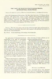
PROC. ENTOMOL. SOC. WASH. 101(1). 1999, pp. 64-68 THE LARVA OF PELECINUS POLYTURATOR (DRURY) (HYMENOPTERA: PELECINIDAE) Norman F. Johnson, Luciana Musetti, James B. Johnson, and Kerry Katovich (NFJ, LM) Department ofEntomology, The Ohio State University, 1315 Kinnear Road, Columbus, OH 43212-1192, U.S.A. (e-mail: [email protected];[email protected]. ohio-state.edu); (JBJ) Division of Entomology, Department of Plant, Soil, and Entomo- logical Sciences, University of Idaho, Moscow, ID 83844-2339, U.S.A. (e-mail: [email protected]); (KK) Department of Entomology, 237 Russell Laborato- ries, University of Wisconsin, Madison, WI 53706, U.S.A. — Abstract. The first instar larva and exuviae of the last instar larva of Pelecinus po- lyturator (Drury) are described, illustrated, and compared with published descriptions of other larvae of Proctotrupoidea. Key Words: larval morphology, Pelecinidae, Proctotrupoidea The application ofcharacters from larval with a diversity of forms without any un- morphology to the study of relationships derlying organizing principle. within the Hymenoptera is very uneven. The superfamily Proctotrupoidea s. str. is There are some good examples of the use- comprised of ten extant families of internal fulness of immatures as an additional parasitoids. The hosts for three of these source of data (e.g., Evans 1987), but for (Renyxidae, Austroniidae, and Peradeni- many superfamilies little or nothing is idae) are unknown, and only a bare mini- mum known. Progress in this area is hindered for of information is available for the several reasons. Most Apocrita are parasit- Monomachidae and Roproniidae. The only oids of other insects, and the hosts for a families for which immature stages have number of groups are very poorly known. been described are the Diapriidae (a large Many species are internal parasitoids; thus group of nearly 2,000 recognized species), it is difficult to obtain early instars and of- Proctotrupidae (331 described species), and ten the larvae are highly simplified in struc- Heloridae (a relict group of 10 extant spe- cies). In total, the larvae ofonly six species ture. Within the Hymenoptera a remarkable ar- from this complex have been described. We ray of larval body plans is found. Clausen report here on the larvae of another family, (1940) outlined fourteen types among the the Pelecinidae, a small group (only one parasitic Hymenoptera alone. Some ofthese species currently recognized) of uncertain are highly simplified, "embryonic," forms, affinities (Rasnitsyn 1980, Dowton et al. 1997). while others possess well-developed, ex- aggerated mandibles and caudal appendag- Materials and Methods es. Later instars usually converge upon a generalized, hymenopteriform type. Little Five parasitoid larvae were dissected phylogenetic pattern has been found amidst from larvae of Scarabaeidae (Coleoptera), this variety; at present we are confronted and preserved in ethanol. The specimens VOLUME 101, NUMBER 1 65 were found in the posterior two-thirds of mx) supported anteriorly by narrow stipital the abdomen of the host. Three final instar sclerite, otherwise lobelike, membranous; exuviae were found attached to scarab re- maxillary palp, labium, and labial palp un- mains from which Pelecinus had pupated. differentiated; head supported internally by Specimens are stored in the collections of extensive, strongly pigmented tentorium JBJ; the Ohio State University; the Insect (Fig. 4, tn) in shape of central plate with Research Collection, University ofWiscon- anterior extensions continuous with labral sin; and El Colegio de la Frontera Sur, San sclerite, lateral arms surrounding base of Cristobal de las Casas, Chiapas. Illustra- mandibles, and broad posterior bilobed tions were made using a camera lucida of plate in labial region, a central ovoid fora- whole specimens under alcohol and exuviae men visible, anterior to this with more in temporary slide mounts in glycerine jel- strongly pigmented triangular prominence, ly- anterior apex of triangle produced into small costa extending into labrum; no pro- Pelecinus polytur—ator (Drury) legs visible; body with indeterminate num- Material examined. USA. Michigan, ber of segments, without setae, apex of ab- Newaygo Co., 18 April 1974, host in soil domen acute; no spirac—les visible. of oak forest, ex Phyllophaga, one first in- Final instar (Fig. 5). Head capsule with star; Branch Co., 23 May 1974, hosts in soil posterior sclerotized, pigmented band, oth- of oak-hickory forest, four first instars, erwise largely membranous; mandible (md) three from large 5 cm long larvae, probably very small, weakly articulated with head; Phyllophaga, one from small 2.5 cm larva, antenna, labrum, maxilla, maxillary palp in- possibly Serica sp. Wisconsin, Marquette distinguishable; labium (lb) visible as me- Co., 11 August 1992, in sandy soil offorest dial triangular raised surface behind man- meadow, ex larva of Phyllophaga drakei dibles, with large circular field correspond- (Kirby) one final instar exuviae; Jackson ing to each labial palp (//?), a small central Co., 4 June 1992, ex P. drakei, one final area presumably representing opening ofla- instar exuviae; Oconto Co., 28 May 1996, bial gland (Ig) between palpi; mouthparts ex Phyllophaga rugosa (Melsheimer), one unsupported by sclerotized pleurostoma or final instar exuviae. MEXICO. Chiapas, Te- hypostoma; body with 7 pairs of spiracles nejapa, Balun Canal, 2,300 m, 14 February visible; tracheae well-developed. 1997, ex Phyllophaga obsoleta (Blanchard) Discussion third instar, one final inst—ar exuviae. First instar (Figs. 1-4). Length 3.3-5.3 The exuviae of the last instar larvae are mm; mandibulate larva (Clausen 1940); associated with pharate adult Pelecinuspo- head capsule well-developed, covering dor- lyturator, and their identity is unequivocal. sal and lateral sides ofhead, margins darkly The early instar larvae, however, are strik- pigmented, sclerotization extending beyond ingly divergent in structure from the exu- margins, gradually disappearing posterior- viae. Our determination of them was based ly; epicranial suture (Fig. 2, es) well-devel- on the fact that they were internal parasit- oped; no indication of eyes; antenna (Figs. oids dissected from larvae of Phyllophaga 1-3, a) indicated by small paired submedial Harris (Coleoptera: Scarabaeidae), the only papilla; clypeolabral area (Fig. 3, cl) largely recorded host in the United States and Can- membranous, supported by ovoid sclero- ada. The specimens also were collected in tized ring, dorsal portion of this ring some- an area in which Pelecinus was very abun- times incomplete; labrum with two medial dant. Muesebeck (1979) recorded Tiphia tubercles (Fig. 1, It); mandible (Figs. 1, 3, berbereti Allen, T. tegulina Malloch, T. md) strongly developed, falcate, bearing a transversa Say, T. vulgaris Robertson, and small subapical tooth; maxilla (Fig. 1, 3, T. intermedia Malloch (Tiphiidae); Myzin- PROCEEDINGS OF THE ENTOMOLOGICAL SOCIETY OF WASHINGTON 66 Figs 1-5 Pelecinuspoh'turator, larva. 1-4, Head of first instar 1. Lateral view. 2, Dorsal view. 3. Frontal view, specimen with mandibles closed. 4, Frontal view, specimen with open mandibles exposmg tentonum. 5 Mouthparts from final instar exuviae, right mandible detached. Abbreviations: a = antenna; cl - clypeolabral area; es = epicranial suture; lb = labium; Ig = opening of labial gland; Ip = labial palp; It = labral tubercle; md = mandible; mx = maxilla; tn = tentorium. Scale in mm. VOLUME 101, NUMBER 1 67 um quinquecinctiim (Fabricius) (Tiphiidae); scribed in H. anomalipes, B. tritoma, and and Ophion nigrovarius Provancher C silvestril. Phaenoserphus viator lacks a (Ichneumonidae) as parasitoids of Phyllo- coinplete head capsule, but does have a phaga. Woodruff and Beck (1989) listed a sclerotized ring surrounding the mouth- second species of Ophion as well as a num- parts. Distinct antennal lobes are found in ber ofadditional species oftiphiids and sco- the helorid, Basalys spp. and the proctotru- liids. We ruled out the aculeates because pids, larger and more prominent than the they are external parasitoids. Ichneumo- structures found in Pelecinus. We observed noids usually are characterized by the pos- no prolegs on any of the first instar larvae; session of a hypostomal spur (Short 1978), these structures have been reported for H. a structure that was not observed in these anomalipes, P. viator, B. parvulus, and an specimens. unidentified proctotrupid (Clausen 1940; Determination ofthe number oflarval in- presumably Nothoserphus scymni Ash- stars of internal parasitoids requires large mead). Two of our specimens have paired, numbers of observations of cohorts of nipple-like protuberances beneath the pos- known age in order to detect structural terior portion of the head capsule (visible changes associated with molting. This has in Fig. 1). Because of their position, we not been done yet for any species of proc- hesitate to call these prolegs or to homol- totrupoid, and no one has yet been able to ogize them with the labial palpi. The num- rear Pelecinus through its life cycle. We ber of observed spiracles reported varies could not determine the age ofthe observed from three {B. tritoma, C. silvestrii) to ten larvae directly or infer their age from pub- pairs {P. viator). lished observations ofrelated species. Clau- The most striking feature we observed in sen (1940) stated that the characteristics the first-instar larva was the large tentorial that set apart mandibulate larvae are lost at endoskeleton. A similar structure was very the first molt. This was confirmed by Clan- briefly described in P. viator by Eastham cy (1946) in his studies ofHelorus, another (1929), suggesting that it may not have proctotrupoid. Therefore, we concluded that been as apparent or strongly pigmented as the larvae dissected from the hosts must be in Pelecinus. The tentorium is not men- late first instars. tioned in the other descriptions. Very little information on the immature The larval specimens were dissected stages of proctotrupoids exists to form a from hosts in the spring (18 April, 1974; 23 context in which to discuss the structural May, 1974) in Michigan. Therefore, it ap- features ofPelecinus. Larvae have been de- pears that the species overwinters as late scribed and illustrated for Helorus anom- first instars within the Phyllophaga larvae. alipes (Panzer) (Heloridae; Clancy 1946), No more than a single larva was found in an unidentified species of Basalys West- any one host. Three specimens were recov- wood (Diapriidae; Simmonds 1952), Basa- ered from large (5 cm) hosts, presumably lys tritoma Thomson (Diapriidae; Wright et the final instar of the beetle. A fourth was al. 1946), Coptera silvesthi (Kieffer) (Dia- found in a much smaller larva, either a priidae; Pemberton and Willard 1918), Par- much younger specimen or a different ge- acodrus apterogynus (Haliday) (Proctotru- nus, perhaps Serica MacLeay (Coleoptera: pidae; Zolk 1924), Phaenoserphus viator Scarabaeidae). Host size may contribute to (Haliday) (Proctotrupidae; Eastham 1929), the large variation in size of adult Peleci- and Brachyserphus parvulus (Nees ab nus. Esenbeck) (Proctotrupidae; Osborne 1960). Acknowledgments Large, sickle-shaped mandibles have been reported in the first instar for all these spe- Thanks to Lorena Ruiz-Montoya (San cies. A sclerotized head capsule was de- Cristobal de las Casas, Mexico) and Daniel 68 PROCEEDINGS OF THE ENTOMOLOGICAL SOCIETY OF WASHINGTON K. Young (Madison, WI) for the loan of Institution Press, Washington, DC. Vol. 1, 1198 specimens. This material is based in part pp. upon work supported by the National Sci- Osborne, P. 1960. Observations on the natural enemies of Meligethes aeneus (F.) and M. viridescens (F.) ence Foundation under Grant No. DEB- [Coleoptera: Nitidulidae]. Parasitology 50: 91- 9521648. 110. Pemberton, C. E. and H. F Willard. 1918. A contri- Literature Cited bution to the biology of fruit-fly parasites in Ha- waii. Journal of Agricultural Research 15: 419- Clancy, D. W. 1946. The insect parasites ofthe Chry- 466. sopidae (Neuroptera). University of California Rasnitsyn, A. P. 1980. [The origin andevolution ofthe Publications in Entomology 7: 403-496. Hymenoptera.] Trudy Paleontologicheskogo Insti- Clausen, C. P. 1940. Entomophagous Insects. Mc- tuta 174: 1-190. Graw-Hill Book Company, Inc., New York. 688 Simmonds, F J. 1952. Parasites of the frit-fly, C>.sr/- pp. nellafrit (L.), in eastern North America. Bulletin Dowton, M., A. D. Austin, N. Dillon, and E. Bar- ofEntomological Research 43: 503-542. towsky. 1997. Molecular phylogeny of the apo- Short, J. R. T 1978. The final larval instars of the critan wasps: the Proctotrupomorpha and Eva- Ichneumonidae. Memoirs of the American Ento- niomorpha. Systematic Entomology 22: 245—255. mological Institute No. 25, 508 pp. Eastham, L. E. S. 1929. The post-embryonic devel- Woodruff, R. E. and B. M. Beck. 1989. The scarab opment of Phaenoserphiis viator Hal. (Proctotry- beetles ofFlorida (Coleoptera: Scarabaeidae). Part poidea), a parasite ofthe larva ofPterostichusni- II. The May orJune beetles (genus Pliyllophaga). ger(Carabidae), with notes on the anatomy ofthe Arthropods of Florida and Neighboring Land Ar- larva. Parasitology 21: 1-21. eas 13. 226 pp. Evans, H. E. 1987. Order Hymenoptera, pp. 597-710. Wright, D. W., Q. A. Geering, and D. G. Ashby. 1946. //; Stehr. F. W., ed.. Immature Insects,Vol. 1. Ken- The insect parasites ofthe carrot fly, Psila rosae. dall/Hunt Publishing Company, Dubuque, Iowa. Fab. Bulletin ofEntomological Research 37: 507- 754 pp. 529. Muesebeck, C. E W. 1979. Pelecinoidea, pp. 1119- Zolk, K. 1924. Pciracodrus apterogymis Halid. biolo- 1120. //; Ki-ombein, K. V., P D. Hurd, Jr, D. R. gia kohta. Zur Biologic von Poracodrus aptero- Smith, and B. D. Burks, eds. Catalog of Hyme- gynus Halid. Tartu Ulikooli Entomoloogia-Katse- noptera in America north ofMexico. Smithsonian jaama Teadaanded 5: 3-10.
