The integration of a micropipette in a closed microfluidic chip with optical tweezers for investigations of single cells: erratum. PDF
Preview The integration of a micropipette in a closed microfluidic chip with optical tweezers for investigations of single cells: erratum.
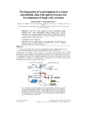
The integration of a micropipette in a closed microfluidic chip with optical tweezers for investigations of single cells: erratum Ahmed Alrifaiy1,2,* and Kerstin Ramser1,2 1Department of Computer Science, Electrical and Space Engineering, Luleå University of Technology, Luleå, 97187, Sweden 2Centre for Biomedical Engineering and Physics, Luleå University of Technology and Umeå University, Sweden *[email protected] Abstract: In July 2011 a new concept of a closed microfluidic system equipped with a fixed micropipette, optical tweezers and a UV-Vis spectrometer was presented [Biomed. Opt. Express 2, 2299 (2011)]. Figure 1 showed falsely oriented mirrors. To clarify the design of the setup, this erratum presents a correct schematic. © 2012 Optical Society of America OCIS codes: (350.4855) Optical tweezers or optical manipulation; (170.3880) Medical and biological imaging; (300.1030) Absorption; (280.2490) Flow diagnostics; (220.4000) Microstructure fabrication; (110.0180) Microscopy. References 1. A. Alrifaiy and K. Ramser, “How to integrate a micropipette into a closed microfluidic system: absorption spectra of an optically trapped erythrocyte,” Biomed. Opt. Express 2(8), 2299–2306 (2011). In July 2001 we presented a new concept of integrating a micropipette within a closed microfluidic system equipped with optical tweezers and a UV-Vis spectrometer [1]. In Fig. 1 of that paper a schematic of the setup was illustrated. The mirrors in the figure were wrongly oriented and hence depicted as semitransparent or transparent glasses. In Fig. 1, below, a new schematic with correctly oriented mirrors is given. Fig. 1. Inverted microscope that incorporates the following techniques: Gastight lab-on-a-chip with an integrated micropipette coupled to a pump system, optical tweezers for 3D steering of the single cells comprising of an IR laser, a beam expander, mirrors and a dichroic mirror and an IR blocking filter to block the IR laser. UV-Vis spectrometer with an integrated optical fiber to record the oxygenation states of the RBC, CCD camera to monitor the trapping dynamics of the cells within the micro-channel system. #161358 - $15.00 USD Received 11 Jan 2012; published 12 Jan 2012 (C) 2012 OSA 1 February 2012 / Vol. 3, No. 2 / BIOMEDICAL OPTICS EXPRESS 295
