The Benign Common Acquired Nevomelanocytic Proliferations PDF
Preview The Benign Common Acquired Nevomelanocytic Proliferations
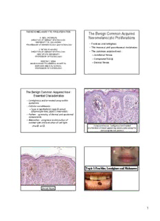
THE NEVOMELANOCYTIC PROLIFERATION The Benign Common Acquired A. NEIL CROWSON Nevomelanocytic Proliferations DIRECTOR OF DERMATOPATHOLOGY UNIVERSITY OF OKLAHOMA • Freckles and lentigines PROFESSOR OF DERMATOLOGY and PATHOLOGY • The mucosal and paramucosalmelanoses CYNTHIA M. MAGRO • The common acquired nevi : DIRECTOR OF DERMATOPATHOLOGY OHIO STATE UNIVERSITY –JunctionalNevus PROFESSOR OF PATHOLOGY –Compound Nevus MARTIN C. MIHM –Dermal Nevus MASSACHUSETTS GENERAL HOSPITAL HARVARD MEDICAL SCHOOL PROFESSOR OF PATHOLOGY The Benign Common Acquired Nevi : Essential Characteristics • Lentiginousand/or nested array within epidermis • Cellular constituents : –Type A (epithelioid), type B (small lymphocyte-like), type C (neuroidal) • Pattern : symmetry of dermal and epidermal components • Maturation : progressive diminution of nuclear size and evolution of cell type (A⇒B ⇒C) Compound nevus: Lentiginousand nested proliferation of bland appearing melanocytesalong the dermal epidermal junction Topic I:Freckles, Lentiginesand Melanoses Dermal Nevus 1 The Freckles and Lentigines • Not classified as a form of precancerous melanocyticproliferation • Their recognition is important because : Multiple punctatelentigines characterize LEOPARD –Clinical appearance resembesmelanoma syndrome Peutz-Jeghers –Multiple lentiginesmay be a sign of a Syndrome: macules systemic disease associated with non- on lips + buccal melanocyticcancers. mucosa CASE 1: A 55 year old woman has an irregular black macular lesion of the introitusthat measures 3.0 x 2.5 cm Diagnosis : Vulvar Melanosis 2 The Genital MelanocyticProliferations: The Mucosal Lentigines Main Forms The Mucosal Lentiginesencompass: (cid:131) Mucosal lentigo • Labial melanoticmacule(lower lip; an (cid:131) Vulvarmelanosis: identical lesion is seen in the oropharynx) (cid:131)With or without melanocytichyperplasia • Genital Lentiginesup to 15 mm in diameter (cid:131) Common acquired nevus • Vulvar,urethral, oral + nasal melanosis: (cid:131) Dysplasticnevus multiple variably coalescing flat lesions (cid:131) Malignant melanoma resembling radial growth phase melanoma; (cid:131) The prototypic genital nevus or atypical all may be melanoma precursors melanocyticnevus of genital type • Conjunctivalmelanosis Vulvar Melanosis • Clinical presentation: presents as a solitary (up to or several centimeters in size) or multiple intensely pigmented macule(s); similar phenomenon on penis is called penile melanosis. • Clinical differential diagnosis: radial growth phase mucosal LMM, patch type bowenoidpapulosis, and pigmented Bowen’s disease Vulvar Melanosis : Histomorphology • Hypermelanosis • Mild (cid:57)in basal layer melanocytenumbers • Acanthosis • No melanocyticatypia • Differential diagnosis: atypical lentiginous melanocytichyperplasia which is a precursor lesion to melanoma 3 Practice point: Uniform homogeneous basilar pigmentation : A clue to benignancy Penile Melanosis : Differential Diagnosis • The appearance of a periurethral pigmented maculein mid to late life may be the only sign of urethral melanoma • Such patients require careful urological examination and biopsy Mucosal LentigoFromSimulators of Malignant Melanoma Drs. Kerland Cerroni, University of Graz Freckles : brown maculeson sun-exposed skin The Freckles and Lentigines • Freckles: light brown maculestypically less than 6 mm on sun exposed skin reflecting increased melanizationdue to an increase in the functional activity of the melanocyte • Lentigines: pigmented maculeless than 1 cm in diameter; typically oval, < 5mm. • Café au laitmacules: –Solitary lesions not uncommon –>5 lesions >0.5cm strongly correlate with a diagnosis of neurofibromatosis –Manifest hypermelanosis, slight increase in melanocytes, giant melanosomes(>2 um) 4 Cafe au Laitmaculesin an adult Cafe au Laitin a toddler Practice point: •Freckles disappear in winter and re-appear in summer. •If these lip lesions did not show that behavior, one would suspect Peutz-Jegherssyndrome Cafe au Laitmaculein an 18 yr Cafe au Laitin an infant old with dysplasticnevi syndrome Lentigines and Freckles • Albright’s syndrome: basis is a postzygoticmutation of the gene GNAS1 that encodes the alpha subunit of a signal transducingheterotrimericguanine nucleotide binding protein. The syndrome comprises: –Polyostoticfibrous dysplasia, –Melanoticmacules, and –Hyperfunctionalendocrinopathy • Sporadic mutation of the tyrosinasegene which is the pathophysiologicbasis of the skin pigmentation • Borders of the lesions are irregular • Skin biopsies show hypermelanosis; there is no increase in the number of melanocytes Practice point: Any unusual pigmented lesion approaching the natal cleft should prompt consideration of underlying abnormalities of spinal cord or spine Nevus Spilus Nevus Spilus: Incipient junctionalnevus super- imposed on a background of lentigosimplex 5 Becker’s Nevus Solar Lentigo • Multiple maculeson sun damaged skin with predilection for face and dorsa of hands • Elongated retiato zones of atrophy, chronic photoactivationof melanocytescan mimic lentigomaligna • Differential Diagnosis : –Incipient lentigomaligna –Superficial pigmented actinic keratosis (SPAK) Solar Lentigines 6 EpithelioidPhotoactivation Chronic Photoactivation • The proliferation is lentiginous(i.esingly disposed cells) • Rarely confluent • Nesting is not seen • The cells have either an epithelioid morphology or demonstrate nuclear angulationand hyperchromasia(i.eso called “lentiginous” cytomorphology) Lentiginous Photoactivation CASE 2: TOPIC II: THE DERMAL DENDRITIC A 27 year old man has a 12 x 12 MELANOCYTIC PROLIFERATION/ cm pigmented lesion with a dusky DERMAL MELANOCYTOSIS bluish hue over the right cheek and eyelid 7 The Nevi of Ota and Ito (cid:131) Clinical features • Both are defined by a unilateral light gray Ota’s Nevus maculepresenting in 1st-2nddecades, up to 5 cm diameter which darkens with age in : •Trigeminal nerve distribution (1/2>3) (OTA, 1939) •Acromioclavicularnerve distribution (ITO, 1954) • Asiatics>African>Caucasians • Darken as patient ages The nevi of Ota and Ito (cid:131) Histomorphology • Band-like disposition of melanophagesand dendritic melanocytesin the upper 1/3rd of the reticular dermis; epidermis spared Practice point: Scleralnevus of Ota is • Dendriticcells cytomorphologicallysimilar to those of associated with choroidalnevi and blue nevus melanoma; ophthalmic examination is • Cellularityis sparse and fibrosis is slight required • Sclerallesions also involve nerves and may be more cellular (Ota) •Ota’s nevus may involve the tympanum and the nasopalatineganglia 8 The Dermal Melanocytoses (cid:131) Nevi of Ota and Ito (cid:131) Sun’s nevus (cid:131) Dermal melanocytichamartoma (cid:131) Mongolian spot (cid:131) Blue nevus (cid:131) Blue nevus with hypercellularity (cid:131) Cellular blue nevus Blue Nevus 2 The Blue Nevus • The Blue nevus of Jadassohn-Tieche • Mainly a lesion of childhood • Most common location: dorsum of hands and feet, less often the buttocks, head and neck; can occur on mucosal surfaces Blue Nevus 3 • Typically 3 to 4 millimeters • Can be multiple and inherited as an autosomaldominant trait Blue nevus in oral cavity 9 EpithelioidBlue Nevus: Seen in 10% of Carney’s complex: spotty pigmentation; cardiac, mammary + cutaneousmyxomas; adrenal hyperplasia; Sertolicell tumor; pituitary adenoma HypomelanoticBlue Nevus Blue Nevus With Hypercellularity • Clinically blue nevi often with a central areas of hypopigmentationand/or firmness with surrounding dense blue coloration • Histologicallyhas blue nevus components with cellular intradermalnidiof spindle cell fascicles Cellular fascicles of spindle cells surrounded by classic blue nevus component 10
Description: