Sierra College Journal of Microbiology PDF
Preview Sierra College Journal of Microbiology
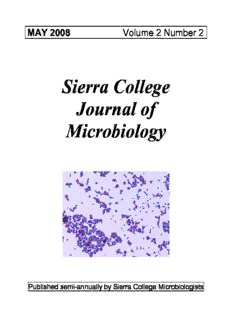
MAY 2008 Volume 2 Number 2 Sierra College Journal of Microbiology Published semi-annually by Sierra College Microbiologists SIERRA COLLEGE JOURNAL OF MICROBIOLOGY VOLUME 2 ♦ MAY 2008 ♦ NUMBER 2 Editors: SASHA WARREN and HARRIET WILSON Contributors: Biology 4 & 8B students – Spring Semester, 2008 The editorial staff wishes to thank Elaine Atnip and Jim Wilson for their support and assistance throughout the semester. http://biosci.sierracollege.edu Editors’ Disclaimer: All papers contained in this journal represent original work by the authors. Editorial staff did no revisions prior to publication. Cover photo: A Gram-stained smear of Micrococcus varians, isolated by Iryna Vlasik and Amber Andronico (pg. 24). This isolate was obtained from tap water that had been filtered with a PUR filter. Magnification: 1000x. (Photomicrograph: S. Warren) SIERRA COLLEGE JOURNAL OF MICROBIOLOGY VOLUME 2 MAY 2008 NO. 2 TABLE OF CONTENTS Isolation and Identification of an Unknown Organism in a Water Sample Obtained from an Outdoor Children’s Playset in Auburn, California Jennifer Agudelo, Leisha Baez Flores, and Cherece Mitchell .…………………. 1-4 Isolation of Common Bacteria Found on an Out-patient Exam Table and the Effectiveness of the Approved Disinfectant Noraline D. Bailey, Camille Beck, and Lluvia Esparza ………………………….. 5-8 Isolation and Identification of Potential Pathogen on Children’s Playground Equipment Becky Lea and Taffee Hoffee ………………………..………………………….. 9-12 Isolation of Kocuria rhizophila from Shopping Cart Handles Shauna Bassett and Liz Taylor-Weber ……………….………………………….. 13-17 Isolation and Identification of Staphylococcus pasteuri from a Presumed Clean Fingernail Corina Kendrick, Shellie Rogers, and Kaitlend Hill ..…………………………… 18-23 Isolation of Micrococcus varians from Tap Water Filtered With a PUR Filter Iryna Vlasik and Amber Andronico ………………..……………………………. 24-27 Isolation of Bacteria Taken from a Water Sample from Lake Wildwood, California Christain Reinheimer ……...……..………………………………………………. 28-31 Isolation and Identification of a Gram-negative Organism found in Creek Water at Empire Mine in Grass Valley, California Dana Perry .....................…………………………………………………………. 32-36 Isolation and Identification of a Gram-Negative Organism Discovered from an Air Plate Edi Kramer …………………………….……..……………………………….…. 37-42 Identification and Analysis of Bacteria found in an Argentine Tegu’s Mouth Jennifer Hayes …….……………………………………………………………... 43-46 Isolation and Identification of Kocuria rhizophila From Air Jill Kearney………………………………………………….…………………… 47-50 Careful Where You Cook; You Never Know What’s Lurking in Your Kitchen Air. Kim Lindstrom, Lynn Sex, and Alaina Stoltenburg ……………….…………….. 51-55 Isolation and Identification of a Gram-positive Bacillus, Corynebacterium auriscanis from an Air Plate Kim Kelderman …………………….....………………………………………….. 56-59 Isolation and Identification of Two Different Types of Microorganisms Found in Arrowhead Bottled Water Jessica Vega and Nick Garcia ……..…………………………………………….. 60-63 Isolation of Bacteria From the Sierra College Salad Bar Cirby Chitty, Vanessa Marconi, and Nanako Yip ……………………………….. 64-70 Isolation and Identification of Selected Gram-positive Bacteria from Household Air in Lincoln, California Sarah Blake …..……………...…………………...……………………………… 71-74 Vol. 2, No. 2 Isolation and Identification of an Unknown Organism in A Water Sample Obtained from an Outdoor Children’s Playset in Auburn, California. JENNIFER AGUDELO, LEISHA BAEZ FLORES, AND CHERECE MITCHELL Microbiology Laboratory, Sierra College, Rocklin, CA 95677 Received 2 May 2008/Accepted 9 May 2008 A water sample was obtained from an outdoor children’s playset in Auburn, California. This sample was used to isolate and identify an unknowm organism we hypothesized to be Escherichia coli. Tests were done to establish the characteristics of the organism, identify it, and determine the potential health concerns related to the unknowm organism. The isolated organism was found to be a gram negative cocco-bacilli. Test results were compared to Enterobacter aerogenes, Escherichia coli, and Klebsiella pneumoniae which were it’s closest relative, according to the National Center for Biotechnology Information website. These tests proved the unknown organism to be Enterobacter aerogenes, which is an opportunistic pathogen that can be associated with certain intestinal disorders. The information used for this project was obtained from the Microbiology Lab Syllabus, Bergey’s Manual of Systematic Bacteriology, and the National Center for Biotechnology Information website. INTRODUCTION A small pool of water had settled in an outdoor children’s playset, located in Auburn, California. The children playing with this toy will have physical contact with organisms in this water. Due to concerns about the health risks associated with children playing with outdoor toys, a water sample was collected for lab testing. The goal is to isolate and identify an unknown organism from the water sample. We hypothesized to find E. coli since it’s commonly found in water supplies and looked for when testing water. If E. coli or any coliform is found on an outdoor playset, it raises an alarm that fecal matter from humans or animals is present. MATERIALS AND METHODS Using a sterile loop we streaked a portion of a water sample, from an outdoor playset, on a TSA plate for colony growth and an EMB plate to test for E. coli. Both plates were incubated for about 24 hours at 37oC. We chose a white colony versus an off white colony to re-streak on a TSA plate for isolation because it looked shiny and pretty (Wilson, 2007). That plate was then placed in the incubator for 24 hours at 37oC. An indirect stain was performed to visualize cell morphology(Wilson, 2007). A gram-stain and KOH test was done to determine if the organism was gram negative or gram positive for cell wall morphology(Wilson, 2007). A colony sample was used to isolate chromosomal DNA for analysis by vortex and heat. The goal of using PCR to amplify rDNA is to isolate the 16S ribosomal DNA using primers 1 Vol. 2, No. 2 Agudelo, Flores, and Mitchell Bacteria 8-forward and 1492-reverse. PCR was then run on a 1% agarose gel and the 1500 bp product was purified, using the QIA quick Gel Purification Kit. Purified rDNA was sent to the Division of Biological Sciences sequencing facility in UC Davis and sequenced using primer Bacteria-8-forward. This gave us one electropherogram back. The sequence from the isolated organism was compared to those in the database at National Center for Biotechnology Information website using the Basic Local Alignment Search Tool. Based on sequencing results we performed SIM, MRVP, urease, citrate tests (Wilson, 2007). RESULTS There were two different types of colonies that grew on the TSA plate we first streaked. One was a shiny white color and the other an off white color. The EMB plate had dark red colonies with no color change of metallic green. We isolated the white colony in a new TSA plate and observed shiny, white, circular, entire, opaque and convex colonies that were about 1- 3mm in diameter. The indirect stain revealed our organism to be cocco-bacilli (Wilson, 2007). The gram stain showed us pink organisms, and the KOH test gave us a “snotty” result (Wilson, 2007). After comparing our rDNA sequence to National Center for Biotechnology Information using the Basic Local Alignment Search Tool our rDNA closely matched Enterobacter aerogenes (accession # AF395913.1). The rDNA nucleotide sequence showed a 97% similarity, with a score of 1096, a ratio of 638/815 of matching nucleotides. There were two other organisms which had a 97% similarity, and they were Escherichia coli and Klebsiella pneumoniae (NCBI, 2008). We performed more tests in order to differentiate which species our rDNA matched. The SIM test resulted in a yellow tube agar with growth away from the stab line (Wilson, 2007). The MR test result was golden brown and the VP test result was cherry red (Wilson, 2007). The urease test stayed peach (Wilson, 2007). The citrate test turned blue (Wilson, 2007). Figure 1- SIM tube result. 2 Vol. 2, No. 2 SIERRA COLLEGE JOURNAL OF MICROBIOLOGY DISCUSSION The water sample collected for this project was first streaked on a TSA plate for colony isolation of an unknown organism. An EMB plate was also streaked due to our hypothesis of finding Escherichia coli. There was no metallic green color anywhere in the EMB plate to indicate that Escherichia coli was present. A white colony was chosen for isolation over an off- white colony and was re-streaked on a new TSA plate. An indirect stain was done to determine cell morphology which was cocco-bacilli. A gram stain and KOH test confirmed that the organism is gram negative and lysed when KOH was added due to that gram negative cell wall structure. Results from the NCBI BLAST revealed that Enterobacter aerogenes, Escherichia coli, and Klebsiella pneumoniae were closely matched to our rDNA sequence (NCBI, 2008). The SIM test indicated that our organism didn’t make indole or H S, and was motile (Wilson, 2 2007). The MRVP revealed that the organism performed butanediol fermentation (Wilson, 2007). The citrate test showed that the organism can utilize citrate and the urease test confirmed that the organism doesn’t have the enzyme urease (Wilson, 2007). These results concluded that our organism was more closely matched to Enterobacter aerogenes (Wilson, 2007). TEST Enterobacter Escherichia Klebsiella Our Isolate aerogenes coli pneumoniae Indole (SIM) - + - - Motility (SIM) + + - + Methyl Red - + + - Voges + - - + Proskauer Citrate + - + + Urease - - + - Table 1- Comparison of test results from unknown organism and closest matching species. These results proved our hypothesis to find Escherichia coli wrong. Enterobacter aerogenes is an opportunistic pathogen, a fecal indicator, and is found in soil, human and animal feces, and also dairy products (Richard, 1984). The pathogenicity of the Enterobacter group is of a low order, but they can be associated with certain intestinal disorders and other syndromes (Bailey, 1966). ACKNOWLEDGEMENTS Sierra College Foundation North Valley and Mountain Biotechnology Center at American River College. LITERATURE CITED Bailey, W.R., P.H.D. and E.G. Scott, M.S., M.T. (ASCP). Diagnostic Microbiology, Second Edition. The C.V. Mosby Company, 1966. 3 Vol. 2, No. 2 Agudelo, Flores, and Mitchell National Center for Biotechnology Information (NCBI). 7 April 2008. Basic Local Alignment and Search Tool (BLAST). 7 April 2008. http://www.ncbi.nlm.nih.gov/BLAST. Richard, C. Genus Enterobacter Hormaeche and Edwards 1960, 72AL; Nom. Cons. Opin. 28, Jud. Comm. 1963, 38. In N.R. Krieg, J.G. Holt, R.G.E. Murray, D.J. Brenner, M.P. Bryant, J.W. Moulder, M. Pfennig, P.H.A. Sneath and J.T. Staley (Eds.), Bergey’s Manual of Systematic Bacteriology, First Edition, Vol. 1, pp.465-469. Williams & Wilkins, 1984. Wilson, Harriet. Microbiology Laboratory Syllabus, Exercises and Questions, Biological Sciences 4. Sierra College Biological Sciences Department, 2007. 4 Vol. 2, No. 2 Isolation of Common Bacteria Found on an Out-patient Exam Table and the Effectiveness of the Approved Disinfectant NORALINE D. BAILEY, CAMILLE BECK, LLUVIA ESPARZA Microbiology Laboratory, Sierra College, Rocklin, CA 95677 Received 2 May 2008/Accepted 9 May 2008 A study was conducted to test the sterility of patient exam tables in outpatient clinics. Swab samples were taken from two doctor’s exam tables before and after they were sterilized. The results show the presence of bacterial growth prior to sanitizing and no growth following sanitizing, thus the disinfectant agent PDI Sani- cloth germicidal disposable wipes removed all viable cells. From the cultures grown in lab, a bacterium was isolated and analyzed from our contaminated plate (prior to sanitizing). Through utilizing PCR, DNA sequencing and the Bergey’s Manual, the isolated bacterium was identified as Bacillus subtilis, an organism that is not pathogenic to humans. INTRODUCTION In everyday life humans are surrounded and exposed to bacteria. There are some bacteria that act in favor to the host they survive on, in contrast to the good there is also a bad. Some bacteria that individuals are exposed to can lead to various life-threatening or life changing diseases. The severity of illness is dependent upon many factors. For example, the quantity or the strain of bacteria that an individual comes in contact with may have an adverse effect. Therefore, we set out to discover what types of organism are thriving on exam tables in an outpatient clinical setting. Based on literature review regarding contamination of exam tables in patient rooms, one may predict that the results of the bacteria collected and analyzed will show a presence of many common bacteria (Environment Protection Agency, 1997). On average there are 4 to 10 patients seen between the cleaning of the exam tables. The only barrier between the patient and the exam table is a paper lining. In some situations the beds are cleaned by wiping with PDI Sani-cloth immediately following an exam when the patient has a known or suspected infectious disease such as MRSA or HIV. We would like to test the effectiveness of the PDI Sani-cloth germicidal disposable wipes. The bacteria most likely to show up in the samples gathered and tested in this experiment will be normal flora such as; Staphylococcus aureus, Staphylococcus epidermis, Corneybacterium diphtheria, and Micrococcus luteus (Microbial Flora of Skin, 2008). In addition, there are common opportunistic pathogens that are often found in healthcare environments, such as: Streptococcus pneumoniae, Klebsiella pneumoniae, Escherichia coli, Haemophilus influenza, and Acinetobacter baumannii (Maley, 2000). Are patients really safe from exposure to the contaminated surfaces of these tables? The goals of this work are: to isolate bacterial organisms from patient exam tables and to discover the effectiveness of the disposable sanitizing wipes. 5 Vol. 2, No. 2 Bailey, Beck, and Esparza MATERIALS AND METHODS A patient exam table in an out patient clinic was sampled after six patients were seen and came in contact with the exam tables. The exam table was sanitized prior to the six patients. After the six patients were seen, the edge of the table (which has the most exposure to contact) was swabbed with a sterile Q-tip like applicator. The applicator was then submerged into a small sterile tube containing sterile water. The next steps were to plate out the water/organism mixture onto TSA plates and incubate them at 37°C for 48 hours. After the 48 hour incubation period there were approximately 10 different organisms present on our TSA plate. We chose to work with one organism in particular because of its colony morphology. The following tests were used to identify the cellular morphology: Indirect stain, Gram stain, Endospore stain, Acidfast stain, Capsule stain, KOH test, and a Wet Mount. The next step was to isolate the DNA. We isolated the chromosomal DNA by conducting a PCR reaction. Our goal in using the PCR reaction was to amplify and make a lot of copies of our organism 16s ribosomal RNA gene (rDNA). We used the primer Bacteria 8-F and 1492 reverse. Once we had many copies of the rDNA gene, a gel containing 1% agarose could be set up and the 1500Bp product was ready to be purified removing everything (primer, taq polymerase…) but the gene. The purification step was done by using the QIAquick kit which is made by Qiagen. After using this kit our end product was many copies of the purified 1500Bp rDNA gene and nothing else. The purified gene was then sent to the Division of Biological Science Sequencing Facility at UC Davis. The gene was then sequenced using the primer Bacteria 8-F. We then received an electropherogram of our organisms DNA sequence and we compared it to other known sequences in the National Center for Biotechnology Information (NCBI) database using Basic Local Alignment Search Tool (BLAST). Based on the results from the DNA sequencing we conducted the following tests: Indirect Stain, Gram Stain, Endospore Stain, Acidfast, Capsule, KOH, Wet mount, and Catalase. RESULTS The DNA results show our organism is 99% identical to Bacillus subtilis accession # A4825035.1 with 99% accuracy. The ratio of identical matching nucleotides is 872/878 and the Bit Score is 1589. The colony morphology of the organism was: filamentous form, undulate margin, umbonate elevation, opaque optical character, whitish beige pigment, dry and wrinkled surface texture, and a size range from 3mm x 3mm – 6mm x 7mm. All of the colonies also displayed a white ridged design in the middle. Our isolate had long single celled rods that are motile and are 1.5 micrometers x 1 micrometer in size, and is catalase positive. We preformed the following tests and obtained the following results: KOH: no snot, KOH negative. Gram stain: purple, Gram +. Acidfast stain: blue, no mycolic acid, and non-acid fast. Endospore stain: spores present, the organism forms endospores under starvation and other extreme environments/conditions. Capsule stain: capsules present; the organism does have capsules. After comparing our organism to the Bergey’s Manual, all of the cellular, colony and physiological (catalase) characteristics matched (Garvie1872). 6
Description: