Self-assessment colour review of reptiles and amphibians PDF
Preview Self-assessment colour review of reptiles and amphibians
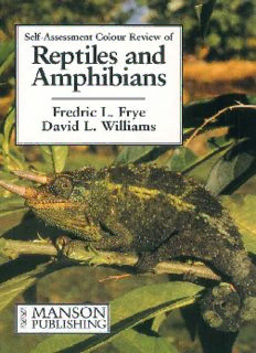
Self-Assessment Colour Review of Reptiles and Amphibians Fredric L. Frye BSc, DVM, MSc, CBiol, FIBiol, FRSM Davis, California David L. Williams MA, VetMB, CertVOphthal, MRCVS Royal Veterinary College, London Manson Publishing/The Veterinary Press Copyright © 1995 Manson Publishing Ltd ISBN 1–874545–32–4 All rights reserved. No part of this publication may be reproduced, stored in a retrieval system or transmitted in any form or by any means without the written permission of the copyright holder or in accordance with the provisions of the Copyright Act 1956 (as amended), or under the terms of any licence permitting limited copying issued by the Copyright Licensing Agency, 33–34 Alfred Place, London WC1E 7DP. Any person who does any unauthorised act in relation to this publication may be liable to criminal prosecution and civil claims for damages. A CIPcatalogue record for this book is available from the British Library. For full details of all Manson Publishing Ltd titles please write to Manson Publishing Ltd, 73 Corringham Road, London NW11 7DL, UK. Design and layout:Patrick Daly Colour reproduction; Reed Reprographics,Ipswich,UK. Printed by Grafos S.A., Barcelona,Spain . Cover illustration: A young adult male Jackson’s Chameleon, Chamaeleo Jacksoni, Acknowledgements The senior author wishes to express his heartfelt appreciation to Brucye Frye, who offered many constructive suggestions and edited the senior author’s original manu- script as the cases were selected and the text was written. Since its inception, this project has been a joy for both of us. We hope that those who peruse these pages and learn from these cases will gain the vital information that is necessary when diagnosing and treating the diverse reptilian and amphibian patients who often differ markedly from more conventional veterinary patients. It took 30 years to accumulate these cases. We urge our readers to make their cases available to others so that we may all increase our knowledge. Michael Manson, managing director of Manson Publishing Limited, consistently showed great interest and enthusiasm in this project, and it was a genuine pleasure dealing with him and his entire editorial and production staff during the prepublica- tion preparation of this book. Several colleagues generously contributed images or specimens which illustrate this book. In alphabetical order, the following permitted the senior author to repro- duce the following photographs and radiographs (identified by question numbers): Dr. Alonso Aguirre: 191. Dr. Stephen L. Barten: 47(page 36); 225(pages 163 and 164); 258. Dr. Ralph F. Claxton: 130(page 93). Dr. Nathan W. Cohen: 76. Dr. James H. Corcoran: 48(page 35, bottom left). Dr. James Detterline: 46;194. Mr. Robin Houston: 2. Dr. Douglas R. Mader: 36;218. Dr. Scott McDonald: 83;123;131. Mr. and Mrs. J. E. Merrill: 166. Ms. Wendy Townsend: 116. The remaining images are the work of the authors. 3 Dedication To Brucye and Jennie, Lorraine, Erik, Bice, Noah and Ian. 4 Introduction This text was written with a retrospective view and deep appreciation of the history of comparative medicine and comparative pathology. Pioneers, such as the 18thcentury’s John Hunter, and Rudolph Virchow, who died early this century, contributed mightily to our fund of knowledge; they were among the first physician-pathologiststo realise the rationale of the commonsense approach of considering not only the solitary lesion but, more importantly, the intimate relationship of one condition, organ, or organ system to the well-being or disease of the entire organism. Of equal importance is that both of these giants of comparative medicine appreciated the value of teaching this approach to their contemporary health care professionals when explaining physi- ological interdependencies. This was revolutionary, even heretical, to many ears and was diametrically opposed to the dogma of that era. Hunter’s and Virchow’s philoso- phy was particularly enlightened when one considers that pathogenic bacteria, fungi, actinomycetes and viruses were only relatively recently discovered as the causes of infectious disease. The patient must be seen as a whole, not merely the sum of his or her many dis- parate body parts. Balzac* observed that ‘There are conflicts between diseases and physicians…of which physicians alone have any knowledge and whose reward in cases of success is never found in the paltry price of their labours nor, indeed, under the patient’s roof but in the sweet gratification…bestowed upon true artists by the satisfaction they feel…in having acomplished a worthy work.’ Those sentiments were as germane to veterinary surgeons as they were to human physicians and barber sur- geons; and they are as appropriate today as they were well over a century-and-a-half ago (perhaps even more so) because today the healing arts have at their disposal so many more means for arriving at an accurate diagnosis and novel methods for treat- ing diseases. Only after carefully assembling a history, keenly observing the patient’s physical signs and conducting specialised investigations can a list of differential diag- noses be constructed; then, like kernels of grain, that list must be winnowed gradual- ly until only one or, at most, a very few possibilities remain. From these, a prognosis is formulated; only then can a rational course of therapy or corrective surgery be established and carried out. Because of economic considerations, valuable clinical laboratory investigations may not always be executed, but most of these cases will not be diminished if enough required information has been gathered from the client and if the patient has been examined meticulously. By completing each task conscien- tiously and consistently, the probability of success is enhanced. In this self-assessment guide we have provided sufficient explicit information from which to formulate dif- ferential diagnoses, decide upon treatment plans and arrive at prognoses. The clues to the solution of clinical puzzles are often subtle. When examining radiographs and electrocardiograms, you may find surprises lurking, ready to snag the unwary. In some instances, the diagnosis is obvious, but the prognosis or treatment might not be so clear. This approach was taken because in real life situations the diagnosis and treatment of patients are not always trouble-free or easily achieved. We have system- atically decreased the number of provisional diagnoses by proposing various *Balzac, Honoré de. (1840). Pierrette.Ives, GB transl. (1897), Geo Barrie & Sons, Philadelphia, 215-216. 5 reasonable explanations, when necessary. Several things must be understood: not all clients tell the whole truth; some may not actually lie about their animals, but also they may not always volunteer details that might be vitally important to the final diagnosis and outcome of their particular case; others may, for their own reasons, conceal facts that might be germane. As mentioned before, economics often plays a major role in how an animal responds to treatment – or whether it is even examined and treated professionally – particularly if the definitive diagnosis relies upon expen- sive tests or procedures. We have selected cases which are instructive and probably would/could be seen in clinical practice. The majority are common everyday cases; a few are exotic and may have been observed only once. For those readers who have never observed even a single example of one of the common everyday cases, the pur- chase of this text is justified; the rarer cases are included to whet the intellectual appetites of the more experienced of our readers. Perhaps, just perhaps, a similar puzzling case will arrive on your doorstep one day; having experienced the question and answer vicariously in the pages of this guide may make your case all the more memorable! The popularity of certain reptiles as pets or study animals is reflected in the selection of cases for this book; it is for this reason that so many iguanas are included as representatives of reptilian disease. Some people who possess large, showy and expressive lizards are often willing and able to incur the considerable expense of having their pets properly diagnosed and treated; others who own another species of herbivorous lizard perceived as being of less value may elect not to have it examined and evaluated. However, we are confident that information regarding the physiological responses to disease in iguanas is sufficiently similar to disease process- es in other herbivorous lizards. Bacterial infections, parasitism and many metabolic disorders in North American chelonians closely mimic the same conditions observed in European, African or Asian turtles, terrapins and tortoises and vice versa. We have included more than a single case of some particular conditions (but each has differed somewhat) because we believe that repeating the discussion of these common, but significant, conditions is important due to their ramifications; in these instances we have endeavoured to include special features that will maintain your interest. We wish to enunciate one caveat: ‘normal’ values are only rarely cited in this guide. Unlike humans and domestic animals, from which thousands of data have been col- lected and collated, reptiles and especially captive reptiles (1) may or may not be nor- mal (or even healthy) when their body fluids are sampled, and (2) the very nature of captivity and the stress incurred during restraint in order to obtain the samples intro- duce variables that are reflected in the laboratory results. We recognise these short- comings and urge the reader to consider these undeniable facts when judging whether a laboratory finding is meaningful or out of ‘normal’ range. (We used only the most current ‘accurate’ values available, but advise you to keep your sceptic’s salt-shaker at the ready!) Fredric L Frye David L Williams January 1995 6 English and Latin names African bullfrog Pyxicephalus adspersus African clawed frog Xenopus laevis African leopard tortoise Geochelone pardalis African pancake lizard Malococherus tornei Agama lizard Agamasp. American alligator Alligator mississippiensis American bullfrog Rata catesbiana Anaconda snake Eunectes marinus Argentine horned frog Ceratophrys oranata Asian box turtle Cuora amboinensis Asian red-tailed rat snake Gonysoma oxycephala Asian water dragon lizard Physignathus concincinus Australian bearded dragon lizard Pogona vitticeps Australian taipan Oxyuranus scutellatus Axolotl Ambystomasp. Blanding’s turtle Clemmys blandingi Blood python Python curtus Boa constrictor Boa constrictor constrictor Box turtle Terrapene carolina Burmese python Python molurus bivittatus California king snake Lampropeltis getulus californiae Caribbean rhinoceros iguana Cyclura nubila lewisii Carolina anole lizard Anolis carolinensis Children’s python Liasis childreni Chilean tortoise Geochelone chilensis Chuckwalla lizard Sauromalus obesus Collared lizard Crotaphytus collaris Common iguana Iguana iguana Copperhead snake Agkistrodon contortrix Corn snake Elaphe guttata Desert tortoise Xerobates agassizzi Diamondback terrapin Malaclemys terrapin East African chameleon Chamaeleo dilepsis East African Fischer’s chameleon Chamaeleo fischeri East African pancake tortoise Malacochersus tornieri Emerald tree boa Corralus caninus European green lizard Lacerta viridis Fence lizard Sceloporous occidentalis Gaboon viper Bitis gabonica Galapagos tortoise Geochelone elephantopus Garter snake Thamnophis sirtalis Giant blue-tongued skink Tiligua gigas Gila monster lizard Heloderma suspectum Gopher snake Pituophis melanoleucus catenifer Gopher tortoise Gopherus polyphemus Green sea turtle Chelonia mydas Ground iguana Cyclura cornuta Hermann’s tortoise Testudo hermanni Hog-nosed snake Heterodan platyrhinos Indigo snake Drymarchon corais 7 Jackson’s chameleon Chamaeleo jacksoni Javanese file snake Acrochordus javanicus King snake Lampropeltis getulus Leopard gecko Eublepharis mascularis Malagasy tree boa Sanzinia madagagasarensis; Boa mandrita Mangrove monitor lizard Varanus indicus Map turtle Graptemyssp. Mata mata turtle Chelus fimbuatus Mexican dwarf python Loxocemus bicolor Nile monitor lizard Varanus niloticus North American banded king snake Lampropeltis alterna Pacific pond turtle Clemmys marmorata Rat snake Elaphesp. Rattlesnake Crotalus atrox Red tegu lizard Tupinambis rufescens Red-eared slider turtle Trachemys scripta elegans Red-eyed frog Agalychnis callidryas Red-legged tortoise Geochelone carbonaria Reeve’s turtle Chinemys reevesi Reticulated python Python reticularis Rhinoceros iguana Cyclura nubila Rhinoceros viper Bitis nasicornis Rosy boa Lichanura trivirgata Royal python Python regius Russell’s viper Vipera russelli Savannah monitor lizard Varanus exanthematicus Snapping turtle Chelydra serpentina Soft-shelled turtle Apalonesp. Solomon Island skink Corucia zebrata Spectacled caiman Caiman sclerops Spiny-tailed iguana Ctenosaurussp. Tegu lizard Tupinambis teguixin Texas tortoise Xerobates berlandieri Timor monitor lizard Varanus timorensis Tokay gecko Gekko gekko Tree boa Eucratessp. Tree frog Pseudachris (hyla) regilla Vine snake Oxybelis aeneus Water monitor lizard Varanus salvator Water snake Natrix cyclopion Western painted turtle Chryemys picta Western terrestrial garter snake Thamnophis sirtalis terrestris Western toad Bufo boreas halophilus 8 1, 2 & 3: Questions 1 i. What are the two oph- thalmic conditions affecting the eye of this elderly tortoise? ii. How would you manage this case? 2 All of the snakes in a large collection are found to be infested with this small dark brown-to-black invertebrate that crawls about on the sur- face of the snakes’ skin. i.What is this creature? ii.What is its significance? iii. How would you treat this infestation? 3 During mask induction of anaesthesia many reptiles engage in prolonged breath- holding and, under varying environmental conditions, they can survive even very low oxygen saturation in their inspired air. i.Describe the metabolic processes by which reptiles are able to achieve these ‘oxygen debts’. ii.What is the significance of these processes? 9
Description: