Prokaryotic Profiles: The Bacteria and Archaea PDF
Preview Prokaryotic Profiles: The Bacteria and Archaea
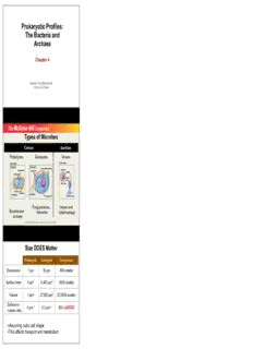
Prokaryotic Profiles: The Bacteria and Archaea Chapter 4 Adapted from McGraw Hill by Dr. G Cornwall Types of Microbes CCeelllluullaarr Acellular Prokaryotes Eukaryotes Viruses (a) Cell Types (b) VirusTypes Prokaryotic Eukaryotic Chromosome Ribosomes NucleusMitochondria EnvelopeCapsid Ribosomes Nucleic acid AIDS virus CellwallCell Flagellum membrane Flagellum Cell membrane Bacterial virus Fungi,protozoa, Viruses and Bacteria and helminths bacteriophage archaea Copyright © The McGraw-Hill Companies, Inc. Permission required for reproduction or display. Size DOES Matter Prokaryote Eukaryote Comparison Dimensions 1 µm 30 µm 30X smaller Surface Area 6 µm2 5,400 µm2 900X smaller Volume 1 µm3 27,000 µm3 27,000X smaller Surface-to- 6 µm-1 0.2 µm-1 30X LARGER volume ratio •Assuming cubic cell shape •This affects transport and metabolism 4.1 Prokaryotic Form and Function Structures common to bacterial cells Common to ALL •Cell membrane •Cytoplasm •Ribosomes •One (or a few) chromosomes Found in most •Cell wall •Surface coating or glycocalyx Structures found in some bacterial cells •Flagella •Pili •Fimbriae •Capsules •Slime layers •Inclusions •Actin cytoskeleton •Endospores Prokaryote Cell 4.2 External Structures •Appendages: Cell extensions •Common but not present on all species •Can provide motility (flagella and axial filaments) •Can be used for attachment and mating (pili and fimbriae) Flagella • Three parts: Filament, hook (sheath), and basal body • Vary in both number and arrangement • Polar arrangement: flagella attached at one or both ends of the cell • Monotrichous- single flagellum • Lophotrichous- small bunches or tufts of flagella emerging from the same site • Amphitrichous- flagella at both poles • Peritrichous- dispersed randomly over the structure of the cell (not polar) Flagellar Function & locomotion •Flagellated bacteria can detect and move in response to chemical stimuli - Chemotaxis •positive chemotaxis = movement in the direction of a favorable chemical stimulus (usually a nutrient) •negative chemotaxis = movement away form a repellant (usually harmful) compound •mechanism for detecting chemicals is linked to flagellar function •Some photosynthetic bacteria exhibit Phototaxis - movement in response to light. Bacterial Motility • Move by runs and tumbles • counterclockwise spin of flagella propels forward • clockwise the cell stops and tumbles (changes course) • in peritrichous forms the flagella sweep towards one end, and rotate together • Attractant molecules may inhibit tumbles and permit progress • Repellant molecules cause numerous tumbles allowing bacterium to redirect itself Flagella of Spirochetes Spirochetes have an unusual wriggly movement • Axial filaments (periplasmic flagella) located in the periplasm • between cell wall and cell membrane • Bundles of many flagella • contractions cause the bacterium to corkscrew • motion can be seen in the spirochetes of syphilis Appendages for Attachment & Mating • Fimbria - used for attachment and sometimes motility • attach to each other and surfaces • biofilms • Pili (sex pilus)- used for attachment and genetic exchange (conjugation) • rigid tubular structure made of protein • so far only found in gram negative Concept Check What type of flagellar arrangement has appendages at both poles of a rod-shaped cell? A. Monotrichous B. Amphitrichous C.Lophotrichous D.Peritrichous The Glycocalyx • Develops as a coating of repeating polysaccharide units, protein, or both • Protects the cell • Sometimes helps the cell adhere to the environment • Differ among bacteria in thickness, organization, and chemical composition • Slime layer- a loose shield that protects some bacteria from loss of water and nutrients • Capsule- when the glycocalyx is bound more tightly to the cell and is denser and thicker Functions of the Glycocalyx •Capsule is formed by many pathogenic bacteria- protect the bacteria against phagocytes •Slime layer is important in formation of biofilms •Capsule- important for pathogenesis, prevent phagocytosis 4.3 The Cell Envelope: The Boundary layer of Bacteria •Majority of bacteria have a cell envelope •Lies outside of the cytoplasm •Composed of two or three basic layers •Cell wall •Cell membrane •In some bacteria, the outer membrane Differences in Cell Envelope Structure • The differences between gram-positive and gram- negative bacteria lie in the cell envelope • Gram-positive • Two layers • Cell wall and cytoplasmic membrane • Gram-negative • Three layers • Outer membrane, cell wall, and cytoplasmic membrane Structure of the Cell Wall •Helps determine the shape of a bacterium •Provides strong structural support •Most are rigid because of peptidoglycan content •alternating glycans G & M) •peptide cross-bridges • Keeps cells from rupturing because of changes in pressure due to osmosis • Target of many antibiotics- disrupt the cell wall, and cells have little protection from lysis • Gram-positive cell wall • A thick (20 to 80 nm), homogeneous sheath of peptidoglycan • Contains tightly bound acidic polysaccharides • Gram-Negative Cell Wall • Single, thin (1 to 3 nm) sheet of peptidoglycan • Periplasmic space surrounds the peptidoglycan Gram Staining Nontypical Cell Walls • Some aren’t characterized as either gram-positive or gram-negative • Some don’t have a cell wall at all • Archae- unusual and chemically distinct cell walls • Mycoplasmas and some archea - lack cell wall entirely • membrane is stabilized by sterols and is resistant to lysis • Very small bacteria (0.1 to 0.5 µm) • Range in shape from filamentous to coccus • Important medical species: Mycoplasma pneumonia •Mycobacterium and Nocardia- unique types of lipids •Contain mycolic acid (a wax) •Modified Gram-positive structure •Includes important pathogens •Tuberculosis •Leprosy •Opportunistic wound infections Concept Check You have isolated a bacterium from your skin. Chemical analysis shows that it contains proteins, peptidoglycan, lipids, DNA, and acidic polysaccharides (teichoic acid). What sort of bacteria is this? A. Gram positive B. Gram negative C. Cannot tell from this information The Gram-Negative Outer Membrane • Similar to the cell membrane, except it contains specialized polysaccharides and proteins • Uppermost layer- contains lipopolysaccharide (LPS) • Innermost layer- phospholipid layer anchored by lipoproteins to the peptidoglycan layer below • Outer membrane serves as a partial chemical sieve • Only relatively small molecules can penetrate • Access provided by special membrane channels formed by porin proteins Cell Membrane Structure • Also known as the cytoplasmic membrane • Very thin (5-10 nm) • Contain primarily phospholipids and proteins • The exceptions: mycoplasmas and archae • Functions • Provides a site for functions such as energy reactions, nutrient processing, and synthesis • Regulates transport (selectively permeable membrane) • Secretion Practical Considerations of Differences in Cell Envelope Structure • Outer membrane- an extra barrier in gram-negative bacteria • Makes them impervious to some antimicrobial chemicals • Generally more difficult to inhibit or kill than gram- positive bacteria • Cell envelope can interact with human tissues and cause disease • Corynebacterium diphtheriae - diphtheria • Streptococcus pyogenes - strep throat 4.4 Bacterial Internal Structure •Contents of the Cell Cytoplasm •Gelatinous solution •Site for many biochemical and synthetic activities •70%-80% water •Also contains larger, discrete cell masses (chromatin body, ribosomes, granules, and actin strands) Bacterial Chromosome •Single circular strand of DNA •Aggregated in a dense area of the cell- the nucleoid Plasmids •Nonessential pieces of DNA •Double-stranded circles of DNA •Often confer protective traits such as drug resistance or the production of toxins and enzymes
Description: