Primary Aberrant Oculomotor Nerve Regeneration From a Posterior Communicating Artery Aneurysm PDF
Preview Primary Aberrant Oculomotor Nerve Regeneration From a Posterior Communicating Artery Aneurysm
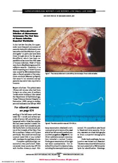
CLINICOPATHOLOGICREPORTS,CASEREPORTS,ANDSMALLCASESERIES SECTIONEDITOR:W.RICHARDGREEN,MD Human Subconjunctival Infection of Macacanema formosana: The First Case of Human Infection Reported Worldwide In the last few decades, the appar- ently more frequent occurrence of zoonotichelminthinfectionsinman haveattractedtheattentionofpara- sitologists.1Inparticularhavebeen those brought about by the genus Dirofilaria, which has been re- portedtocausemorethan900cases of human infection. Most of them wereduetoDirofilariarepensandDi- rofilaria immitis. Therefore, it is worthreportingonarecenthuman casecausedbyMacacanemaformo- sana,afilarialparasiteofthecatar- Figure1.Thesubconjunctivalwormunderslitlampbiomicroscope.Itwasmotileandactive. rhinemonkeys(Macacacyclopsis), thatcametoourattentionandap- parentlyhasneverbeenreportedin humans. ReportofaCase.Thepatientwasa 19-year-old woman who had been livinginanurbanareaofnorthern Taiwansincechildhood.Shevisited theDepartmentofOphthalmology, TaipeiVeteransGeneralHospitalin November 1999 owing to sudden painandrednessinherlefteye.Mild For editorial comment see page 634 swellingoftheleftlowereyelidwas notedfor1monthandseveralepi- sodesofsevereleftperiorbitalpain happenedwithinthesefewmonths. ShehadnevertraveledoutsideTai- Figure2.Thewholenematodemeasured7.5(cid:1)0.5cm. wanandneverhadanimalsaspets. Herfamilyhadaroutineannualpic- lampexaminationdisclosedasub- normal.Nomicrofilariawasfound nicattheriversideoftheHsin-Tien conjunctivalactivewormatthenasal inbloodandurinesamples.Nole- ShiinnorthernTaiwanuntil3years sideofherlefteyewithmarkedcon- sionwasseenonchestradiographs previously.Shecouldnotrecallany junctivalcongestion(Figure1).In- or brain computed tomographic unusual insect bite or any contact direct ophthalmoscopic examina- scans. withmonkeysduringthesepicnics. tion revealed a normal fundus. Underlocalanesthesia,thecon- Completeophthalmologicex- Findingsonexaminationoftheright junctivawasincisedanddissected. amination was performed. Best- eye were unremarkable. The re- Thewormwasremovedaliveandin- correctedvisualacuitywas6/4OU sultsofbloodandbiochemicaltests, tactandpreservedin5%formalde- with Snellen distance chart. Slit- includingtheeosinophilcount,were hyde for identification. Histologi- (REPRINTED)ARCHOPHTHALMOL/VOL120,MAY2002 WWW.ARCHOPHTHALMOL.COM 643 ©2002AmericanMedicalAssociation.Allrightsreserved. Downloaded From: https://jamanetwork.com/ on 02/19/2023 arowofapproximately7caudalpa- pillae,asymmetricallyarranged,on eithersideofthecloaca. Theesophaguscouldbevisu- alizedthroughthesemitransparent bodywall.Itwasdividedintoashort muscularportionandamuchlonger glandularportion.Thelateralchords contained dark, granular material that could be seen throughout the lengthofthechords(Figure5). Inthetransversesectionofthe worm (Figure 6), the multilay- eredstructureofthecuticlewasevi- dentaswastheunderlyingthinhy- podermis. The large lateral chords displayed clusters of pigmented granulesthatwerescatteredinthe hypodermisaswell.Themusclecells werecoelomyarianandnumerousin Figure3.Thecephalicendofthenematode(originalmagnification(cid:1)60). eachquadrantofthebody.Thepseu- docoelewasvirtuallyfilledwiththe large genital tube packed with de- veloping spermatozoa (spermato- cytes).Thedigestivetubewassmall and round and lined with a rela- tively thick endothelium. On the basis of these morphological char- acteristics, the nematode can be identifiedasanadultmaleofMaca- canemaformosana(Nematoda,On- chocercidae, Dirofilariinae). The morphologicalcharacteristicstodif- ferentiate M formosana from other nematodes (Filarioidea, Ascari- doid, Oxyuroidea, Strongylid, Spi- rurid,Strongyloid,Rhabditoid,and Trichinellae)werewelldescribedby OrihelandAsh.1SchadandAnder- son2hadreportedthedifferentialdi- agnosisofMformosanawithother Onchocercidae, in particular with Edesonfilariamalayensis,thenema- Figure4.Thecaudalendofthenematodewherethe2asymmetricalspicules(S)andsomepapillae(P) canbeseen(originalmagnification(cid:1)60). todemostsimilartoMformosana. calsectionswerepreparedforfurther therconfirmationoftheidentity.It Comment.Macacanemaformosana studyoftheparasite.Periorbitalpain was threadlike and approximately wasfirstidentifiedanddescribedby and conjunctival congestion re- 7.5cmlongwithamaximumdiam- SchadandAndersonin1963.2Itwas solvedafterthesurgery.Therewas eterof510µm(Figure2).Thean- anematodeofnewgenusandspe- nosignofrecurrentinfectionafter teriorextremity(Figure3)wassub- cies of the family Onchocercidae, 2years’follow-up. conical with a small, round oral subfamilyDirofilariinae.Theyfound opening without lips and sur- thefilariaintheperitrachealtissue Parasitologic Findings. The para- rounded by 2 pairs of circumoral and diaphragm of Macaca cyclop- site was sent to the Department of papillae.Thecuticlewasthinwith sis, a monkey native to Formosa Public Veterinary Health and Ani- fine transverse striations. The tail (Taiwan). Like all filariae, the mi- malPathology,UniversityofBolo- (Figure4)wasshortwith2spic- crofilariaewerefoundintheblood gna,Bologna,Italy,andtheDepart- ulesofunequallength.Thelonger ofthehost.Littlewasknownabout mentofTropicalMedicine,Tulane one(ontheworm’sleftside)mea- thisparasiteexceptforareportpub- UniversityMedicalCenter,NewOr- sured 512 µm and the shorter (on lished in 1968 by Bergner and leans, La, for identification. It was the worm’s right side) one, par- Jachowski.3 They found the para- later sent to the Museum of Natu- tially protruding from the cloaca, site in the peritracheal and man- ral History, Paris, France, for fur- wasabout120µmlong.Therewas dibularintermuscularconnectivetis- (REPRINTED)ARCHOPHTHALMOL/VOL120,MAY2002 WWW.ARCHOPHTHALMOL.COM 644 ©2002AmericanMedicalAssociation.Allrightsreserved. Downloaded From: https://jamanetwork.com/ on 02/19/2023 sues.Therewasnosubconjunctival infection reported in the monkey. TheprevalencerateofMformosana in the Taiwanese monkey was re- portedtobeashighas42%innorth- ern Taiwan where the patient had picnicked.Culicoides(Insecta,Dip- tera, Ceratopogonidae), a cosmo- politan genus of biting midges of- tencausinggreatannoyancetoman and animals, was proposed as the mostprobabletransmittingvectorof theparasite.ManyspeciesofCuli- coides were identified in northern Taiwan.Thislargereservoirofthe parasiteandtheexistenceofthevec- tors made human infection pos- sible.Toourknowledge,thisisthe firstcaseofhumaninfectioncaused by M formosana to be reported worldwide.However,othercasesin Figure5.ThedetailofasegmentofMacacanemaformosanawherethescatteredbrownishgranules thesameareamaywellhavetaken withinthelateralchords(L)canbeseen(originalmagnification(cid:1)60). place,butunobserved,asseemedto happen with D repens in endemic zones.4Theprepatentperiodofthe wormhasnotyetbeendetermined. Basedonthesizeoftheworm,the patientwouldhavebeeninfectedfor at least 1 year and symptoms ap- peared only after the worm mi- grated into the subconjunctival space.ThereportedhumanDirofi- lariainfectionsusuallyconsistedof asingleworm,5andtreatmentforthe sorenessconsistedoftheworm’ssur- gical removal. There was no evi- denceofrecurrentinfectioninour patientafter2years’follow-up. CasesofsubconjunctivalDre- pensinfectionoccurwidelythrough- outEuropean,African,MiddleEast- ern,andAsiancountries.However,it hasneverbeenreportedinTaiwan. Fromthecasesreportedandourob- Figure6.Thetransversesectionofthenematode(partial).Cindicatescuticle;I,intestine;M,muscular servation,subconjunctivalDrepens fibers;T,testicle;andarrowheads,brownishgranulesinthelateralchord(trichrome-Masson-Goldner and M formosana infections shared stain,originalmagnification(cid:1)250). the characteristics of sudden onset andweretreatedsolelybysurgicalre- nematodes,suchasMansonellaper- sible zoonotic infection should be movaloftheworm.Fromthesizeof stansandEdesonfilariaspecies,and keptinmind. the worms recovered, they might intheselfsameMformosana.Their have been migrating in subcutane- origin is obscure and further stud- Ling-IngLau,MD ous space for a period of time and ies are necessary to understand Fenq-LihLee,MD causedsymptomsonlywhentheyap- theirnature. Wen-MingHsu,MD peared in the subconjunctiva. The Thisis,toourknowledge,the Taipei,Taiwan episodic periorbital pain preceding firstcasereportofhumaninfection SilvioPampiglione,MD,PhD subconjunctivalinfectioninourpa- caused by M formosana although MariaLetiziaFioravanti,PhD tientcouldbecausedbytheworm’s other cases may have gone by un- Bologna,Italy migrationwithinthisarea. observedorunidentified.Sincethe ThomasC.Orihel,PhD Thepresenceofbrownishgran- Taiwan monkeys (Macaca cyclop- NewOrleans,La ules in the lateral chords is a phe- sis) have been largely involved in nomenon that has been observed laboratory studies and have close We would like to thank Shyh-Haw on other occasions both in other contact with human beings, pos- Tsay,MD,DepartmentofPathology, (REPRINTED)ARCHOPHTHALMOL/VOL120,MAY2002 WWW.ARCHOPHTHALMOL.COM 645 ©2002AmericanMedicalAssociation.Allrightsreserved. Downloaded From: https://jamanetwork.com/ on 02/19/2023 TaipeiVeteransGeneralHospital,and tinellymphnode(SLN)biopsysoon sioninhisleftuppereyelid,andthe F. Rivasi, PhD, histopathologist at afterthediagnosisofhisprimarytu- areaoftheneckdissectionandpa- ModenaUniversity,Emilia,Italy,for mor. An SLN was identified and rotidectomyhadhealed.Findingson preparingthehistologicalsectionsof showedhistologicevidenceofMCC. thepatient’sophthalmologicexami- the parasite; and Kin-Mu Lee, PhD, To our knowledge, this is the first nationwereessentiallynormal,with Department of Parasitology, Na- reportedcaseofapositiveSLNsec- no clinical evidence of local or re- tionalYangMingUniversity,Taipei, ondarytoMCCoftheeyelid. gionalrecurrenceofcancer.Hissys- and Odile Bain, PhD, Museum of temic workup, including com- Natural History, Paris, France, for ReportofaCase.A61-year-oldman putedtomographyoftheheadand theirkindsuggestionsontheidentity notedanerythematouslesiononhis neck,abdomen,andpelvis,chestra- oftheparasite. left upper eyelid in May 2001. He diography, and magnetic reso- Presentedasaposteratthe18th went to his local ophthalmologist, nance imaging of the brain, was CongressoftheAsia-PacificAcademy whoexcisedthelesionbutdidnot negative for tumor. A multidisci- of Ophthalmology, Taipei, Taiwan, examine it histologically. In June plinaryteam,includinganophthal- March10-14,2001. 2001, the lesion recurred. The re- micsurgeon,aheadandnecksur- Corresponding author: Fenq- current lesion measured approxi- geon, a head and neck medical Lih Lee, MD, Department of Oph- mately12mmindiameter.Thepa- oncologist,andaheadandneckra- thalmology,TaipeiVeteransGeneral tient sought an opinion from an diation oncologist examined the Hospital,No.201,Section2,Shih-Pai oculoplastic surgeon, who per- patient and recommended that he Road, Shih-Pai, Taipei 11217, Tai- formedawidelocalexcisionofthe receive adjuvant external beam wan (e-mail address: fllee@ms3 lesionwithfrozensectioncontrolof radiation therapy to the eyelid, .hinet.net). the margins. The histologic find- parotidnodes,anddeepercervical Reprints:Ling-IngLau,MD,De- ings were consistent with MCC of nodes. The team also recom- partment of Ophthalmology, Taipei theeyelid(Figure,AandB).Sen- mendedthatthepatientbegiven4 VeteransGeneralHospital,No.201, tinellymphnodebiopsywassched- coursesofchemotherapywitheto- Section 2, Shih-Pai Road, Taipei uledbuthadtobedelayedbecause poside and cisplatin after the 11217,Taiwan(e-mail:lilau@ms41 thepatientdevelopedacuteappen- completionofradiationtherapy. .hinet.net). dicitis,necessitatinganemergentap- pendectomy. In July 2001, the pa- Comment. In patients with MCC, 1. OrihelThC,AshLR.ParasitesinHumanTis- tient underwent SLN biopsy using the regional lymph nodes are sues.Chicago,Ill:AmericanSocietyofClinical Pathologists;1995. acombinationofisosulfanbluedye thoughttobethemostcommonand 2. SchadGA,AndersonRC.Macacanemaformo- andTc99m–labeledsulfurcolloid. earliestsiteofmetastasis;thus,ad- sanan.g.,n.sp.(Onchocercidae:Dirofilariinae) Theafferentlymphaticswereiden- juvant treatment of the regional fromMacacacyclopsisofFormosa.CanJZool 1963;41:797-801. tified before surgery using radio- lymphnodeshasbeenadvocatedby 3. BergnerJF,JachowskiLA.Thefilarialparasite, nucleotide imaging. Intraopera- many investigators.3,5,6 Jean et al7 Macacanemaformosana,fromtheTaiwanmon- keyanditsdevelopmentinvariousarthropods. tively,acombinationofradiolabeled successfully identified 1 or more FormosanSci.1968;22:1-68. sulfurcolloidandisosulfanbluedye SLNsin19of20patientswithstage 4. PampiglioneS,CanestriTrottiG,RivasiF.Hu- wasusedtoidentifySLNs.Anarea IMCCwhounderwentSLNbiopsy mandirofilariasisduetoDirofilaria(Noch- tiella)repens:areviewofworldliterature.Paras- offocalradioactiveuptakewasiden- atthetimeofinitialwidelocalex- sitologia.1995;37:149-193. tified in the left preauricular (pa- cision. The authors found that 5 5. Ruiz-MorenoJM,Bornay-LlinaresFJ,PrietoMaza rotid)areausingahandheldgamma (26%) of the 19 patients in whom G,MedranoM,SimonF,EberhardML.Subcon- junctivalinfectionwithDirofilariarepens:sero- probeandwasmarkedontheskin. SLNs were successfully identified logicalconfirmationofcurefollowingsurgery. The corresponding SLN was re- had at least 1 histologically posi- ArchOphthalmol.1998;116:1370-1372. moved and analyzed histologically tiveSLN.OtherisolatedcasesofSLN using serial sectioning and immu- biopsyforMCChavealsobeenre- nohistochemicalstaining.Thenode ported.8 Merkel Cell Carcinoma was found to be positive for MCC Merkel cell carcinoma of the of the Eyelid With a (Figure,CandD).Thepatientsub- eyelidisthoughttoaccountfor10% Positive Sentinel Node sequentlyunderwentatotalparoti- ofallcasesofMCC.2Inareviewof dectomy and completion neck allpreviouslyreportedcasesofMCC Merkelcellcarcinoma(MCC)ofthe dissection.Theparotidectomyspe- of the eyelid, Kivela and Tarkkat- eyelid is a rare but aggressive ma- cimenincluded1additionallymph nen2concludedthatuptotwothirds lignancythatmetastasizesearlytore- nodethatwaspositiveforMCC.The of patients eventually develop re- gionallymphnodes.1Mostclinical node was located in the deep lobe gionalnodalinvolvement.Thisrate seriessuggestarateofregionalnodal oftheleftparotidglandandshowed is higher than the rate reported in involvement between 21% and extracapsularextension. mostsingleseriesofMCCoftheeye- 66%.2-4Earlydetectionofoccultre- In September 2001, the pa- lid.Inthelargestsingleseriestodate, gionalnodaldiseasemayallowfor tientself-referredtotheUniversity Peters et al3 reported clinical re- earlyinstitutionofadjuvanttherapy. of Texas M. D. Anderson Cancer gionalnodalinvolvementin3(21%) WedescribeapatientwithMCCof Center(Houston)forfurtherman- oftheir14patients. the eyelid with clinically unin- agementofhistumor.Atthistime, Sentinellymphnodebiopsyal- volvednodeswhounderwentsen- he had a well-healed area of exci- lowsforearlydetectionofoccultre- (REPRINTED)ARCHOPHTHALMOL/VOL120,MAY2002 WWW.ARCHOPHTHALMOL.COM 646 ©2002AmericanMedicalAssociation.Allrightsreserved. Downloaded From: https://jamanetwork.com/ on 02/19/2023 A B C D A,HistologicsectionofMerkelcellcarcinoma(MCC)oftheuppereyelid(primarylesion)demonstratesadiffuse,poorlycohesiveproliferationofsmallcellswith finelydispersedchromatin,lackingprominentnucleoli,andwithoutsignificantinterveningstroma(originalmagnification(cid:1)4).B,High-poweredviewshowsround cellswithfinelydistributedchromatin.Manyofthecellsdisplayhyperchromaticnucleiconsistentwithapoptoticbodies(originalmagnification(cid:1)40).C,Histologic sectionofthesentinellymphnodeshowssmallfociofroundcellssimilarinappearancetotheprimarylesion(originalmagnification(cid:1)20).D,High-poweredview ofthesamenodeimmunostainedwithantibodiesagainstcytokeratinshowspositivecytokeratinexpressionsupportingthediagnosisofmetastaticMCC(original magnification(cid:1)40). gional lymph node metastasis and moid carcinoma), using radiola- tion of micrometastasis in the re- thus,moreaccuratestagingofMCC beled sulfur colloid alone. These gional nodes allows for immediate andthepossibleinstitutionofearly authorsreportedsuccessfulidenti- institution of adjuvant therapy, adjuvanttherapy.AlthoughSLNbi- fication of at least 1 SLN in 5 pa- whichmayincludecompletionneck opsyhasrecentlybecomethestan- tients,althoughthetechniqueused dissection,externalbeamradiation dard of care for most solid tumors in the latter report is believed by therapy, and adjuvant chemo- throughoutthebody,SLNbiopsyin someinvestigatorsnotlikelytolead therapy. theperioculararearemainsinvesti- to the correct identification of the gational.9Toourknowledge,there sentinel nodes.12 Neither of the 2 BitaEsmaeli,MD havebeenonly2previousreportsof previousreportsofSLNbiopsyfor AresuNaderi,MD application of SLN biopsy tech- conjunctival and periocular tu- LillieHidaji,BS niquesforconjunctivalandperiocu- morsfoundapositiveSLN.Toour GeorgeBlumenschein,MD lar tumors.10,11 We described suc- knowledge, ours is the first re- VictorG.Prieto,MD,PhD cessful identification of SLNs in a ported case in which an SLN was Houston,Tex single patient with a conjunctival successfully identified in a patient melanoma,usingacombinationof withaneyelidtumorandwasalso Corresponding author and reprints: radiolabeledsulfurcolloidandiso- foundtobehistologicallypositive. BitaEsmaeli,MD,OphthalmologySec- sulfanbluedye.10Wilsonetal11at- Thiscaseunderscoresthefeas- tion, Department of Plastic Surgery, temptedidentificationofSLNsin5 ability and potential usefulness of Box443,M.D.AndersonCancerCen- patientswithperioculartumors(2 SLNbiopsyasamethodforidenti- ter,1515HolcombeBlvd,Houston,TX melanomas,2meibomianglandcar- fyingoccultmetastaticdiseasefrom 77030(e-mail:besmaeli@mdanderson cinomas,and1caseofmucoepider- anMCCoftheeyelid.Earlydetec- .org). (REPRINTED)ARCHOPHTHALMOL/VOL120,MAY2002 WWW.ARCHOPHTHALMOL.COM 647 ©2002AmericanMedicalAssociation.Allrightsreserved. Downloaded From: https://jamanetwork.com/ on 02/19/2023 1. SinghAD,EagleRCJr,ShieldsCL,ShieldsJA. relatedmaculardegenerationwith- Clinic,Rochester,Minn,onMarch Merkelcellcarcinomaoftheeyelids.IntOph- out observing deleterious retinal 8,2000,becauseofagrowingpig- thalmolClin.1993;33:11-17. 2. KivelaT,TarkkanenA.TheMerkelcellandas- complications.5Theencouragingre- mentedchoroidallesionintheleft sociatedneoplasmsintheeyelidsandperiocu- sultsinpilotstudieswithTTTinthe eye that had been observed to in- larregion.SurvOphthalmol.1990;35:171- management of occult choroidal crease in thickness from 2.3 to 187. 3. PetersGB,MeyerDR,ShieldsJA,etal.Man- neovascularmembraneshasledto greaterthan4mmduringaninter- agementandprognosisofMerkelcellcarci- the development of a multicenter valof9years. nomaoftheeyelid.Ophthalmology.2001;108: prospectiverandomizedclinicaltrial Thevisualacuitywas20/20OD 1575-1579. 4. VictorNS,MortonB,SmithJW.Merkelcellcan- (Transpupillary Thermotherapy and20/25+3OS.Therighteyewas cer:isprophylacticlymphnodedissectionin- [TTT]ofOccultSubfovealChoroi- normal.Resultsofexaminationofthe dicated?AmSurg.1996;62:879-882. 5. FenigE,BrennerB,KatzA,etal.Theroleof dal Neovascularization in Patients lefteyeshowedanormalanteriorseg- radiationtherapyandchemotherapyinthe With Age-Related Macular Degen- mentandapigmented,elevatedcho- treatmentofMerkelcellcarcinoma.Cancer. erationTrial)inwhichpatientswith roidallesion,measuring9(cid:1)9mmin 1997;80:881-885. 6. BoyleF,PendleburyS,BellD.Furtherin- subfoveal choroidal neovascular base dimension, located approxi- sightsintothenaturalhistoryandmanage- membranes are randomized to a mately4mmsuperonasaltothedisc mentofprimarycutaneousneuroendocrine (Merkelcell)carcinoma.IntJRadiatOncolBiol shamtreatmentoratreatmentwith (Figure 1). The ultrasonographic Phys.1995;31:315-323. a single 60-second exposure of in- studiesdemonstratedasolid,dome- 7. JeanM,SolorzanoC,RossM,etal.Roleforsen- fraredlightfromthediodelaser(810 shapedtumor(B-scan)withlowin- tinelnodebiopsyinMerkelcellcarcinomapa- tients.ProcAmSocClinOncol.2001;20:358a. nm)usingabeamdiameterof3mm ternal reflectivity (A-scan) consis- 8. WasserbergN,FeinmesserM,SchachterJ,Fenig and 800 mW of power (Elias Rei- tentwiththediagnosisofmelanoma. E,GutmanH.Sentinel-nodeguidedlymph- chel,MD,oralandwrittencommu- Thethicknessofthelesionwas4.4 nodedissectionforMerkelcellcarcinoma.Eur JSurgOncol.1999;252:444-446. nication, January 12, 2000). The mm.Asubretinalfibroticplaqueover- 9. EsmaeliB.Sentinellymphnodemappingfor uniqueopportunityaffordedbyapa- lying the central portion of the tu- patientswithcutaneousandconjunctivalmela- tientscheduledforenucleationfor morandasecondaryretinaldetach- noma.OphthalmolPlastReconstrSurg.2000; 16:170-172. amalignantmelanomalocatedinthe mentoverlyingthenasalportionof 10. EsmaeliB,EicherS,PoppJ,etal.Sentinellymph nasal choroid led to this experi- thetumorwereseen.Subretinalfluid nodebiopsyforconjunctivalmelanoma.Oph- thalmolPlastReconstrSurg.2001;17:436-442. mentinwhichinfraredlightfroma extendedapproximately1discdiam- 11. WilsonMW,FlemingJC,FlemingRM,Haik diode laser was directed to the eterbeyondthenasalperipheryofthe B.Sentinelnodebiopsyfororbitalandocular macula through a contact lens us- mass. Small accumulations of exu- adnexaltumors.OphthalmolPlastReconstrSurg. 2001;17:338-344. ingthevariablesidenticaltothose datesatthenasalboundaryofthetu- 12. EsmaeliB.Indiscussionof:WilsonMW,Flem- recommended in the TTT of Oc- morwerealsoseen.Theretinainthe ingJC,FlemingRM,HaikB.Sentinelnodebi- cult Subfoveal Choroidal Neovas- central macular region appeared opsyfororbitalandocularadnexaltumors.Oph- thalmolPlastReconstrSurg.2001;17:338-345. cularization in Patients With Age- subtlythickenedonbiomicroscopy RelatedMacularDegenerationTrial. findings. Thediagnosisofactivelygrow- Report of a Case. A 65-year-old ingmalignantmelanomawasmade, The Effect of Transpupillary womanwasreferredtotheDepart- and definitive therapy was recom- Thermotherapy on the mentofOphthalmologyoftheMayo mended.Therapeuticoptionswere Human Macula Transpupillarythermotherapy(TTT) wasintroducedbyinvestigatorsfrom theNetherlandsin1995asanalter- nativetreatmentforchoroidalmela- noma.1,2 Since that time, TTT has been used to treat small choroidal melanomas,andpreliminaryresults indicatingthatTTTcancontrolsmall melanomas with follow-up of 5 or more years have been published.3,4 However, localized retinal destruc- tion,vascularocclusions,andnerve fiberbundledefectsarecommonlyas- sociatedwitheffectivetreatmentof small melanomas with TTT. De- spitetheseobservedretinalcompli- cations,someinvestigatorshavere- centlyreportedthatTTT,usingthe samelaserintensitytotreatchoroi- dalmelanoma,maysuccessfullytreat Figure1.Colorfundusphotographshowingthelocationofachoroidalmelanomainthelefteye. occult subfoveal choroidal neovas- Aserousdetachmentofthesensoryretinawasclinicallyconfinedtotheregionoverlying cularizations in patients with age- thenasalportionofthetumor. (REPRINTED)ARCHOPHTHALMOL/VOL120,MAY2002 WWW.ARCHOPHTHALMOL.COM 648 ©2002AmericanMedicalAssociation.Allrightsreserved. Downloaded From: https://jamanetwork.com/ on 02/19/2023 A B C D Figure2.Prelaserexposure.A,Colorfundusphotographofposteriorpoleshowinganormal-appearingmacularregion.B,Earlyarteriovenousphaseduring fluoresceinangiography.C,Arteriovenousphaseduringfluoresceinangiographyshowingsubtlehyperfluorescenceintheposteriorpoleandearlycystoidedema. D,Recirculationphaseshowingpatchydiffuseintraretinaldyestainingandpatternofsectorcystoidmacularedema. discussed. Brachytherapy was en- the macula of the left eye was ex- dehyde. The retina and choroid in couraged,butthepatientwascon- posed to a 3-mm beam of infrared themaculaweredissectedenbloc, cernedaboutthepotentialforcon- laserlight(810nm)using800mW fixedinsolution,andembeddedfor tinuingproblemswiththeeyeand ofpower.Thelaserbeamwasdeliv- transmission electron microscopy. stronglydesiredanenucleation. ered to the posterior pole through Lightmicroscopyofthisregionwas We obtained institutional re- astandardfunduscontactlens(Carl not performed. The remaining tis- viewboardapprovalfortheexperi- Zeiss,Inc,Thornwood,NY),expos- suewasexaminedbymeansoflight ment. The patient was fully in- ingthemaculatolightfromthein- microscopy. formedregardingtheuseofinfrared frared laser for 60 seconds. Visual laser light in the management of acuity was measured with a stan- Histopathologic Findings. Color maculardiseaseandthepossibility dardSnellenchart,andthecentral photographsofthemacularregion thatthetreatmentcouldcauseanal- fieldwasevaluatedwithanAmsler showed no obvious abnormality terationofthepigmentepithelium grid3minutesafterlightexposure. (Figure2A).Thepretreatmentfluo- and retina overlying the choroidal Fivedaysafterlaserexposure,were- resceinangiogramshowedanearly targettissue.Sheagreedtohaveher examinedtheeye.Bestcorrectedvi- pattern of complex vascular loops retinaexposedtolightfromthein- sualacuitywasdetermined,andthe withinthetumorandpatchyleak- frared laser for 60 seconds using a central visual field was evaluated ageofdyefromthesesitesleading powerof800mW.Shealsoagreed with an Amsler grid. Biomicros- toearlypatchytissuestainingofthe toundergocolorfundusphotogra- copyandophthalmoscopywereper- tumor.Laterframesshoweddiffuse phyandfluoresceinangiographyof formed, and the appearance of the stainingoftheretinaoverlyingthe theretinabeforeandafterlightex- posteriorpolewasdocumentedwith tumor.Dyeleakagefromfluorescein- posure. colorfundusphotographyandfluo- incompetentcapillariesintheperi- Afterdocumentingtheappear- rescein angiography. The eye was fovealregionwasseen,whichpro- anceofthetumor(Figure1)andthe enucleated approximately 3 hours duced an incomplete pattern of maculabymeansofslitlampbiomi- laterandfixedina0.1Mphosphate- cystoid edema visible in the late croscopy color photographs and buffered solution containing 4% framesofthestudyandarclikear- fluoresceinangiographybeforeTTT, paraformaldehydeand1%glutaral- easofdiffuseintraretinalstainingsu- (REPRINTED)ARCHOPHTHALMOL/VOL120,MAY2002 WWW.ARCHOPHTHALMOL.COM 649 ©2002AmericanMedicalAssociation.Allrightsreserved. Downloaded From: https://jamanetwork.com/ on 02/19/2023 A B C Figure3.Fivedaysafterinfraredlaserexposure.A,Color fundusphotographs5daysafter60-secondinfraredlaser exposureofthemaculatoa3-mmbeamofinfrared lightfromthediodelaserusing800mWofpower. B,Arteriovenousphaseduringfluoresceinangiography showingsubtlehyperfluorescenceinposteriorpoleandearly sectorcystoidedema.C,Recirculationphaseshowing diffuseintraretinaldyeleakageandsectorcystoidmacular edema.Thefindingsareunchangedfromthosedocumented inFigure2. periortothefovea,temporaltothe changeintheappearanceofthefun- shapes rather than the usual oval- fovea,andtoalesserextentinfero- dus. The fluorescein angiogram at shapedmelanosomegranules(Fig- temporaltothefovea(Figure2B-D). the 5-day follow-up visit demon- ure4C).Focaldisruptionofcellu- Thelargestdimensionofthestain- strated findings identical to those lar membranes and dispersion of ingsitehadadiameterofapproxi- seen in the pretreatment angio- pigmentgranulesamongouterseg- mately3mm(beforeandafterlaser gram (Figure 3). ments of photoreceptor cells were exposure). Lightmicroscopyoftheenucle- seen.Vacuolationanddistensionof During and immediately after atedeyeshowedamalignantmela- theoutersegmentsofthephotore- transpupillaryexposureofthemacula nomalocatedintheposteriorcho- ceptorswerealsoobserved,withpar- tolightfromtheinfraredlaser,nodis- roid, nasal to the optic disc, that tial disintegration of the lamellar cerniblealterationintheophthalmo- formed a mass measuring 10(cid:1)10 structure with a rare thumbprint- scopicappearanceofthefunduswas (cid:1)3 mm and consisted predomi- likeconfiguration(Figure4D).The seen.Three minutes after light ex- nantlyofepithelioidcells.Aserous underlyingchoriocapillarisshowed posure,thepatientnoticedabluish detachment of the sensory retina congestion,butthevesselswerenor- discolorationinthecentralfieldof was seen overlying the nasal por- mal,withintactwallsandnormalen- visionthatsheoutlinedontheAm- tion of the tumor. A plaque of fi- dothelial cells. The larger choroi- sler grid. The best-corrected dis- brous tissue was visible over the dal vessels also appeared normal tance visual acuity 3 minutes after central portion of the tumor, and (Figure4E). lightexposurewas20/100.Whenthe cystoiddegenerationwaspresentin patientreturned5dayslater,thecen- the overlying retina (Figure 4A Comment.Transpupillarythermo- tralvisualacuityhadrecoveredtothe andB). therapyhasbeenusedinrecentyears pretreatmentlevel(20/25),butshe Results of the ultrastructural as a therapeutic alternative in the still recognized a faint bluish dis- examination of the macular and management of some choroidal colorationinthecentralpartofher paramacularregion,whichhadbeen melanomas.1-4Wehaveshownthat visiononAmslergridtesting.Shere- dissected and embedded for trans- effectivetreatmentofselectedsmall ported that this dyschromatopsia mission electron microscopy, choroidal melanomas almost al- hadbeendecreasinginintensityeach showed retinal pigment epithelial ways leads to a profound field de- day since the exposure. Examina- cells with numerous cytoplasmic fect because of destruction of the tionofthefundus5daysafterlaser granulesoflipofuscinandmelano- photoreceptorsandnervefibersin exposure showed no discernible fuscin with round and irregular theretinaoverlyingthetreatedtu- (REPRINTED)ARCHOPHTHALMOL/VOL120,MAY2002 WWW.ARCHOPHTHALMOL.COM 650 ©2002AmericanMedicalAssociation.Allrightsreserved. Downloaded From: https://jamanetwork.com/ on 02/19/2023 A B C D E Figure4.A,Low-powerlightmicroscopyshowingchoroidal malignantmelanomawithalayerofheavilypigmentedspindle cellsnearitssurface.Afibrousplaqueisvisibleoverthe centralportionofthetumor,andtheoverlyingretinashows markedcystoiddegeneration(hematoxylin-eosin,original magnification(cid:1)20).B,High-powerlightmicroscopyshowing thatthisneoplasmiscomposedofapredominanceof epithelioidcells.Nomitosesornecrosisisseen (hematoxylin-eosin,originalmagnification(cid:1)200).C,Electron microscopyofthemacularregionatthesiteoflaserexposure showingretinalpigmentepitheliumcontainingcytoplasmic granulesoflipofuscin(arrowhead)andmelanofuscin(arrow) withroundandirregularshapes.Focaldisruptionofcellular membraneswithdispersionofpigmentgranulesintoouter segmentsofphotoreceptorcellsisseen(leadcitrate,original magnification(cid:1)25000;barindicates2µm).D,Electron microscopyshowingvacuolizationanddistensionoftheouter segmentsofthephotoreceptorswithpartialdisintegrationof thelamellarstructuresandfocalthumbprintlikeconfiguration ofsomelamellae(leadcitrate,originalmagnification(cid:1)25000; barindicates2µm).E,Electronmicroscopyshowing congestedbutotherwisenormal-appearingvesselswithintact wallsinthechoroidunderlyingthemacularretinaatthesiteof laserexposure(leadcitrate,originalmagnification(cid:1)48000; barindicates3µm). mor.4Whenusedfortreatmentofa multicenterrandomizedstudyofoc- generation(800mW,60secondsof choroidalmelanoma,thesamevari- cult choroidal neovascular mem- exposure, and 3-mm beam diam- ablesrecommendedbytheongoing branes in age-related macular de- eter)areusuallyassociatedwithex- (REPRINTED)ARCHOPHTHALMOL/VOL120,MAY2002 WWW.ARCHOPHTHALMOL.COM 651 ©2002AmericanMedicalAssociation.Allrightsreserved. Downloaded From: https://jamanetwork.com/ on 02/19/2023 tensiveretinaldamage,causingalo- pearssimilartotherelativeabsence lar region by means of ultrastruc- calized scotoma and, frequently, a ofatakethatwehaveobservedwhen turalstudies. wedge-shaped field defect as a re- attemptingtoprophylacticallytreat Theabsenceofrecognizablede- sult of nerve fiber bundle destruc- normal-appearingtissueadjacentto struction of the retina and retinal tion. Therefore, some concern ex- a pigmented choroidal melanoma. vasculatureobservedinthissingle ists that these variables have been Takesareoftennotwellseeninthe experimentdoesnotensurethatvas- chosen for treatment of choroidal normal-appearing tissue. For ex- cular closure and retinal destruc- neovascular membranes. Our pa- ample, a 3-mm beam of laser light tionwillnotoccurwhenTTTisused tient with a malignant melanoma thatisplacedsoastoequallystraddle totreatoccultchoroidalneovascu- locatedinthenasalchoroid,sched- theedgeofapigmentedchoroidaltu- larmembrane.However,inthiscase, uled for enucleation, consented to mor and clinically normal tissue withclinicalandangiographicevi- have her macula exposed to light adjacenttoitoftendramaticallyout- denceofmildretinaledemaandno fromthediodelaserusingthevari- lines the ophthalmoscopically rec- retinal or subretinal blood, a 60- ablescitedabove.Inthispatient’saf- ognizableperimeterofthetumor.The secondexposureof800mWusing fectedeye,subtleintraretinaledema tumor,theoverlyingretina,andthe a3-mmbeamdiameterdidnotcause involvedtheposteriorpole,presum- retinalpigmentepitheliumbecome clinicallyrecognizabledamagetothe ablyrelatedtotheactivelygrowing gray white, whereas the adjacent macularretina,theretinalvessels,or melanoma located in the superior retina overlying normal-appearing theunderlyingchoriocapillarisand nasalfundus.Thisintraretinalede- choroid frequently remains clini- otherchoroidalvessels. ma was recognized in the macula callyunchangedoronlyminimally beforeexposuretothelaserlight.The gray. We believe that the pigment DennisM.Robertson,MD melanomaitselfshowedcomplexvas- withinthetumorislargelyrespon- DivaR.Saloma˜o,MD cularpatternsonfluoresceinangi- sible for generating a considerable Rochester,Minn ography with extensive leakage of amountofheat,whichcausesthetu- dyethatdiffusedintotheoverlying mor,theoverlyingpigmentepithe- retina,whereheavyfluoresceinstain- lium, and retina to turn gray more Thisstudywassupportedinpartbya ingwasseeninthelatepartofthe readily.Inaddition,thechoriocap- grantfromResearchtoPreventBlind- fluoresceinstudy. illarisandlargervesselsinthecho- ness,Inc,NewYork,NY,andinpart Laser exposure of the macula roidarealteredinthepresenceofa bytheMayoFoundation,Rochester, failedtoproduceaclinicallyrecog- melanoma,thusdecreasingtheabil- Minn. nizable reaction in the retina dur- ityofthechoroidtoplayitsroleasa WethankBonnieRonkenforher ing the treatment, and no changes heatsinktodisperseenergy.Theab- secretarialsupportandCherylHann, wererecognizedonresultsofacare- senceofaheavyconcentrationofpig- MS,forhersupportwiththeelectron ful clinical examination 5 days af- mentandthepresenceofanormal microscopystudies. ter exposure. A fluorescein angio- choroid, providing a normal heat Corrresponding author and gram5daysafterthelaserexposure sink, can facilitate dispersement of reprints: Dennis M. Robertson, MD, showednodifferencefromthefluo- energy,therebyminimizingthepo- Department of Ophthalmology, resceinangiogramobtainedimme- tential for thermal damage to the Mayo Clinic, Rochester, MN 55905 diatelybeforethelaserexposure.Al- overlyingretina. (e-mail:[email protected]). thoughthecentralvisualacuitywas Inthisstudy,wewereunable 1. OosterhuisJA,Journee-deKorverJG,Kake- reducedto20/100immediatelyaf- toidentifyTTT-inducedadverseef- beeke-KemmeHM,BleekerJC.Transpupillary terexposure,5daysafterTTT,the fectsintheretinaorthepigmentepi- thermotherapyinchoroidalmelanomas.Arch centralvisualacuityhadrecovered theliumbymeansofclinicalexami- Ophthalmol.1995;113:315-321. 2. Journee-deKorverJG.TranspupillaryThermo- tothepretreatmentvisualacuity. nationorfluoresceinangiography. therapy:ANewTreatmentofChoroidalMela- Wewereunabletoseeagray- However,weobservedhistological noma.TheHague,theNetherlands:Kugler Publications;1998. ingoftheretinaafterexposuretola- andultrastructuralabnormalitiesin 3. ShieldsCL,ShieldsJA,CaterJ,etal.Trans- serenergieswiththesamedosagethat thetissueaftertheeyewasenucle- pupillarythermotherapyforchoroidalmela- ordinarilycausesagraying(“take”) ated. We believe that these abnor- noma:tumorcontrolandvisualresultsin100 consecutivecases.Ophthalmology.1998;105: whendirectedtoachoroidalmela- malities can be explained by the 581-590. noma.Thesubtleedemaofthemacu- presence of the preexisting retinal 4. RobertsonDM,BuettnerH,BennettSR.Transpu- larretinainthiscasewassimilarto edema. However, some of the ob- pillarythermotherapyasprimarytreatmentfor smallchoroidalmelanomas.ArchOphthalmol. the mild retinal edema frequently served abnormalities could have 1999;117:1512-1519. seen with occult choroidal neovas- beencausedbylightalone,asshown 5. ReichelE,BerrocalAM,IpM,etal.Transpupil- larythermotherapyofoccultsubfovealchoroi- cular membranes. However, we do by Robertson and Erickson6 and dalneovascularizationinpatientswithage- notbelievethatthepresenceofreti- GreenandRobertson.7Althoughwe relatedmaculardegeneration.Ophthalmology. nal edema could explain the ob- lookedforevidenceofvascularclo- 1999;106:1908-1914. 6. RobertsonDM,EricksonGJ.Theeffectofpro- servedabsenceofatakeintheretina sure or coagulative necrosis in the longedindirectophthalmoscopyofthehuman and/or retinal pigment epithelium smallcapillariesinthechoroid,we eye.AmJOphthalmol.1979;87:652-661. during or 5 days after the laser ex- were unable to demonstrate such 7. GreenWR,RobertsonDM.Pathologicfindings ofphoticretinopathyinthehumaneye.AmJ posure. This absence of a take ap- changesinthechoroidinthefoveo- Ophthalmol.1991;112:520-527. (REPRINTED)ARCHOPHTHALMOL/VOL120,MAY2002 WWW.ARCHOPHTHALMOL.COM 652 ©2002AmericanMedicalAssociation.Allrightsreserved. Downloaded From: https://jamanetwork.com/ on 02/19/2023
Description: