Plant Anatomy PDF
Preview Plant Anatomy
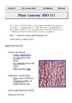
Welcome to the BIO 311 Plant Anatomy website. Students can use this site to review the cell and tissue types seen in lab. The photomicrographs included in the 'Lab Review Slides' are accompanied by a brief descriptions and questions about the specimens. Many terms are linked to an online glossary. BIO 311 will next be offered in the Fall semester, 2002. Alison Roberts ([email protected]) SITE CONTENTS Course information Contact information Syllabus (from Fall 2002) Lab schedule (from Fall 2002) Other anatomy links Laboratory review slides Text listing of all slides Thumbnail gallery of slides listed by lab Lab manual (from Fall 2002) Glossary of terms To view photomicrographs of plant anatomy slides, you should visit the "Lab review slides" section. These images (saved in GIF format) are displayed in a window similar to the one shown below. A few notes... 1. "Previous Slide" and "Next Slide" arrows: click these buttons to view the previous and next photomicrographs in the series. Images are arranged in the order that they are presented in lab. Note that these button are not the same as the "Back" and "Forward" buttons on your browser! The previous slide and next slide buttons are only available on pages with photomicrographs. 2. Image title 3. Image: in GIF format 4. Description and questions: click the underlined text to see glossary definitions 5. Navigation buttons: click these buttons to visit different areas with the site. (These are duplicates of the buttons at the top of the page.) If you find problems with this site or have suggestions for improvements, please contact Alison Roberts at [email protected]. Alison Roberts ([email protected]) © 1999-2002 AWR IMAGE LISTS The slides listed below are organized by lab topic. Click on the slide numbers to view a photomicrograph and sample questions. You can also choose to view this list as thumbnail images organized by lab. (Thumbnail images may take longer to load.) Information about how the images were prepared for the web is available at the bottom of this page. LAB TOPICS Lab 1 Introduction to plant structure Lab 8 Anatomy of stems Lab 2 Plant cells Lab 9 Anatomy of leaves Lab 3 Meristems, growth, & differentiation Lab 10 Anatomy of roots Lab 4 Dermal tissue system Lab 11 Organ modification Lab 5 Ground tissue system Lab 12 Vascular cambium Lab 6 Vascular tissues: xylem Lab 13 Secondary growth Lab 7 Vascular tissues: phloem LAB 1 Introduction to plant structure (No slides) Return to top of page LAB 2 Plant cells View Mesophyll cells from Zinnia View Section of red pepper View Starch grains from bean embryo View Epidermal peel of Tradescantia showing purple pigment in the vacuoles View Cross section of a pine needle View Bright field and polarization micrographs of a stem section of Aloe View Bright field and polarization micrographs of a stem section of Myriophyllum View Section of pear fruit View Circular-bordered pits from pine wood View Electron micrograph of plasmodesmata Return to top of page LAB 3 Meristems, growth, & differentiation View Longitudinal section of Elodea shoot apical meristem View Longitudinal section of Equisetum shoot apex View Longitudinal section of Lonicera shoot apex meristem View Cross section of Salvia shoot apex View Developmental sequence of leaf initiation in Coleus View Longitudinal section of Zea shoot apical meristem View Cross section of Syringa shoot apex View Longitudinal section of Allium root tip View Longitudinal section of Botrychium root tip View Longitudinal section of Zea root tip View Capsella embryo Return to top of page LAB 4 Dermal tissue system View Clivia leaf View Castalia leaf View Coleus leaf View Nerium leaf with guard cells and stomatal crypts View Zea mays leaf x.s. View Begonia leaf x.s. View Orchid aerial root View Epidermal peel of sedum View Epidermal peel of sorghum View Trichomes of Eleagnus leaf View Salvia shoot apex View Venus fly trap View Glands on the stem of Limonium Return to top of page LAB 5 Ground tissue system View Syringa leaf cross section View Castalia leaf cross section View Myriophyllum stem cross section View Storage parenchyma in bean cotyledon View Storage parenchyma in bean cotyledon View Persimmon endosperm View Succulent leaf of tumble weed View Section of avocado View Cross section of Ranunculus root View Cross-section of castor bean stem View Bean stem cross section View Sambucus stem cross section View Fibers (extraxylary) in Yucca leaf View Cross-section of Hoya stem View Cross-section of Castalia leaf View Bean seed coat cross section View Maceration of bean seed coat Return to top of page LAB 6 Vascular tissues: xylem View Squashed vascular bundle from Zinnia stem View Major vein in cleared Zinnia leaf View Minor vein in cleared Zinnia leaf View Tracheids from Pinus, a gymnosperm View Large vessel in ash wood showing several vessel View Vessel elements of Liriodendron sectioned in two different planes View Vessel element from Ephedra, an unusual gymnosperm View Tracheids, fiber tracheids, and xylary (libriform) fibers Return to top of page LAB 7 Vascular tissues: phloem View Cross section of Cucurbita phloem View Longitudinal section of Cucurbita View Cross section of Vitis phloem View Longitudinal section of Vitis phloem View Sieve elements of Cucurbita stained with IKI and aniline blue Return to top of page LAB 8 Anatomy of stems View Stem x.s. of Helianthus (sunflower) View Stem x.s of Medicago (alfalfa), a typical dicot View Stem x.s. of Asparagus, a typical monocot View Stem x.s. of Psilotum, a fern View Stem x.s. of Adiantum (a fern) View Stem x.s. of Polypodium (a fern) View Stem x.s. of Trifolium (clover) View Stem x.s. of Cucumis (cucumber) View Stem x.s. of Lycopersicon (tomato) Return to top of page LAB 9 Anatomy of leaves View Leaf x.s. of Syringa (lilac) View Paradermal section of Syringa palisade mesophyll. View Paradermal section of Syringa spongy mesophyll View Paradermal section of Syringa minor vein View Leaf x.s. of Zea mays View Leaf x.s. of Bouteloua, a C4 grass View Leaf x.s. of Poa, a C3 grass View Leaf x.s. of pine View Leaf x.s. of Taxus (yew, a conifer) View Leaf x.s. of Podocarpus (a conifer) View Immature leaves of Syringa View Higher magnification view of immature Syringa leaf Return to top of page LAB 10 Anatomy of roots View Cross-section of immature Ranunculus root (buttercup) View Higher magnification view of immature Ranunculus stele View Cross-section of mature Ranunculus root View Higher magnification view of mature Ranunculus stele View Root x.s. of Zea mays (corn) View Root x.s. of Salix (willow) View Higher magnification view of Salix root View Branch root with vascular connections to primary root View Stem x.s. of Lycopersicon (tomato) Return to top of page LAB 11 Organ modification Return to top of page LAB 12 Vascular cambium View Vascular bundle from stem of Ranunculus View Vascular bundle from the stem of Medicago (alfalfa) View Stem cross-section of Phaseolus (common bean) with secondary growth View One-year-old stem of Tilia View Annual rings in two-year-old stem of Tilia View Xylem rays in three-year-old stem of Tilia View Mature primary root stele of Salix (willow) View Secondary root of Salix View Secondary stem of Metasequoia showing pith. View Secondary root of Metasequoia View Cambium from Pinus with fusiform and ray initials View Cambium from Juglans (walnut) with fusiform and ray initials View Cambium from Robinia (black locust) Return to top of page LAB 13 Secondary growth View Pine wood with annual rings and resin ducts View Pine wood rays View Pine wood showing bordered pits View Wood from Chaemaecyparus, a gymosperm View Chaemaecyparus wood with axial parenchyma, tracheids and bordered pits View Magnolia wood View Magnolia wood with axial vessels and rays View Magnolia wood with scalariform perforation plates View Ash wood View Ash wood View Ash wood with axial vessels, simple perforation plates, and rays View Red oak wood with axial fibers, axial vessels, rays, annual rings View Red oak wood with uniseriate and multiseriate rays View White oak wood with ray and axial parenchyma View Three year old Tilia stem View Pelargoniumstem with primary growth only View Pelargonium stem showing initiation of cork cambium View Pelargonium stem with cork cambium and cork View Pelargoniumstem with cork and cork cambium View Lenticel of Sambucus Return to top of page This site was prepared for the web by Eric Roberts. Alison Roberts ([email protected]) © 1998-2002 AWR LAB MANUAL INFORMATION The table of contents, lab schedule and an electronic copy of the lab manual in Adobe Acrobat pdf ('portable document format') are available for viewing online. The lab manual is also available for downloading and printing. In order to view or print the pdf file, you must have the Acrobat Reader application and web browser extension installed on your computer. These programs, and instructions for installing them, are free and available from the Adobe Systems. View lab schedule for Fall, 2002 l View lab manual table of contents. l Visit Adobe Systems to download the Acrobat Reader and the Acrobat l web browser extension. View BIO 311 manual in pdf format. l Adobe, and Acrobat are trademarks of Adobe Systems Incorporated. LAB SCHEDULE FALL 2002 LAB MONTH DATES READING * TOPIC 1 Sept. 10 15-22 Introduction to plant structure 2 17 42-7, 52-6 QUIZ, Plant cells Meristems, growth, & 3 24 84-96 differentiation 4 Oct. 1 107-109 QUIZ, Dermal tissue system 5 8 62-67 Ground tissue system
