Pernatal skeletal injuries in two Balaenopterid whales PDF
Preview Pernatal skeletal injuries in two Balaenopterid whales
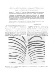
PERINATALSKELETALINJURIES IN TWO BALAENOPTERIDWHALES ROBERTA. PATERSON AND STEPHEN M. VAN DYCK Paterson,R.A.&VanDyck,S.M. 19960720:Perinatalskeletalinjuriesintwobalaenopterid whales. Memoirsofthe QueenslandMuseum39(2): 333-337. Brisbane. ISSN0079-8835. Two recently born balaenopterid whales {Balaenoptera acutorostrata and Megaptera novaeangliae) stranded on the coast ofsouthern Queensland exhibited similar pericranial andriblesionsconsideredtoresultfromcompressioninjury.Birthtrauma,perhapsassociated withcaudalpresentation,isconsideredthemostlikelycauseofthelesions. Balaenopterid whales, skeletalinjuries, birthtrauma. RobertA. Paterson & Stephen M. Van Dyck, QueenslandMuseum, P.O. Box3300, South Brisbane, Queensland4101, Australia; received22 February 1996. DESCRIPTIONS The Queensland Museum (QM) cetacean col- lection contains skeletal material of 11 juvenile QMJM7301. A 2.9 m long 9 minke whale balaenopterids of which 7 are minke whales stranded at the Big Sand Hill, Moreton Island Balaenoptera acutorostrata, 3 are humpback (27°13'S, 153°22'E) on ll.vi.87. Its flipper and whales Megaptera novaeangliae and 1 a blue bodycolourationwastypicalofthediminutiveor whale B.musculus musculus. Among these, Type 3 form (Best, 1985) and was illustrated in pericranial and rib lesions are evident in 2 very Paterson (1994). The umbilicus was healed. Su- immature specimens, 1 minke whale and 1 perficial 'cookie-cutter' lesions similar to those humpback whale. This paper describes the described in otherexamples ofthis species (Wil- pathology and speculates on its cause. liamson, 1975;Arnoldetal., 1987)werenotedas FIG. 1. Minke whale QMJM7301. Radiograph demonstrating numerous bilateral ventral rib fractures. The sternum has not been included. 334 MEMOIRSOFTHEQUEENSLAND MUSEUM F1G. 2. Minke whale QMJM7301 (Left). Dorsal view of skull (98cm long) demonstrating periosteal new bone formation on the lateral aspectofthe supra-or- bitalprocessoftheleftfrontalbone.(Right)Close-up profile viewofthesame region. tological examination (Fig. 5) demonstrated well as predatory rakes near the dorsal fin. periosteal new bone formation superficial to a Bilateral rib fractures were palpable but there sub-periostealcystconsistentwithtraumaseveral were no wounds or scars superficial to them. weeks priorto death (J. Musgravepers.comm.). Abundant callus (some was lost during prepara- tion) was evident both by direct inspection and DISCUSSION radiological examination (Fig. 1). Afterprepara- tionoftheskullaraisedareaofshell-likeossifica- Therearevery limiteddataconcerningthetime tion 6.0 cm long and 2.5 cm in greatest diameter and place ofbirth ofType 3 minke whales. The wasnotedonthelateralaspectofthesupra-orbital two smallest examined in South Africa by Best process ofthe left frontal (Fig. 2). (1985) were 1.92 m and 2.54 m long and they stranded at latitude 34°S in May andJuly respec- QM JM7303. A 4.2 m long 8 humpback whale tively. The former had a raw and completely stranded at Moon Point, Fraser Island (25°14'S, unhealed umbilicus and was considered to be a 153°00'E) on 17.10.89. It was frozen soon after very recent live birth (Best, 1985). QMJM7301 death and transported to the QM. The umbilicus was2.9mlong,andstrandingoccurredat27°Sin was healed. Numerous bites and rakes, con- June. Its skeleton was extremely immature com- sidered to be due to shark attack, were noted. pared with the 6 other juvenile minke whale QM They includedalarge fresh rightaxillary wound. skeletons in the collection and this suggests Rib fractures (Fig. 3) were not as severe as those a very young age, probably less than 8 weeks. in QMJM7301. During dissection a cystic There are extensive data concerning the time pericranial lesionmeasuring 16.0cmlongand7.0 and place of birth of Southern Hemisphere cm in greatest diameter was noted on the lateral humpback whales. The modal length at birth is aspect of the supra-orbital process of the right 4.3 m (Chittleborough, 1958) and most births frontal (Fig. 4). There were no soft tissue or occurin the vicinityoflatitude20°Sbetweenlate cutaneousabnormalities superficial tothe lesion. July and mid September(Townsend, 1935; Chit- Its raised periosteal edge was biopsied and his- tleborough, 1965; Paterson & Paterson, 1989). PERINATALSKELETALINJURIES IN BALAENOPTERIDWHALES 335 FIG. 3. Humpback whale QMJM7303. Radiograph demonstrating healing undisplaced bilateral ventral rib fractures. The sternum has not been included. (The vertical 'tracks' in some ribson the leftofthephotograph resultfrom drillingto insertnumberingwires). QMJM7303 was 4.2 m long and stranded at (Hunt & Schwab, 1992). Such an injury in 25°S in October on the western side of Fraser humans isoften life-threatening and may require Island, a region frequented by humpback whales assisted ventilation. However, some healing was during the southern migration (Corkeron et al., evident in the fractures of QMJM7301 and its 1994). QMJM7303 was probably less than 6 death was not due to acute chest trauma. weeks old when it died. The lateral aspect of the supra-orbital process Radiological opinion was sought in an attempt of the frontal would be susceptible to injury, todate the ribfractures. They were consideredto particularlyinanimmatureanimal,ifdorsolateral have occurred approximately 6-8 weeks before compressionoccurred. Pericranial injury,similar death and were likely tohaveresulted fromcom- tothat in QMJM7301 andQMJM7303,occurs in pression and not from blunt trauma (J.P. Masel, c.2% of human neonates and is termed cephal- pers. comm.) The position and extent of the haematoma. It usually results from cranial bilateral rib fractures are consistent with com- moulding during parturition and, as an isolated pression injury described in human neonates by finding, is not associated with mortality or per- Caffey (1973). The extensive fractures in sistent morbidity (Caffey, 1973). The degree of QMJM7301 areconsideredtorepresenta 'central new bone formation in the pericranial lesion of flair atermusedinhumantraumawhen multiple QMJM7301 (Fig.2) suggests a longer period of rib fractures occur on both sides of the sternum survival than QMJM7303 in which new bone 336 MEMOIRS OFTHEQUEENSLAND MUSEUM thebroadest foetal part, makesan abrupt [pelvic] engagement. Caudal presentation hasbeen noted in a humpback whale (Dunstan, 1957) and such presentation inmysticetescouldbeassociatedon occasions with the difficulties described by Ar- thur (1964), although the pelvic structures of cetaceansandlargeterrestrialanimalsdiffercon- siderably. Hartley (1983) listedcontusions tothe cranial periosteum and rib fractures among the postmortem findings in a large series of foal perinatal mortalities. Weconcludeonthebasisofavailableevidence thatthepericranialandribinjuriesinQMJM7301 and QMJM7303 were sustained during parturi- tion. However, itremainsconjecturaliftheycon- tributed to the premature deaths ofthese whales. ACKNOWLEDGEMENTS We received valuable assistance from many persons. Patricia Paterson assisted with the dis- section and retrieval ofQMJM7301. Vic Hislop retrieved QMJM7303, arranged its freezing, and transported ittothe QM.John Masel, Directorof Metropolitan Paediatric Radiology in Brisbane, dated the rib fractures and John Musgrave of Sullivan, Nicolaides and Partners prepared and diagnosed the histological specimens from QMJM7303. Deidre Pyecroftgaveusthebenefit of her veterinary experience and searched the relevantliterature. BruceCowell andJeffWright QM of the prepared the photographs. Sophie Kupisand StephenMarmo,oftheJindaleeMedi- cal Centre and Royal Brisbane Hospital respec- FIG.4. HumpbackwhaleQMJM7303. Dorsalviewof tively, took the radiographs. skull (which measures 1 13cm in length)demonstrat- ing periosteal new bone formation on the lateral LITERATURECITED aspectofthesupra-orbitalprocessoftheright frontal bone. ARNOLD, P., MARSH, H. & HEINSOHN, G. 1987. The occurrenceoftwo forms ofminke whales in formation was limited to the edge of the lesion eastAustralianwaterswithadescriptionofexter- (Fig- 4). nal characters and skeleton of the diminutive or Most data concerning foetal presentation and dwarfform. Scientific ReportsoftheWhalesRe- the mechanismofparturition in cetaceans derive search Institute,Tokyo 38: 1-46. ARTHUR, G.H. 1964. 'Wright's veterinary obstetrics from studies of captive odontocetes. Caudal & (including diseases of reproduction)'. (Bailliere, presentation is usual (McBride Kritzler, 1951; Tindall & Cox London). : Slijper, 1962), a presentation considered disad- BEST,P.B. 1985.Externalcharactersofsouthernminke vantageous in large domestic animals. Arthur whales and the existence of a diminutive form. (1964) noted that in such animals the foetus is Scientific Reports of the Whales Research In- wedge-shaped when the presentation is cephalic stitute,Tokyo 36: 1-33. and this serves to progressively dilate the birth CAFFEY, J. 1973. 'Paediatric X-ray diagnosis'. (Year BookPublishers New York). canal during its passage, whereas in caudal CHITTLEBOROUGH,: R.G. 1958. Breeding cycle of presentation compression ofthe foetal abdomen the female humpback whale Megaptera nodosa causes expansion of the ribs and the costal arch (Bonnaterre). Australian Journal of Marine and engages abruptly. Also, the foetal occiput, often FreshwaterResearch9: 1-18. PERINATALSKELETALINJURIESINBALAENOPTERIDWHALES 337 FIG.5.HumpbackwhaleQMJM7303.Histologicalsection(x100)fromedgeofsupra-orbitallesiondemonstrated in Fig. 4. Thickened periosteum (A) is seen superiorly; spicules ofvertically arrangedosteoid seams (B) with interveningconnectivetissue are seencentrally and a layeroffibroustissue (C) linesasub-periostealcyst(D) at the lower left of the photograph. The appearances are those ofperiosteal separation with some new bone formation superficial toapost-traumaticsub-periostealcyst. 1965. Dynamics of two populations of the MCBRIDE, A.E. & KRITZLER, H. 1951. Observa- humpback whale Megaptera novaeangtiae tionsofpregnancy, parturition and post-natal be- (Borowski). Australian Journal of Marine and haviour in the bottlenose dolphin. Journal of FreshwaterResearch 16: 33-128. Mammalogy. 32(3): 251-266. CORKBERRYODNE,N,P.J.M,.BMR.OW19N9,4.M.H,umSpLbAaDcEk, wRh.aWles&, PATERSON, R.A. 1994. An annotated list of recent additions to the cetacean collection in the Megaptera novaeangliae (Cetacea : Balaenop- QueenslandMuseum.MemoirsoftheQueensland teridae), in Hervey Bay, Queensland. Wildlife Museum. 35(1): 217-233. Research. 21:293-305. DUNSTAN, D.J. 1957. Caudal presentation at birth of PATERSON,R.&PATERSON,P. 1989.Thestatusof a humpback whale, Megaptera nodosa (Bon- therecoveringstockofhumpbackwhalesMegap- naterre).NorskHvalfangst-Tidende.46:553-555. tera novaeangliae in east Australian waters. HARTLEY, W.J. 1983. Some aspectsoffoal mortality Biological Conservation. 47: 33-48. in New South Wales. Proceedings postgraduate SLIJPER,E.J. 1962. 'Whales'.(Hutchinson London). cSyodmnmeiyt.te6e5:in207v-et2e1r2i.nary science, University of TOWNSEND, C.H. 1935. The distribution:ofcertain HUNT,D.M.&SCHWAB,F.J. 1992.Chesttrauma.Pp whalesasshownbylogbookrecordsofAmerican 77-100. In Rosen, P., Doris, P.E., Barkin, R.M., whaleships. Zoologica 19: 1-50. Barkin, S.Z. & Markovchick, V.J. (eds), 'Diag- WILLIAMSON, G.R. 1975. Minke whales offBrazil. nosticradiologyinemergencymedicine'.(Mosby Scientific Reports of the Whales Research In- : St. Louis). stitute,Tokyo 27: 37-59.
