Patient Name: Marston, John J Patient ID: 247836 CT CT ABC AND PELVIS NO CONTRAST PDF
Preview Patient Name: Marston, John J Patient ID: 247836 CT CT ABC AND PELVIS NO CONTRAST
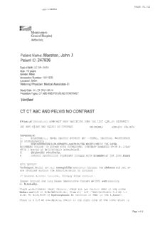
PAGE 01/02 Montgomery General Hospital Med5tar HMlt/j Patient Name: Marston, John J Patient ID: 247836 Date of Birth: 02·18~1939 Age: 73 years Gender: Male Accesslcn Number: 1511670 Location: MGH Referring Physician: Medical Associates Er Study Date: 01~25·2013 09:31 Procedure Types: CT ABD AND PELVIS NO CONTRAST Verified CT CT ABC AND PELVIS NO CONTRAST CUnic:al Indication: LOW BACK PAIN RADIATING DOWN THE LEFT LQWi,Ft EXTREMITY CAT 4152 - CT ABD AND PELVIS NO CONTRAST OJ./25/2013 4384152 1511670 XMPRESSION: 1. BILATERA):., RENAL CALCULI WITHOUT ANY ~~TERAl, CALCULUS. NO EVIDENCE OF HYDRONEPHROSIS. 2. SUBCAiNTlMETER LOW-DENSITY l,AiSION IN THE: RIGHT LOB£ OF THE LlVER, RECOMMEND FOLLOW- UP EITHER WITH ULTRASOUND, CONTRAST ENHANCED CT OR R~,l?EAT CT IN 3 MONTHS AS IS CLINICALLY APPROPRIATE. 3. ENLARGED PROSTATE. 4. CHRONIC OBSTRUCTIVE PULMONARY DISEASE WITH SCARRING AT THE LUNG BASES. FULL RESULT! 'l'echniquel Serial axj.a,l tomograJ;lhic section.s through the abdomen and pel vi~ j are obtained without the administration of contrast. CT Abdomen Without Contrast, Urinary Stone Protocol~ Images through the lung bases demonstrate changes of COPD with scarring bi h.terally. There bilateral renal calculi, there are two calculi seen in the righe ar\'.!l kidney and one in 1:-he left kidn.ey, 1!~sy all' are -'Approximately 0.2 em j.n size. No evid,ence of hydronephrosis. No calculus is seen in the 1,.l;reter~. There is a 0.8 em low-density l®sion in the right lobe of the liver which is Page 1 of 2 PAGE 02/02 Patient: John Marston 10: 247838 Study Date; 01·25·2013 09:31 not fully evaluateQ due to laok of cQucrast, it is located near tne gall.bl~dder fossa. Follow-up is recommenQed with ultra$ound or. contr.ast enhanoed CT or. with repeat CT in 3 months. The sp Leen is unremarkel.ble. Thill pancreas the adrenal gl.;!l.nds and the reeroper.i tioneum are 1.m.remar.Kable. I I Atherosclerotic calcification iJ> seen in the abdominal aorta. Th.ere is no bowel obstruction. CT Pelvis Without Contr~.sti Ur.inary Stone Protocol: There j,s maz ked distention of the urinary bll:l.dder. There is no pelvic mass or ascites. No calcul~s is seen in the distal ureters or in the urinary bladder. The prostate g]'and is mildly enlarged. Multiple phleboliths are noted in the pelvis. Mukul Das MD 01-25"2013 09:43 Page 2of2 1. "~""HV.LLJ v Volol + > ;JUl'l'/48715 Page 1 of 2 ' .• O J.:\J1.V 1Y 1 an i 1'R I'l'CE l'lTII .11' DRTVr. STE T-20 ... lQLOCY OTSEY,1\.ID 20R12 .. (101) 774-:1400 ASSOCJ.ATES COURTESY COpy TO: Patient: MARSTO" .TOH:\, ARTHUR SCHOE.NGOLD MD OOB: 02/Hlf1939 Age: 73 18111 PRINCE PHILIP DR MRN: no 128606 Sex: Male STE Phone #: 301-987-2685 OLNEY, MD 20832 Accession: 2753931 DATE OF SERVICE: 01/3112013 EXAM: MRI LUMBAR SPINE WITHOUT CONTRAST HISTORY: 73-year-old male with lumbar radiculopathy, particularly in the left leg. TECHNIQUE: Sagittal T1- and T2-weighted images as well as axial T2-weighted images are performed through the lumbar spine without the intravenous administration of contrast. In addition, sagittal STIR or sagittal fat-suppressed T2-weighted sequence was also performed. COMPARISON: Prior x-ray of the lumbar spine of 10/15/2008. FINDINGS: Dextroscoliosis of lumbar vertebrae. Evidence of spondylolysis of pars interarticularis of LS vertebra leading to grade I spondylolisthesis of L5 over S1 vertebral body. As a result, there is foraminal stenosis noted bilaterally at LS-S1 level in craniocaudad dimensions leading to impingement of the right L5 nerve root in the foramen and encroachment on the left: LS nerve root in the foramen. Right foraminal stenosis also identified at L3-L4 at L4-L5 levels secondary to osteophytic encroachment from the endplates and facet joint causing impingement of the right L4 nerve root in the neural foramen. At L 1-L2 level, mild bulge of the annulus identified causing flattening along the anterior border of the dural sac. Hypertrophic changes noted in ligamentum flavum causing mild degree of narrowing of the dural sac with bilateral lateral recess stenosis. At L2-L3 level, mild bulge of the annulus identified without distortion ofthe dural sac. At L3-L4 level, diffuse bulge of the disk identified encroaching along the anterior border of the dural sac. Along with hypertrophic changes noted in ligamentum flavurn, there is a mild-to-moderate degree of acquired central spinal canal stenosis with bilateral lateral recess stenosis worse on right side compared to left with complete obliteration of the fat planes in the right lateral recess causing impingement of the right L4 nerve root. Mild right foraminal stenosis with encroachment noted on the right L3 nerve root. At L4-L5 level, diffuse asymmetrical bulge/protrusion of the disk identified lateralized to right side extending in right lateral recess and in the right neural foramen encroaching on the anterior border of the dural sac, along with bilateral lateral recesses worse on right side compared to left and extending in right neural The attache!l document» contain l,"ntidcntiul health inr. ••. matron. If)'lIu believe ynu have received tl,is information in cr"IIr, please enntuct the sender at. the phone number stilted ubovc nnd dcst. •.•• }' (tI •• nnt simpl~' Iliscal~l) the info.mnt.iun hnmediutely, 1d.\l81l~li<..: Resonance Imaging (T\,fRT). Opcri'l'v1RI • Computed Tomographv (CT). Nuclear Medicine rET:CT. Marnmographye Hltrasound e l)EXA • f1uonl"'opy. Interveruionul e X-Ray Providing JC{tdh)log.).' Services at n dllesda Bov .. -ie Snulh C'ht!vy (,has~ Cfinton Frederick South Germantown {Ircenbelt T . eixurc World MCI\,t1C (\:lRr) Olne.y Rod,~ill" Waldor! White O"k ' • '~'WHV.L'-' V VJ. 1. -1 0U 1'/748715 Page 2 of 2 01 ,:\F.Y 1 R111 "PRT"1CF PTTTT ,W DRTVr. STF T-20 O1.:\EY, MD 2()R:l2 (101) 774-.1400 Coutinucde Page 201'2 Patient: MARSTO:'\, .TOH:'\ DOB: 02118/1939 A~e: 73 MRN: 128606 Sex: Mille Phone #: 301-987-2685 Accession: 2753931 DATE OF SERVICE: 01/3112013 foramen causing encroachment on the right L4 nerve root. Along with hypertrophic changes noted in ligamentum flavum and facet joint, there is moderately severe acquired central spinal canal stenosis, At LS-S 1 level, pseudoprotrusion ofthe disk is lateralized more to the right side compared to the left in the right lateral recess. No distortlon of the dural sac, The remainder of the neural foramina show intact perineural fat planes. Conus medullaris is at its normal location and appears to be within normal limits. Normal marrow signal activity of the vertebral bodies. IMPRESSION: Lumbar spondylosis. Spondylolysis of pars interarticularis of L5 vertebra leading to grade I spondylolisthesis with bilateral foraminal stenosis and impingement of the right L5 nerve root in the neural foramen and encroachment on the left L5 nerve root in the neural foramen. Acquired central spinal canal stenosis at L3-L4 and L4-L5 levels, and to a lesser extent at L 1-L2 level with right foraminal stenosis and impingement of the right L3 and L4 nerve roots in the foramina. Dilip Arwlndekar MD Electronically Signed: 1/31/13 10:12 am DA/jt Referring Phys: Dr, KEVIN MCMAHON Phone Number: 301-774-8958 The attached ('neuments contuin •. ,"onfitlcntial health infurrnatmu. If you hdievc, .YUtl have received t.his information in ern,.·, please cnntnct the sender lit the phone number stated uhnvc 1I011.1(!stro)' (tin nnt. simply (lismnl) the informat.ion imm c ..tiutcly. Magnetic R~'01l:I1",e 1"''''I:\;ng (MRT) • Open MRT. Computed Tomography (CT). Nuclear Medicine Pf.T;CT. Muml11ography. Ultrasound 0 DEX;\ • Fluoroscopv .111ler\,enl;nnal. X-Ray Providing Rm/io/Og), Services ai nelhe.sd<l nOW1{~ Bowie Soulh Chevy Cha,~ Chnlnn Freder-ick Frederick South Germantown Greenbelt I .eisure World MCI\.fTC (\·mT) Olney Rockville ~i:~\'ell T .ocks Waldorf While Oak T'-Ian 08 07: 13:36 2013 Page 2 of 3 ROBERT R.L. SMITH. M.D. Medical Director J.972810020842 SFECIMEN COLLECTED: 01/07/2013 08:04 COMPLETED REPORT: 01/08/2013 07:13 ARTHUR SCHOENGOLD,M.D. (R-78506) MARSTON,JOHN STE '1'10 (K1,M 9813 SAILFISH TER 18111 PRINCE PHILIP DR ) MONTGOMERY VILLAGE,MD 20886 OLNEY MD 20832 DOB: 02/18/1939 PT/PH#: 1-301-987-2685 Acn: 1972810020842 Original report sent to ordering physician: CARDIAC ASSOCIATES, CARDIAC ASSOCIATES, 18109 PRNC PHILIP DR #125, OLNEY, MD CHEMISTRY: AST ( 17 U/L 10-35 GLUCOSE-- 91 MG/DL ( 65-99 16 U/L 9-60 BUN------ 16 MG/DL( 7-25 ) ALT SGOT) CREATININE 0.95 mg/dL(0.70-1.18) ( BU/CR RATIO N /A ( 6-22 ) SGPT) CALCIUM--- 9.9 MG/DL( 8.6-10.3) SODIUM--- J.42 mmol/L( 135-146 ) POTASSIUM- 4.6 mmol/L( 3.5-5.3 ) CHLORIDE- 105 mmol/L( 98-110) CO/2----- 26 mmo1/L( 21-33 ) CHOLESTEROL---------------------- 147 MG/DL ( 125-200 TRIGLYCERIDE--------------------- 111 MG/DL «150) ~or patients >49 years of age, the reference limit for Creatinine is approximately 13 higher for people identified as African-American. Bun/Creatinine ratio is not reported when the BUN and creatinine values are within normal limits. *HDL CHOLESTEROL------------------ 3 MG/DL (> or = 40) LDL CHOLESTEROL, CALCULATED------ 8 MG/DL «130) 8 7 SIGNA,URE DAn: REPORn:D rre "too"" reccr atorv .Ii..<iles were pertonrec Py ocesi Diaorostic~. 1901 SLlpl'"r Spnr o RoM. Baltlll'ore. MD 21227 200201 (41) 03/04/2012 07;40:55 PM Page 5 of 15 PATIENT: MARSTON, JOHN J MR#: 247-836 BILLING #: 00022355812 SEX: M DATE OF BIRTH: 02/18/1939 UNIT: 1 NOR0105A CARDIOLOGY CONSULTATION CONSULT REQUESTED BY: Hospitalist Service DATE OF CONSULTATION: 03103/2012 REASON FOR CONSULTATION: Presyncope and history of coronary disease. HISTORY OF PRESENT ILLNESS: The patient is a 73-year-old gentleman with a significant cardiac history of coronary artery disease, status post CABG, who also has a known history of esophagitis, who presented with a short episode of mild dizziness yesterday in association with a headache. He did not have any nausea or vomiting. He did not have any chest discomfort or shortness of breath. He currently has remained asymptomatic overnight in observation with occasional episodes of mild sinus bradycardia with heart rates in the 40s to 50s. The patient denies any PND or orthopnea. He has not had any syncope at home. His enzymes have ruled out. The patient, to his knowledge, has not had an echo in some time. He is followed routinely by Dr. Tannenbaum. He has not had any prior history of syncope or palpitations. PAST MEDICAL AND SURGICAL HISTORY: 1. Significant for coronary artery disease, status post CABG in 1996. 2. Status post left heart catheterization in 2010 with resulting medical management. 3. Tobacco use. 4. Hypertension. 5. Sinus bradycardia. 6. Hypercholesterolemia. 7. Esophagitis. ALLERGIES: Morphine. CU RRENT MEDICATIONS: 1. Pravastatin 40 mg p.o. at bedtime . • CONSUL. TATION - MEDSTAR MONTGOMERY MEDICAL. CENTER ARTHUR SCHOENGOLD, M.D. • ./o.vUI,,·,r;:7Ul.lia...1. ttecOrdS 03/04/2012 07:41:06 PM Page 6 of 15 MARSTON, JOHN J PATIENT: 247-836 MR#: 2 PAGE #: 2. Pantoprazole 40 mg p.o. daily. 3. Eye drops. 4. Enalapril10 mg p.o b.i.d. 5. Atenolol 25 mg p.o. daily. 6. Aspirin 81 mg. SOCIAL HISTORY: He continues to smoke. He denies alcohol or drug use. FAMILY HISTORY: Negative for premature coronary disease. REVIEW OF SYSTEMS: Negative for nausea or vomiting, fever or chills, weight gain, weight loss, neurological symptoms or rash. The rest of the 11-point review of systems is negative. PHYSICAL EXAMINATION: VITAL SIGNS: Blood pressure 140/54. Heart rate 68. Respirations are 20. Temperature is 98. HEENT: Extraocular movements are intact. Sclerae anicteric. Oropharynx clear. Moist mucous membranes. NECK: No JVP. No lymphadenopathy. No thyromegaly. LUNGS: Clear to auscultation bilaterally, without increased respiratory effort. HEART: Regular rhythm, without murmurs, rubs or gallops. PMI is nondisplaced. ABDOMEN: Soft, nontender, and nondistended. Bowel sounds positive. No hepatosplenomegaly. EXTREMITI ES: No clubbing, cyanosis or edema. Range of motion is full. SKIN: Warm and dry, without evidence of erythema or rashes. NEUROLOGIC: Cranial nerves 2-12 intact. Sensation is intact to light touch. PSYCHOLOGIC: Affect is appropriate. Alert and oriented x3. LABORATORY DATA AND STUDIES: Telemetry, reviewed by myself, does reveal some sinus bradycardia while sleeping, in the 40s, but otherwise normal sinus with heart rates in the 60s to 80s. No AV block. EKG revealed normal sinus rhythm without ST or T wave changes. -CONSULTATION - MEDSTAR MONTGOMERY MEDICAL CENTER ARTHUR SCHOENGOLD, M.D. 03/04/2012 07:41:18 PM Page 7 of 15 MARSTON, JOHN J PATIENT: 247-836 MR#: 3 PAGE #: Labs revealed a negative troponin. Hematocrit is 40.9. White blood cell count is 9.37. INR is 0.98. Electrolytes are a" normal. Creatinine is 0.82. TSH 1.74. HDL 35 and LDL 78. The patient also has a meningioma located on CT scan. IMPRESSION: 1. Presyncopal event. 2. History of coronary disease, status post coronary artery bypass graft. 3. Tobacco use. 4. Sinus bradycardia. At this point, the presyncopal event could be multifactorial. The patient has a 3.6 cm right parietal meningioma, for MR of the brain. RECOMMENDATIONS: 1. Okay to continue atenolol for now. 2. Okay to go home from a cardiovascular standpoint, as there does not appear to be any high-risk arrhythmia or bradycardia at this point. 3. We will plan an echo and event monitor as an outpatient to determine whether or not any medication changes or other interventions are needed and to determine the etiology of the syncope. A loop recorder can be considered if the event monitor is negative. 4. Follow up with Dr. Tannenbaum in 3 to 4 weeks. 5. Neurosurgery followup for the meningioma. 6. Discussed with Dr. Schoengold, his primary Cafe physician. Pending Electronic Signature SEAN BEINART, M.D . • CONSULTATION- MEDSTAR MONTGOMERY MEDICAL CENTER ARTHUR SCHOENGOLD, MD. CARDIAC PAGE 02/02 ASSOCI~T~S Quest Quest Diagnostics Incorporated Diagnostics ® R01H!.RT R.L. SMITIl. M.l). 1901 Sulphur SJ,'Irillg Roud - Baltimore. Marylund 21227-0580 M~j~ul Oitcclor M~1I1 LabOtaltlry 411l-247-910(}· ,D.C. Area 30 H'i2J.6900 Out~idc M/lrylnnd j ·ROc'>-LAB-XCEL 1972a10018587 SPECIMEN COLLECTED: 01/31/2012 08;37 COMPLETED REPORT: 02/0l/2012 04:19 TANNENBAUM,ERIC (R-197281) MARSTO~,JOHN CARDIAC ASSOCI~TES (K1,A) PATIENT IO: 18109 P~C PBXLIP DR #125 ~CCESSION #: 1~7281001S5S7 OLNEY, MD 20832 9813 SAILFISH TER MONTGOMER.Y VILLAGE, MD 20886 , PATIENT PHONE#: 1-301-987-2685 :.:,~,jl·""" ;~,'-' ;';l?~TIENT DeB: 0.2!lS/1~39 r A TrENT NAME DATE LAB NUMBER 1. \B H.FI'OI~ MARSTON, JOHN 01/31/2012 73 ,M ~2322706 I CHEMISTRY: ..!2§L?JJ/U AST----------------------~------- '2.D 17,,/.11/L 10-35 ALT-,.:.---------------------------- 2'2 16' ".oiL 9-60 i:' ,- CHOLESTEROL---------------------- 132 149.yMG/DL ( l25-200 TRIGLYCERIDE-~-----------~----~~- \ 23 «150) *HDL CHOLESTEROL-------~--~--~--~~ ,MG/DL (;:. or '"' ,;y.-: LDL CHOLESTEROL, CALCULATED------ 3f, 6 MG/DL 40) 71.,. ',' 8',v',' " MG/DL «130) Desir.-able range .dOO mg/dL for patien~s,~with9HD.;6r diabetes and -:: 70 mg/dL for .diabetic pat~~tits,',;with, known heart disease. CHOLESTEROL/HDL 3.9 0.0-5,0 RATIO-----------~' j I -:;-:;END OF REPORT - MARSTON,JOHN AB2322706 - TOTAL 1 l?AGE(S»> (COMPLETED) 02/01/2012 04;20 DATE ~EPORTED
Description: