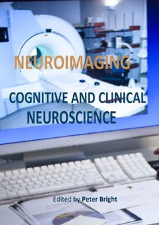
Neuroimaging - Cognitive and Clinical Neurosci. PDF
Preview Neuroimaging - Cognitive and Clinical Neurosci.
NEUROIMAGING COGNITIVE AND CLINICAL NEUROSCIENCE Edited by Peter Bright NEUROIMAGING – COGNITIVE AND CLINICAL NEUROSCIENCE Edited by Peter Bright Neuroimaging – Cognitive and Clinical Neuroscience Edited by Peter Bright Published by InTech Janeza Trdine 9, 51000 Rijeka, Croatia Copyright © 2012 InTech All chapters are Open Access distributed under the Creative Commons Attribution 3.0 license, which allows users to download, copy and build upon published articles even for commercial purposes, as long as the author and publisher are properly credited, which ensures maximum dissemination and a wider impact of our publications. After this work has been published by InTech, authors have the right to republish it, in whole or part, in any publication of which they are the author, and to make other personal use of the work. Any republication, referencing or personal use of the work must explicitly identify the original source. As for readers, this license allows users to download, copy and build upon published chapters even for commercial purposes, as long as the author and publisher are properly credited, which ensures maximum dissemination and a wider impact of our publications. Notice Statements and opinions expressed in the chapters are these of the individual contributors and not necessarily those of the editors or publisher. No responsibility is accepted for the accuracy of information contained in the published chapters. The publisher assumes no responsibility for any damage or injury to persons or property arising out of the use of any materials, instructions, methods or ideas contained in the book. Publishing Process Manager Sandra Bakic Technical Editor Teodora Smiljanic Cover Designer InTech Design Team First published May, 2012 Printed in Croatia A free online edition of this book is available at www.intechopen.com Additional hard copies can be obtained from [email protected] Neuroimaging – Cognitive and Clinical Neuroscience, Edited by Peter Bright p. cm. ISBN 978-953-51-0606-7 Contents Preface IX Chapter 1 Cytoarchitectonics of the Human Cerebral Cortex: The 1926 Presentation by Georg N. Koskinas (1885–1975) to the Athens Medical Society 1 Lazaros C. Triarhou Chapter 2 Images of the Cognitive Brain Across Age and Culture 17 Joshua Goh and Chih-Mao Huang Chapter 3 Neuroimaging of Single Cases: Benefits and Pitfalls 47 James Danckert and Seyed M. Mirsattarri Chapter 4 Functional and Structural Magnetic Resonance Imaging of Human Language: A Review 69 Manuel Martín-Loeches and Pilar Casado Chapter 5 Neuro-Anatomical Overlap Between Language and Memory Functions in the Human Brain 95 Satoru Yokoyama Chapter 6 Neuronal Networks Observed with Resting State Functional Magnetic Resonance Imaging in Clinical Populations 109 Gioacchino Tedeschi and Fabrizio Esposito Chapter 7 Resting State Blood Flow and Glucose Metabolism in Psychiatric Disorders 129 Nobuhisa Kanahara, Eiji Shimizu, Yoshimoto Sekine and Masaomi Iyo Chapter 8 The Memory, Cognitive and Psychological Functions of Sleep: Update from Electroencephalographic and Neuroimaging Studies 155 Roumen Kirov and Serge Brand VI Contents Chapter 9 Neuroimaging and Outcome Assessment in Vegetative and Minimally Conscious State 181 Silvia Marino, Rosella Ciurleo, Annalisa Baglieri, Francesco Corallo, Rosaria De Luca, Simona De Salvo, Silvia Guerrera, Francesca Timpano, Placido Bramanti and Nicola De Stefano Chapter 10 Functional and Structural MRI Studies on Impulsiveness: Attention-Deficit/Hyperactive Disorder and Borderline Personality Disorders 205 Trevor Archer and Peter Bright Chapter 11 MRI Techniques to Evaluate Exercise Impact on the Aging Human Brain 229 Bonita L. Marks and Laurence M. Katz Chapter 12 Human Oscillatory EEG Activities Representing Working Memory Capacity 249 Masahiro Kawasaki Chapter 13 Neuroimaging Data in Bipolar Disorder: An Updated View 263 Bernardo Dell’Osso, Cristina Dobrea, Maria Carlotta Palazzo, Laura Cremaschi, Chiara Arici, Beatrice Benatti and A. Carlo Altamura Chapter 14 Reinforcement Learning, High-Level Cognition, and the Human Brain 283 Massimo Silvetti and Tom Verguts Chapter 15 What Does Cerebral Oxygenation Tell Us About Central Motor Output? 297 Nicolas Bourdillon and Stéphane Perrey Chapter 16 Intermanual and Intermodal Transfer in Human Newborns: Neonatal Behavioral Evidence and Neurocognitive Approach 319 Arlette Streri and Edouard Gentaz Chapter 17 Somatosensory Stimulation in Functional Neuroimaging: A Review 333 S.M. Golaszewski, M. Seidl, M. Christova, E. Gallasch, A.B. Kunz, R. Nardone, E. Trinka and F. Gerstenbrand Chapter 18 Neuroimaging Studies in Carbon Monoxide Intoxication 353 Ya-Ting Chang, Wen-Neng Chang, Shu-Hua Huang, Chun-Chung Lui, Chen-Chang Lee, Nai-Ching Chen and Chiung-Chih Chang Contents VII Chapter 19 Graphical Models of Functional MRI Data for Assessing Brain Connectivity 375 Junning Li, Z. JaneWang and Martin J. McKeown Chapter 20 Event-Related Potential Studies of Cognitive and Social Neuroscience 397 Agustin Ibanez, Phil Baker and Alvaro Moya Chapter 21 Neuroimaging Outcomes of Brain Training Trials 417 Chao Suo and Michael J. Valenzuela Chapter 22 EEG-Biofeedback as a Tool to Modulate Arousal: Trends and Perspectives for Treatment of ADHD and Insomnia 431 B. Alexander Diaz, Lizeth H. Sloot, Huibert D. Mansvelder and Klaus Linkenkaer-Hansen Chapter 23 Deconstructing Central Pain with Psychophysical and Neuroimaging Studies 451 J.J. Cheng, D.S. Veldhuijzen, J.D. Greenspan and F.A. Lenz Preface The rate of technological progress is encouraging increasingly sophisticated lines of enquiry in cognitive neuroscience and shows no sign of slowing down in the foreseeable future. Nevertheless, it is unlikely that even the strongest advocates of the cognitive neuroscience approach would maintain that advances in cognitive theory have kept in step with methods-based developments. There are several candidate reasons for the failure of neuroimaging studies to convincingly resolve many of the most important theoretical debates in the literature. For example, a significant proportion of published functional magnetic resonance imaging (fMRI) studies are not well grounded in cognitive theory, and this represents a step away from the traditional approach in experimental psychology of methodically and systematically building on (or chipping away at) existing theoretical models using tried and tested methods. Unless the experimental study design is set up within a clearly defined theoretical framework, any inferences that are drawn are unlikely to be accepted as anything other than speculative. A second, more fundamental issue is whether neuroimaging data alone can address how cognitive functions operate (far more interesting to the cognitive scientist than establishing the neuroanatomical coordinates of a given function – the where question). The classic neuropsychological tradition of comparing neurologically impaired and healthy populations shares some of the same challenges associated with neuroimaging research (such as incorporation of individual differences in brain structure and function, attribution of specific vs general functions to a given brain region, and the questionable assumption that the shared components operating in two tasks under comparison recruit the same neural architecture. However, a further disadvantage of functional neuroimaging relative to the neuropsychological approach is that it is a correlational method for inferring regional brain involvement in a given task – and interpretation of signal should always reflect this fact. Spatial resolution and sensitivity is improving with the commercial availability of ultra-high field human scanners, but a single voxel (the smallest unit of measurement) still corresponds to many thousands of individual neurons. Haemodynamic response to input is slow (in the order of seconds) and the relationship between this function and neural activity remains incompletely understood. Furthermore, choice of image preprocessing parameters can appear somewhat arbitrary and an obvious rationale for selection of statistical thresholds, correction for multiple corrections, etc. at the analysis stage can X Preface likewise be lacking in some studies. Therefore, to advance our knowledge about the neural bases of cognition, rigorous methodological control, well-developed theory with testable predictions, and inferences drawn on the basis of a range of methods is likely to be required. Triarhou (Chapter 1) provides a translation of Georg Koskinas’ 1926 presentation to the Athens Medical Society in which the neuropsychiatrist described 107 cytoarchitectonically defined cortical areas (plus 60 “transition” areas) in the human brain. In comparison to Brodmann’s (1909) universally recognised system (in which 44 cortical areas are defined), the von Economo and Koskinas system (published as an atlas and textbook in 1925) provided a fourfold increase in cortical specification. The author provides a compelling argument for more widespread adoption of von Economo and Koskinas’ detailed criteria (commonly used in clinical neuroscience) in neuroimaging studies of human cognition. Heterogeneity in brain structure and function across individuals is an important issue in neuroimaging research. Although attempts are made to manage such differences during stages of preprocessing and statistical analyses of datasets (as well as during the participant selection process), there can be a tendency to neglect the importance of individual differences due to the importance in the literature of identifying commonalities in the functioning of our brains. For example, it is quite common in fMRI studies to find participants who have relatively “silent” brains relative to others undertaking the same cognitive task. Age is a well recognised factor affecting brain structure and function, but the importance of cultural differences is relatively poorly understood. Goh and Huang (Chapter 2) present neuroimaging findings associated with age and cultural experience and also consider their interaction. Interestingly, research appears to suggest that culture-specific functional effects present in early adulthood are robust and remain in place despite subsequent age-related neurobiological change. Such observations also suggest that ageing effects in the brain may, in part, be contingent upon the nature of external experiences – raising clinical implications for modulating or offsetting neurocognitive changes associated with increasing age. Danckert and Mirsattari (Chapter 3) consider the viability of fMRI studies of single neurological cases for furthering our understanding of brain-behaviour relationships. With careful attention to methodological issues, the authors present a strong argument for the single case approach (for both clinical and cognitive neuroscience purposes) in which comprehensive neuropsychological assessment and fMRI are employed and the results interpreted in the context of large-scale normative structural and functional MRI data. Chapter 4 (Martín-Loeches & Casado) provides a useful review of recent research on the neural correlates of human language and Chapter 5 (Yokoyama) considers whether (and the extent to which) brain regions responsible for core language processes can be dissociated from those responsible for more general cognitive processes associated with working memory and central executive function. Preface XI Tedeschi and Esposito (Chapter 6) present an excellent consideration of the utility of measuring resting state networks (RSNs) in clinical populations using fMRI. Some authors have questioned whether systematic neuroimaging analysis of the resting state represents an appropriate context for advancing cognitive theory. Nevertheless, this review presents a highly compelling argument for studying RSNs (particularly when combined with MRI tractography) in order to enhance understanding of pathological mechanisms in a range of neurological conditions. Kanahara et al. (Chapter 7) focus their comprehensive review on single-photon emission computed tomography (SPECT) and photon emission tomography (PET) studies of resting state blood flow and metabolism in a range of psychiatric conditions including schizophrenia, major depressive disorder, bipolar disorder and obsessive-compulsive disorder. Sleep deprivation is associated with a wide range of neurocognitive effects, but attention and other aspects of executive function appear particularly vulnerable. Kirov and Brand (Chapter 8) review evidence for the role of sleep in the regulation of cognitive functions, with particular focus on neuroimaging investigations. The distinction between vegetative state (VS) and minimally conscious state (MCS) is clearly expressed in the clinical literature. The former refers to a state of “wakeful unawareness” in which patients are awake, can open their eyes and produce basic orienting responses, but have a total loss of conscious awareness. MCS differs to the extent that patients with this diagnosis are able to produce cognitively mediated behavioural responses. From a clinical perspective however, the distinction can be very difficult and a number of recent neuroimaging studies have provided indirect evidence that some VS patients have been able to communicate answers to orally presented questions. This rather disturbing finding that such patients may be more aware than the clinicians (or family members) may realise is of profound clinical importance given the very different prognosis and treatments indicated in the two conditions. Marino et al. (Chapter 9) review the role of neuroimaging in improving our understanding of coma, VS and MCS while recognising the continuing importance of comprehensive standardised clinical assessment. Impulsive behavior is a major component of several neuropsychiatric disorders including schizophrenia, attention-deficit/hyperactivity disorder (ADHD), substance abuse, bipolar disorder, and borderline and antisocial personality disorders. The temporal, motor and reward related aspects of impulsiveness and decision-making are exemplified by the impulsive behaviors typically evident in ADHD and borderline personality disorder (BPD), with or without comorbidity. Archer and Bright (Chapter 10) consider the role of structural and functional neuroimaging for furthering our understanding of the cause and development of impulsivity in these conditions. Marks and Katz (Chapter 11) carefully evaluate the potential role of MRI for establishing the nature of the relationship between exercise and the integrity (both physiological and cognitive) of the brain. The question of whether (and the extent to which) exercise can offset age-related cognitive decline is one which has attracted a
