NASA Technical Reports Server (NTRS) 20170001349: Dependence of Early and Late Chromosomal Aberrations on Radiation Quality and Cell Types PDF
Preview NASA Technical Reports Server (NTRS) 20170001349: Dependence of Early and Late Chromosomal Aberrations on Radiation Quality and Cell Types
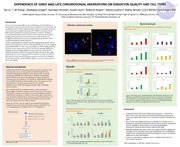
DEPENDENCE OF EARLY AND LATE CHROMOSOMAL ABERRATIONS ON RADIATION QUALITY AND CELL TYPES Tao Lu1,2, Ye Zhang3, Stephanie Krieger4, Samrawit Yeshitla1, Rosalin Goss5, Deborah Bowler6, Munira Kadhim6, Bobby Wilson5, Larry Rohde2, and Honglu Wu1 1NASA Johnson Space Center, Houston, TX, 2University of Houston Clear Lake, Houston, TX, 3NASA Kennedy Space Center, Cape Canaveral, FL, 4KBRwyle, Houston, TX, 5Texas Southern University, Houston, TX, 6Oxford Brookes University, UK Introduction Results (Continued) FISH whole chromosome painting Exposure to radiation induces different types of DNA damage, increases mutation and Radiation type dependence of genomic instability in early and chromosome aberration rates, and increases cellular transformation in vitro and in vivo. The susceptibility of cells to radiation depends on genetic background and growth condition of late time points cells, as well as types of radiation. Mammalian cells of different tissue types and with different genetic background are known to have different survival rate and different Mammary Epithelial (M10) Mammary Epithelial (M10) mutation rate after cytogenetic insults. Genomic instability, induced by various genetic, 0.40 0.40 metabolic, and environmental factors including radiation, is the driving force of s 0.35 s 0.35 n n o o ita 0.30 ita 0.30 tumorigenesis. Accurate measurements of the relative biological effectiveness (RBE) is re re bb 0.25 bb 0.25 A A important for estimating radiation-related risks. lam 0.20 lam 0.20 o o s s o 0.15 o 0.15 m m o o To further understand genomic instability induced by charged particles and their RBE, we rhC 0.10 rhC 0.10 % 0.05 % 0.05 exposed human lymphocytes ex vivo, human fibroblast AG1522, human mammary epithelial 0.00 0.00 Ctrl 0.4 2 4 Ctrl 0.4 2 4 Gy, Proton Gy, Proton cells (CH184B5F5/M10), and bone marrow cells isolated from CBA/CaH (CBA) and C57BL/6 (C57) mice to high energy protons and Fe ions. Normal human fibroblasts AG1522 have Mammary Epithelial (M10) Mammary Epithelial (M10) apparently normal DNA damage response and repair mechanisms, while mammary 0.40 0.40 s 0.35 s 0.35 epithelial cells (M10) are deficient in the repair of DNA DSBs. Mouse strain CBA is radio- n n o o ita 0.30 ita 0.30 re re sensitive while C57 is radio-resistant. Metaphase chromosomes at different cell divisions bb 0.25 bb 0.25 A A lam 0.20 lam 0.20 after radiation exposure were collected and chromosome aberrations were analyzed as oso 0.15 oso 0.15 m m o o rh 0.10 rh 0.10 RBE for different cell lines exposed to different radiations at various time points up to one C C % 0.05 % 0.05 month post irradiation. 0.00 0.00 Figure 2. Examples of whole chromosome FISH (chr. 3, purple and chr. 6, azure) painting. Translocations Ctrl 0.1 0.5 1 Ctrl 0.1 0.5 1 are pointed by yellow arrows, insertions by white arrows, and ring by red arrow. Gy, Fe Gy, Fe AG1522 AG1522 Results 0.14 0.14 Materials and Methods sn sn oita 0.12 oita 0.12 re re b 0.10 b 0.10 b b Peripheral whole blood from two healthy donors was collected in Vacutainer tubes Comparison of chromosomal aberrations in lymphocytes after Fe A la 0.08 A la 0.08 m m oso 0.06 oso 0.06 containing sodium citrate. Peripheral blood mononuclear cells (PBMCs) were and Proton irradiation m m o 0.04 o 0.04 rh rh C C immediately separated by centrifugation, washed twice with PBS, counted and Lymphocytes % 0.02 % 0.02 0.00 0.00 Ctrl 0.4 2 4 Ctrl 0.4 2 4 resuspended in RPMI1640 with 2mM Glutamine and10%FBS. Normal human Gy, Proton Gy, Proton fibroblasts AG1522 were grown in alpha-MEM with 10% FBS and antibiotics in humidified tissue culture chamber at 37˚C with 5% CO . Human mammary epithelial AG1522 AG1522 2 e 0.14 cells (CH184B5F5/M10) were cultured in DMEM medium with supplement of 10% F s 0.14 n s o 0.12 n FBS and antibiotics. Cells were exposed in vitro to Fe ions or protons itareb 0.10 oitare 0.12 b b 0.10 A b (600MeV/nucleon) at NASA Space Radiation Laboratory (NSRL) at Brookhaven lam 0.08 A la 0.08 o m s 0.06 o o s 0.06 m o National Laboratory. o 0.04 m rhC orh 0.04 % 0.02 C % 0.02 After irradiation, PBMCs were stimulated to grow in medium containing 1% 0.00 0.00 Ctrl 0.1 0.5 1 n Ctrl 0.1 0.5 1 Gy, Fe Phytohemagglutinin (PHA) and the epithelial cells were subcultured continuously to o Gy, Fe t o enable growth. metaphase chromosomes were collected at different cell divisions r P using Colcemide and Calyculin-A. Metaphase spreads were subject to fluorescence Figure 5. Percentage of cells containing initial or late chromosome damage in human in situ hybridization (FISH) with whole chromosome probes for chromosomes 3 and fibroblasts and epithelial cells after exposure to protons and Fe ions. Initial damage 6 (MetaSystems). Chromosome aberrations were analyzed on Zeiss fluorescence was assessed at first mitosis post irradiation, and the late damage was assessed at Figure 3: Different retention of chromosome aberration in lymphocytes after proton one month after exposure. Chromosome aberrations at one month post irradiation microscope Axioplan 2 with Leica CytoVison software. exposure at early and late time points. Data are fraction of aberrations in total counted appeared to lack a clear dose response for the epithelial cells. Femoral bone marrow single cell suspension was obtained from CBA/CaH and metaphase spreads. C57BL/6J male mice (8-14 weeks old). Cells were exposed to 600 MeV/u Fe ions (LET: 175 KeV/micron) and 600 MeV/u Protons (LET: 0.26 KeV/micron). Chromosome aberrations in mouse bone marrow stems cells Conclusions in liquid bulk cultures Femoral bone marrow CBA Mice C57 Mice • In lymphocytes, the chromosome aberration frequency at 1 month after Cells irradiated in 15ml centrifuge tubes: exposure to Fe ions was close to the unexposed background, whereas the CBA Day 2 DBA Day 6 C57 Day 2 C57 Day 6 600 MeV/u Fe ions (0, 0.05, 0.1, 0.2, 1.0 Gy) 0.7 0.7 chromosome aberration frequency at 1 month after exposure to protons was or 0.6 0.6 600 MeV/u Proton (0, 0.05, 0.1, 0.2, 1.0Gy) higher. 0.5 0.5 e F 0.4 0.4 • Bone marrow cells isolated from CBA mice showed similar frequencies of 0.3 0.3 0.2 0.2 chromosome aberrations between the early and late time points after proton 0.1 0.1 or Fe ion irradiation, while cells from C57 mice showed different 0 0 0.0 0.1 0.1 0.2 1.0 0.0 0.1 0.1 0.2 1.0 chromosome aberration rates between different time points. CBA Day 2 CBA Day 6 C57, Day 2 C57, Day 6 0.70 0.7 • Mammary epithelial cells have a higher chromosome aberration background 0.60 0.6 and higher rate of initial aberrations than the fibroblasts, but the fibroblasts n 0.50 0.5 o 0.40 0.4 t retained more chromosomal aberrations after long term culture (1 month) in Cells from liquid bulk culture o 0.30 0.3 r at days 2 and 6 were P 0.20 0.2 comparison to their initial damage. harvested and prepared for 0.10 0.1 • Caution must be taken in using RBE values to estimate health risks from cytogenetic analysis 0.00 0 0.0 0.1 0.1 0.2 1.0 0.0 0.1 0.1 0.2 1.0 space radiation exposure. Figure 1. Radiation exposure of femoral bone marrow cells isolated from 8-14 week old Figure 4. Transmissible chromosomal instability in liquid bulk cultures derived from Fe male CBA and C57 mice. ion irradiated haemopoietic stem cells. *Work supported by the NASA Space Radiation Health Program.
