Table Of ContentKINEMATIC AND EMG COMPARISON OF GAIT IN NORMAL AND MICROGRAVITY
John K. DeWitt1, W. Brent Edwards2, Gail P. Perusek3, Beth E. Lewandowski3,and Sergey Samorezov4
1Wyle Integrates Science and Engineering Group, Houston, TX, USA; 2The Iowa State University, Ames, IA, USA; 3NASA
Glenn Research Center, Cleveland, OH, USA; 4ZIN Technologies, Cleveland, OH, USA. email: [email protected]
INTRODUCTION METHODS
Astronauts regularly perform treadmill locomotion Five subjects (2M/3F) completed treadmill walking
as a part of their exercise prescription while at 1.34 m·s-1 and running at 3.13 m·s-1 in NG, and
onboard the International Space Station. Although MG. NG trials were collected on a laboratory
locomotive exercise has been shown to be treadmill at NASA Glenn Research Center. AM
beneficial for bone, muscle, and cardiovascular trials were collected during parabolic flight onboard
health, astronauts return to Earth after long duration a DC9 aircraft at NASA Johnson Space Center. The
missions with net losses in all three areas [1]. These external load provided by bungees (EL) during AM
losses might be partially explained by fundamental was 87.3 ± 6.6 %BW. Data were collected in each
differences in locomotive performance between location on different days; the schedule was not
normal gravity (NG) and microgravity (MG) under the control of the investigators.
environments.
Kinematic data were collected with a video motion
During locomotive exercise in MG, the subject must capture system (SMART Elite, BTS Bioengineering
wear a waist and shoulder harness that is attached to SPA, Milanese, IT) at 60 Hz. The 3-D positions of
elastomer bungees. The bungees are attached to the lower extremity and trunk markers were recorded,
treadmill, and provide forces that are intended to rotated into a treadmill reference frame, and
replace gravity. However, unlike gravity, which projected on to the sagittal plane. All subsequent
provides a constant force upon all body parts, the kinematic calculations were completed in 2-D.
bungees provide a spring force only to the harness.
Therefore, subjects are subjected to two Telemetered EMG (Myomonitor III Wireless EMG
fundamental differences in MG: 1) forces returning System, Delsys Inc., Boston, MA) was used to
the subject to the treadmill are not constant, and 2) obtain muscle activation data of the tibialis anterior,
forces are only applied to the axial skeleton at the gastrocnemius, rectus femoris, semimembranosus,
waist and shoulders. The effectiveness of the and gluteus maximus. Before any motion trials,
exercise may also be affected by the magnitude of subjects performed maximal voluntary isometric
the gravity replacement load. Historically, contractions of each muscle to standardize electrode
astronauts have difficulty performing treadmill placement. All motion capture and EMG data were
exercise with loads that approach body weight synchronized via a global analog pulse that was
(BW) due to comfort and inherent stiffness in the recorded simultaneously by each hardware device.
bungee system.
Hip, knee, and ankle joint range of motion (ROM)
Although locomotion can be executed in MG, the and flexion and extension extremes were computed
unique requirements could result in performance using the angles between adjacent segments with
differences as compared to NG. These differences markers defining their long axes. EMG data were
may help to explain why long term training effects rectified and filtered and then examined to quantify
of treadmill exercise may differ from those found in the time of initial activation and total activation
NG. The purpose of this investigation was to duration during each stride using the methods of
compare locomotion in NG and MG to determine if Browning et al. [2]. Multiple strides were analyzed
kinematic or muscular activation pattern differences for each gravitational location and trial means were
occur between gravitational environments. computed. Effect sizes and their 95% confidence
Kinematic Differences
intervals were computed joint kinematic and EMG 80
scores between each condition. 70
60
RWmEohdSeenU) ,cL noTimnSeb tAiynN-isnDigx aDclolI mSfaCpcaUtoriSrssSo tIneOss twNs e(EreL m, laodceo.m Boetcivaue se Degrees, deg345000
20
our intent was to identify differences between
10
gravitational locations, we will limit our
0
presentation to those variables in which the 95% Hip ROM, Low EL, Walking Max Hip Flexion, High EL, Max Ankle Dorsiflexion, Hip ROM, High EL, Running
Walking High EL, Walking
confidence interval for the effect size did not
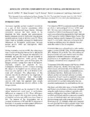
include 0 (see Table 1, Figure 1). EMG Differences
100
Hip ROM during walking was larger in MG during 90
80
tfwhhlieaegxs hL i loEoanrwL gd. e EuTrr Lhiinn ec g Nog MnaGdsG titrthoi otachnnna, en Mam nNGiduG sdt.h u wMer iahnasixg pa i wcmatcaiuvhlmkaieit nevddgeo drews agiirftrllheiee axtrthi eoiernn Initial Activation, % stride 3456700000
the stride in MG during the high EL condition. 20
10
0
Hip ROM was the only kinematic measure during Gastrocnemius, High Gluteus Maximus, Semimembranosus, Gluteus Maximus, Semimembranosus,
EL, Walking Low EL, Running Low EL, Running High EL, Running High EL, Running
running that was differentiated between MG NG
gravitational locations. Subjects achieved greater Figure 1: Kinematic (upper) and EMG (lower)
amounts of hip flexion in MG. During each running dependent variables with effect size differences
condition, the gluteus maximus and between MG & NG.
semimembranosus were activated later in the stride
Our data suggest that there may be kinematic and
in MG.
muscle activation differences during running
between MG and NG that could influence training
Although we tested only a small sample, we have
responses, and may help to better understand why
detected some differences between locomotion in
these deficits occur. Future research is necessary
MG and NG that centralize about the hip, with the
with larger subject sizes to better quantify kinematic
exception of ankle kinematic and musculature
and EMG differences between locations.
effects found during walking with high EL.
Returning astronauts have been found to have a net
REFERENCES
decrease in bone mineral density at the hip after
1.LeBlanc AD, et al. J Musculoskelet Neuronal
longterm spaceflight [1]. Interestingly, hip ROM
Interact, 7, 33-47, 2007.
appears to increase in MG compared to NG. This
2.Browning RC, et al. Med Sci Sports Exerc, 39,
increase may be an adaptation to accommodating
515-525, 2007.
the gravity replacement load.
Table 1: Effect size and 95% confidence intervals for kinematic and EMG dependent variables
Walking ES 95% CI Running ES 95% CI
Low EL
Hip ROM 1.62 [0.19,3.05] Gluteus Maximus Initial Activation 1.80 [0.33,3.26]
Semimembranosus Initial Activation 3.35 [1.43,5.28]
High EL
Gastrocnemius Initial Activation –2.48 [–4.13,–0.83] Hip ROM 1.41 [0.03,2.80]
Max Hip Flexion 1.73 [0.28,3.18] Gluteus Maximus Initial Activation 1.64 [0.21,3.07]
Max Ankle Dorsiflexion –1.48 [–2.88,–0.08] Semimembranosus Initial Activation 5.04 [2.51,7.57]

