Table Of ContentNASA-CR-205373
¢?....
..._.s
Year 2 Annual Report
Grant #: IRP 95-101
Title: Quantitation of Bone Growth Rate Variability in Rats Exposed to Micro- (near zero G)
and Macrogravity (2G)
Investigators:
Timothy G. Bromage (Hunter College)
Stephen B. Doty (Hospital for Special Surgery)
Igor Smolyar (Hunter College)
Emily Holton (NASA)
INTRODUCTION
Aims
Our stated primary objective is to quantify the growth rate variability of rat lamellar bone
exposed to micro- (near zero G: e.g. Cosmos 1887 & 2044; SLS-1 & SLS-2)and macrogravity
(2G). The primary significance of the proposed work is that an elegant method will be established
that unequivocally characterizes the morphological consequences of gravitational factors on
developing bone. The integrity of this objective depends upon our successful preparation of thin
sections suitable for imaging individual bone lamellae, and our imaging and quantitation of growth
rate variability in populations of lamellae from individual bone samples.
Year 1"Recap
Our initial efforts were to be spent on specimen preparation as NASA had hoped that our
equipment vendor would supply us despite the restructuring of our 2 year program into 3 years.
While our vendor could not comply, we nevertheless prepared less sophisticated sections that could
still satisfy" the simpler objectives of Year t. We were ve_ _successful, documenting the iametla
formation rate for the SLS experimental rat. "The most astonishing result was that endosteal
lamellae of the humerus (and we expect in many other locations) of the juvenile rat accrue at the
rate of 1 bone lamella per day (Figure I). In other words, the lamella formation rate follows a
circadian rhythm. This is the first calculation of a lamellar formation rate in the mineralized tissue
sciences.
We also made preliminar?' observatio_:s su_,2.__2.."t__<.<"L_,hat the vitaI labels, calccin ana.l_
demectocycline, used in SI.S experimental protocols, 'na,ve a negative effect on bone formation
rates.
Given the delay in refined specimen preparation, we worked hard in other ways to prepare tbr
the quantitative aspect of our research to begin in Year 2" we configured the series of image
processing steps that provide binary "working" images for growth rate quantification, and we
prepared a preliminary version of a Windows®-based program developed for automating the
Lii;i_i_li
measurement taking and for configuring a 3-D chart illustrating bone growth rate variability with
precision to 24 hours.
Year 2: Expectations
During the first half of Year 2 we explained that quantitative analyses of lamellar growth rate
variability of SLS-2 flight bone and their controls would begin. Backscattered electron microscopy
in the scaIming electron microscope (BSE-SEM) of SLS-1 and SLS-2 bone specimens would be
performed in order to assess mineral density changes owing to microgravity.
Quantitative investigations of lamellar growth rate variability would begin on Cosmos 1887
and 2044 samples during the second half of Year 2, as would the accompanying BSE-SEM of these
bone specimens.
The results of our investigations on the morphological and mineral density changes affecting
the development of lamellae under conditions of microgravity were to be summarized, written up in
more final form for publication, and for presentation to the ammal meeting of the American Society
for Gravitational and Space Biology.
Year 2" Actual
We acquired the necessary equipment and developed our proposed specimen preparation
protocol. This novel protocol permits, for the first time, the same specimen to be employed in
analyses by both conventional light microscopy and scanning electron microscopy. The protocol is
so important and unique that we formally presented this technique to annual meetings of Scanning
97 (Scanning 19(3)179-180, 1997) and the American Society for Bone and Mineral Research (J.
Bone Min. Res. 12(1)$202, 1997).
Having readied our specimen preparation protocol for proposed quantitative analyses of
tamellar growth rate variability, we were then able to assess and prepare all SLS-t flight and control
bone (Figures !-4). All SLS-2 bone has been prepared and is currently" under investigation.
Preliminao. BSE-SEM investigations of SLS-1 flight bone have also been perfomaed (Figure 5).
We produced a penultimate version of our Windows(N-based program (refining of this
program is still underway) for automating the quantitalion of lamellar growth rate variability. This
unique program and the results obtained on the effects ot: microgravity on bone development: were
presented to the annual meeting of the ,.4me:'ican A.s'sociaIio_7 o/P/O,'sicalA.JTt/7.:'ol)o/ogisfs (Am. J.
Phys. Anthropol. Suppl. 2483, 1997).
Our cumulative results have been submitted for presentation to the annual meeting of the
American Society.for GraviIational and Space Biology (ASGSB) to be held this November in
Washington, D.C. Our presentation to the ASGSB and the l'orthconaing publication will include
results arising f;:-omour c,,)n_:inucd work on SLS-2 !amellar 2j-ov,th rate variability and the BS!!:-
.Sr:\,'i <)iflight and c(_:a,_roi_" _
MATERIALS AND METHODS
Animals
Specimens from SLS-1 experiments are reported here:
Experiment: SLS- 1
Experimental groups 19 RAHF Flight and Vivarium Control
Mission Length (ML) 9 day flight (ML=9)
Species: male Harlan (Sprague/Dawley)
Bone Element: labeled humerus
The experimental group had a proscribed vital labeling regime of calcein and demeclocycline
prior to and around Launch, and at Recovery (see Figure 1 for details).
Specimen Preparation
Sections ground from methyl methacrylate embedded samples to ca. 120 microns of section
thickness on a Buehler Petro-Thin (Buehler Ltd., IL) were carefully made superficially anorganic
with Tergazyme (Alconox, NJ) enzymedetergent at 50° C for 24 hours. Sections were
subsequently ground on one side through graded emery papers to 1200 grit on a Handimet II. The
embedded section was placed onto a thin film of acetonitrile for 5 minutes. A histological slide was
etched with 20% hydrofluoric acid for 4 minutes, thoroughly rinsed, and air dried.
The section was mounted onto the etched slide using the All-Bond 2 Universal Dental
Adhesive System (Bisco, IL). The slide was first treated with Silane Porcelain Primer for 30
seconds and air dried. A 50:50 mixture of proprietary Primers A and B was brushed onto the
polished section five coats in succession, allowed to dry, and then polymerized for 30 seconds using
a visible light curing gun. The section was then pressed onto the stide,_ the 1_900 _ori*s_ide ihce
down, in a small pool of Dentine/Enamel Bonding Resin and light cured for 1-2 minutes.
The mounted section was again ground on 1200 grit emery paper and polished with a Buehler
Ecomet III to 1.0 micron diamond and to 0.05 micron alumina slurry on a Buehler Vibromet II
Vibratou Polisher until reaching ca. t00 microns of section thickness. Save for tile embedding and
vibratory polishing procedures, the method outlined above requires only 30 minutes of specimen
handling time versus 1-2 days using more traditional methods.
The embedded polished thin section was coverslipped with 100% ethyl acetate and imaged by
polarizing and fluorescence microscopy. Subsequent to this the section was made electrically
conductive with carbon and imaged by BSE-SEM.
tD,Sal ]ll'ICYt
Images ofcndostcal t'dmcllae were obtained with a Leica DMRX'\I!:; light microscope fitted
with linear polarizing filters and a 100 Watt lluorescence system. Images were retrieved by a
Kodak Megaplus CCD camera and transported to the Leica Quantimet 600, a t:ramestore-based
3
imageanalysissystem.An imageprocessingprogramwaswrittenwhichstandardizegdraylevel
imageenhancemenatndfilteringproceduresT. heresultingimageswerethenpassedtothe
quantificationprogramforanalysis(below).ThesamefieldsofviewwerealsoimagedbyaLEO
$440SEMinBSE-SEMimagingmodeat20kV, 500pa,anda 15mmworkingdistance.
QuantificationSoftware
WedevelopedaWindows®-basedprogram(CcompilerforDOCenvironmentf)or
processinglamellarbone.Theprogram,titledLAMELLA, convertsimagesofthewidthsbetween
lamellaeintoanumericaltable.Thetableisrepresentegdraphicallyintheformofa3-Dchart.
ThechartillustratesthequantitativeresultsbasedonaBooleanfunctionthatwasproposedand
describedinouroriginalproposal.
TheinputforthissoftwareisabinaryimageofboneacquiredfromourQuantime6t 00image
processingroutine.ThisbinaryistransferredtoLAMELLA whichoverlays"N" numberof
specifiedtransectsontothescreenimage.LAMELLA automaticallyinteractswiththebinary,
labelingeachlamella.Measurementbsetweeneachsuccessivleamellaareautomaticallyrecorded
andpresentedinanumericalTableQ. TheSURFER(GoldenSoftware,Inc.)programisemployed
torepresenTtableQintheformofa3-Dchartofthebonegrowthrateindicatinggrowthrateinthe
Zdirection,timeintheX direction,andsamplingerrorintheYdirection(increasingerrorinthe3-
Dchartisrepresentetdowardtherearofthechart.Chartsillustratedinthisreportindicatevery
littleerror,however,astheinitialimagesarerathergood,andsothe3-Dlandscapeappearsrather
uniformfromfronttoback).
ANALYSES
Efi_ctofSpaceFlightonBoneI)eposition
Protocol.TheexperimentaplrotocolisillustratedinFigure1. Thepolarizedlightimageat
topisthesourceimagefromwhichthebinaryiscreated.Thefluorescenceimageatbottomis
annotatedwithdatesofsignificancetothe1991SLS-1mission,includingdatesofadministration
ofvital labels.Launch.andRecoveryAnextractofthebinaryimaaeisalsooverlainonthisin_._,e
providinganaccountofthecircadian,:-iaytho,nflametlaformationdiscoveredinYeari ofour
project.
Flight. An example of the response of bone to microgravity is presented in Figure 2. The
polarized light image is at top left. Lamellar processing of this image produces the binau image
against which time markers derived t-'rein the fluorescence image (particularly the dates of
. °
adm..in__,"s_t,,,___._.ao._"caIce p,ihat arc wc!l dcfi_cd} may be well. positioned. The b-,n•arx.' _ma,.z,cis thcp,
sUq':lCCtto I.AMI "I.!.-_" qtaan:_>"aive pr,:.)c:essiz__,,and SURi_-:':-,"_. to produce a "<-i) outpu.t (as t.lac..._..c..I!-b,u.___.,,,
above).
lhc 3-D chklrt describes the iamellar gro_,v"_hrate. Positions of the calccin and demeclocycline
labels are shown by,green and blue bands, respectively, and Launch and Recovery are indicated tot
reference. (}rowth rate is given by the Z (height) axis and time proceeds from leti to right on the X
4
axis. SamplingerrorwouldberevealedbyincreasingirregularityofthechartintheYaxisfrom
fronttoback,butasthedataisveryisotropic,littlechangeineach2-Dprofileexistsalongthisaxis.
ThereareatfirstmarkedchangesinthegrowthratebetweenthefirstcalceinlabelandLaunch.
Theseresultscontinuetosuggestthattheadministrationofthevital labels,calceinand
demeclocyclineh,indersosteoblasticactivity.Labelsareassociatedwith short-temadownward
trendsintheZdirectionfollowedbyrecoveryinthegrowthrates.
At Launchthe3-Dchartrevealsanimmediatedropinbonedepositionowing,perhapst,othe
stressofliftoff andinitial weightlessnessT.hisisfollowedbyadownwardtrenduntilRecovery.
Recoveryisalsoproceededbyaprecipitousdropinthegrowthrateduetocalceintoxicity. This
dropisfollowedbyanincreaseinthegrowthrateat1Guntileuthanized.
Control. An example of the response of bone subject to the labeling protocol at 1G is
presented in Figure 3 (this figure is represented in the same way as that described for Figure 2,
above). The growth rate is intermittently compromised by the labeling protocol until just prior to
the Launch date. In contrast to flight conditions, the control bone reveals an increase in the lamellar
growth rate following the Launch date except for a transitory drop owing to calcein toxicity.
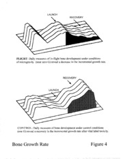
Bone Growth Rate Summary. Figure 4 summarizes the average condition identified from the
SLS-1 experimental sample. The 3-D charts clearly demonstrate that the bone growth rate is
compromised in flight compared to ground controls. Previous gravitational research on the rat has
lacked a certain precision regarding the morphology and character of that specific bone volume
formed during spaceflight as separate from that bone volume laid down prior to or after this period.
No doubt a significant problem has been to recognize the meager lamellar bone volumes added
during short 1-2 week research periods in comparison to high degrees of normal interindividual
gross morphological variation. The novel methods reported here are the first to provide quantitative
results confirming the effects of microgravity with 24 hour resolution.
Effect of Space Flight on Bone Density
Given our success at describing growth rate variability, in the experimental rat, we undertook
BSE-SEM of bone specimens in order to identify, with very high precision (i.e. 24 hours),
differences in bone mineral density that may be dependent upon gravitational factors.
We provide preliminary results on flight specimens in Figure 5. The fluorescence and BSE-
SEM images are provided at top. These images of the same field of view are brought to the same
image size and orientation to ailow for digital comparison. These images were processed together
in order to coni_nn the duration of the experimental period on the BSE-SEM image. Relative bone
density was recorded from within the rectangle on the BSE-SEM image.
The grey level profile from bottom to top, representing the beginning and end of the
experimental period, is portrayed from right to left in the daily density profile below (dates are
indicated above the relatively high densily fractions associated with each bone lamelta). High and
!ow grey levels are associated with high and low mineral densities respectively. There is an initia!
variation in the density between the first ca!cein label and l_aunch. 3"his may be an effect oi:the
three vital labels adlninistered during this time. Launch to Recovery is characterized by a broad
downward lrcnd in density at which point, after i;altering once more, it rises. Toward the end ot'the
experimental period, just one day prior to being euthanized, the relatively low density
5
7!7;'_:.:._/.............. •......._.....................
mineralizationfrontisevident.Thisfrontisveryrecentlyformedbonewhichhasstilltoacquire
thehighermineralizationlevelsofthelamellaebehindit.
YEAR 3
Year 2 work has enabled us to proceed with our anticipated Year 3 objectives which are to
undertake quantitative investigations of lamellar growth rate variability of lamellar bone exposed to
2G and 3G, as well as the related backscattered electron microscopy.
Specimens still lagging in preparation and/or analysis will be processed, and all light and
scanning electron microscopy will be completed. Acquisition of automatic staging this stage in
Year 3 will permit us to make comparisons between LM and BSE-SEM results over significant
bone volumes (the $440 SEM has an automatic stage). We also expect to refine the quantitative
processing to permit the digital marrying of results of bone growth rate variability and density into a
single quantitative portrayal of the bone growth and mineralization deficit during space flight with
24 hour resolution.
The quantitative results of our investigations on the morphological and mineral density
changes affecting the development of lamellae under conditions of micro- and macrogravity will be
summarized, written up in more final form for publication, and for presentation to the annual
meeting of the American Society for Gravitational and Space Biology. A final project report will
be written and presented to NASA scientists and officials.
i_i_i;_i_i_!,!i!i!i!Li!;ii_!;;ii_Lii_:!Ii_i_i!ii_i7ii__;i;_iii_!;i_lLi,:__iW_i'__!ii__!i_i:i_!i_L;_:_:_:_ii_i_;#iii7Ti:iii7'_!_!:i_;!i_!;L_iT¸.iIWii_!ii:iilUT_i!:__iiiWi_i_i!!ii!!_L_i!ii!!::;i;i;_i!i__¸:i7:_!_i!;i_._ii_i:il!ii__i!!i;ii_!i!!i_S!i__ii!;:_!i7ii!_!!_!ii_i!i!i!_!7i__!i_iii!ii__!!!ii!_!__;i!i!ii!!i_iii__Ti!ii!!!_,!!iiii!_!i__!i_!!_i_!i!iiiiiii!ii_!ii!_:!!!7ii_iiii__i_ii;!!i__ii;i!!i__:i;_!!_i!;);ii!;_i!:!!iii_i!!!ii__i!i!!L!i!i__L!!i!!;i!!!i_7_!_:i!:!iii!!_!i_:ii_i:,li_li_ii:iiiiiii_i__i!iLi!!!!!!i!ili!iiiii!ii_i
i ,•i _"_i_
Experimental protocol Figure 1
• •_ i!::i_____"_: 'i •_ :_:_?! _:_!__ii:_i__;'_:"__ _" _...... i_:"¸_¸¸L__::_ili!ii!_i_'L<__i?:i!ii!¸_'_ii:!¸¸_iii!_i!_i17!!ii!_77i_?_U¸¸7;_?i;_7:'I_!I!I!_I_II_II___!_:II_::i_i_ii_L!!_i:i__'i!ii:_ii!i!iiii_i'_!iiii!!i_i!!_i!Liii!__iliii!l;7_ii_iiii!i!il
3iVU H_LAAOU9 _V"]7 =11AlV7 39VlAII 3ONBOS3uon7:I
\ \
AU3AOO3U HONnV7
9NISS::::IOOUd UV7731AIV7 39VlAII IH917 O:IZIUV70d
lOaJu,oo
31V'd HIMO_IO _IV7731AIV7 30VIAll 30N3093Non74
A_3AO03_I
ONISS3OO_Id _IV7731AIV7 30VIAll J.HOI70::lZl_V70d
LAUNCH
RECOVERY
FLIGHT" Daily measures of in-flight bone development under conditions
of microgravity (near zero G) reveal a decrease in the incremental growth rate.
RECOVERY
LAUNCH
CONTROL" Daily measures of bone development under control conditions
(one G) reveal a recovery in the incremental growth rate after vital label toxicity.
Bone Growth Rate Figure 4

