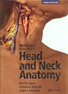
McMinn`s Color Atlas of Head Neck Anatomy 3rd Edition (www.irananatomy.ir) PDF
Preview McMinn`s Color Atlas of Head Neck Anatomy 3rd Edition (www.irananatomy.ir)
MOSBY An imprint of Elsevier Limited. C 2004 Elsevier Limited. All rights reserved. The right of Bari M. logan, Patricia A. Reynolds and Ralph T. Hutchings to be identified as authors of this work has been asserted by them in accordance with the Copyright. Designs and Patents Act 1988 No part of this publication may be reproduced. stored in a retrieval system. or transmitted in any form or by any means, electronic, mechanical. photocopying, recording or otherwise, without either the prior permission of the publishers or a licence permitting restricted copying in the United Kingdom issued by the Copyright licensing Agency, 90 Tottenham Court Road, london WIT 4lP. Permissions may be sought directly from Elsevier'S Health Sciences Rights Department in Philadelphia. USA: phone: (+1) 21S 2387869. fax: (+1) 21S 238 2239, e-mail: healthpermissionsOelsevier.com. You may also complete your request on-line via the Elsevier homepage (http:ltwww.elsevier.com). by selecting 'Customer Support' and then 'Obtaining Permissions'. First edition 1981 0 Yearbook Medical Publishers Second edition 1994 Third edition 2004 ISBN 0 7234 3196S British library Cataloguing In Publication Data A catalogue record for this book is available from the British library Library of Congress Cataloging in Publication Data A catalog record for this book is available from the library of Congress Notice Medical knowledge is constantly changing. Standard safety precautions must be followed. but as new research and clinical experience broaden our knowledge, changes in treatment and drug therapy may become necessary or appropriate. Readers are advised to check the most current product information provided by the manufacturer of each drug to be administered to verify the recommended dose, the method and duration of administration, and contraindications. It is the responsibility of the practitioner. relying on experience and knowledge of the patient, to determine dosages and the best treatment for each individual patient. Neither the publisher nor the authors assume any liability for any injury andlor damage to persons or property arising from this publication. The Publisher your ~ru for boob. JournaI$ and multimedia In the health sclenceJ www.elsevierheolth.com Printed in the UK Contents Preface ix Skull bone articulations 52 Acknowledgements x Facial skeleton 52 Orientation xi Orbital and anterior nasal apertures 53 Orbit 54 Skull and skull bone 1 Roof and lateral wall 54 Floor and medial wall 56 articulations Nasal cavity 58 Roof, floor and lateral wall 58 Skull 2 Maxillary hiatus and nasolacrimal canal 60 From the front 2 Base of the skull 62 Muscle attachments 4 Anterior cranial fossa 62 le Fort facial fractures 6 Middle and posterior cranial fossae 64 From the left 8 External surface, posterior part 66 Muscle attachments 10 pterygopalatine fossa 68 From behind 12 Posterior nasal aperture 70 Vault of skull 14 Fetal skull 72 Base of skull 16 External surface 16 Cervical vertebrae 2 Muscle attachments 18 and neck Infratemporal region and teeth 20 Internal surface 24 Interior of skull, median section 26 Cervical vertebrae 76 Cavities of the skull 28 Atlas 76 Bones of the skull 30 Axis 78 Mandible 30 Third to seventh cervical vertebrae 80 Muscle attachments and age changes 34 Cervical and first thoracic vertebrae 82 frontal bone 36 Other bones 84 Ethmoid bone 38 First rib, manubrium of sternum Sphenoid bone and vomer 40 and costovertebral joints 84 Occipital bone 42 Bones of shoulder girdle 86 Maxilla. nasal bone and lacrimal bone 44 Shoulder girdle and upper Palatine bone and inferior nasal concha 46 thoracic skeleton 88 Temporal bone 48 Parietal bone and zygomatic bone 50 4 Neck 90 Nose. oral region. Surface markings 90 ear and larynx Head, neck and shoulders, superficial muscles 92 Superficial dissection I. Platysma and superficial veins 94 Nose and paranasal sinuses 140 Blood supply and venous drainage 96 Nasal cartilages and nasal cavity 140 Superficial dissection II. Sternocleidomastoid 98 Walls of the nasal cavity 142 Superficial dissection III. Anterior triangle 100 Frontal and ethmoidal sinuses 144 Lymphatic system , 02 Sphenoidal and maxillary sinuses 146 Superficial dissection IV. Posterior triangle 104 Sections of sinuses and nerves of Deep dissection I. Great vessels and nerves the nasal septum 148 and thyroid gland 106 Mouth. palate and pharynx 150 Deep dissection II. Great vessels and Sagittal section of head and neck 1S O thyroid gland 108 Tongue and floor of the mouth 152 Deep dissection 111. Thyroid and Roof and floor of the mouth and the parathyroid glands and root of the neck 110 salivary glands 156 Deep dissection IV. Thyroid gland, Inside of the mouth and the palate 158 thymus and root of the neck 112 External and internal surfaces of the pharynx 160 Deep dissection V. Prevertebral muscles 114 Posterior surface of the pharynx 162 Ear 164 Face. orbit and eye 3 External, middle and internal ear 164 Transverse sections and the auditory ossicles 166 Coronal sections and the auditory ossicles 168 Larynx 170 Face 118 Hyoid bone and laryngeal cartilages 170 Surface markings 118 larynx, pharynx, hyoid bone and trachea 172 Superficial dissection. Parotid gland, Muscles, ligaments, membranes and joints 174 facial nerve and muscles 120 Deep dissection I. Temporalis and Cranial cavity 5 masseter muscles and temporomandibular joint 122 and brain Deep dissection II. Infratemporal fossa and temporomandibular joint 124 Cranial cavity 178 Orbit and eye 128 Sagittal section 178 Eye and lacrimal apparatus 128 Cranial vault, meninges and brain 180 Orbital contents I. From above, Brain and arachnoid mater 182 and extraocular muscles 132 Dura mater and cranial nerves 184 Orbital contents II. Ciliary ganglion Dura mater 186 and dissection from the front 134 Cranial fossae 188 Orbital contents III. Eyes in section Cranial nerves and their connections 190 and the lacrimal gland 136 Cranial fossae, cavernous sinus and trigeminal nerve 192 Cranial cavity, brain and cranial nerves 194 Brain 200 Appendices Brain and meninges 200 Cerebral hemispheres and cerebellum 202 Cerebral veins 204 Cerebral hemisphere 206 Appendix I Dental anaesthesia 248 Blood supply of the cerebral cortex 208 Inferior alveolar and lingual nerve block 248 Brain and brainstem 210 long buccal nerve block 250 Medial surface of the hemispheres Infiltration anaesthesia of the upper teeth 252 and cerebral arteries 212 Posterior superior alveolar nerve block 254 Base of the brain 214 Nasopalatine nerve block 256 Arteries of the base of the brain Greater palatine nerve block 256 and brainstem 216 Mental and incisive nerve block 258 Brainstem. cranial nerves and geniculate bodies 218 Appendix II Reference lists 260 Ventricles of the brain 220 Muscles 260 Internal capsule and basal nuclei 222 Nerves 262 Hemispheres and brainstem in Lymphatic system 264 coronal section 224 Arteries 265 Cerebellum and brainstem 226 Veins 268 Cerebellum. brainstem and spinal cord 228 Skull foramina 269 Cervical vertebral column and Index 273 suboccipital region 230 6 Radiographs Radiographs 234 Vertebral column 234 Carotid arteriogram and venogram of the neck 236 Skull and paranasal sinuses 238 Skull. lateral view 240 Carotid arteriograms 242 Vertebral arteriograms 244 Dural venous sinuses 246 Preface This book was originally penned by the hand of In order to meet readers' demands. we have Professor R. M. H. (Bob) McMinn and first published incorporated new anatomical preparations, in 1981 in response to the need for a specific radiographic images, clinical photographs with anatomical text to suit the educational notes, simple orientation figures to complement the requirements of dental students. It has proved very dissections, and new artworks to aid the popular and this third edition heralds 23 years of understanding and practice of dental anaesthesia. publication in seven languages: English, Japanese, We hope that these new additions will be Portuguese, Spanish, German, Italian and French. appreciated and that the book will continue in its popularity. With the full retirement of Bob McMinn to the Scottish Highlands, a new author, Dr Patricia The name 'McMinn' is retained in the title as a Reynolds, joins the team and, as a senior lecturer in tribute to an outstanding anatomist and our oral and maxillofacial surgery, brings a new level of distinguished c.olleague. clinical expertise to the book. Acknowledgements The authors are indebted to the following: • Dr Ian G Parkin, Clinical Anatomist. University of Cambridge, for expert anatomical knowledge. • Mr Clive Brewis. ENT Registrar, Addenbrooke's Hospital, Cambridge, for advice on the ear. • Mr Martin Watson, Mel lazenby and Lucie Whitehead. Department of Anatomy, University of Cambridge, for preservation of anatomical material. • Mr Ian Bolton, Mr Adrian Newman and Mr John Bashford, Audio Visual Unit, Department of Anatomy, University of Cambridge. for photographic expertise and advice. • Dr Peter Baxter, Dr Brendan Burchell and Dr Simon Poole, of Cambridge. for much appreciated support and guidance. • Inta Ozols, Duncan Fraser. Glenys Norquay, and all the team at Elsevier for their help, advice and support during the preparation of this book. Figure acknowledgements Figures 250, 76F, 126E, 145G, 145H, 1518, 175F, 2118, 225B, 235C, 2350, 235E, 23BA, 240, 242A, 2438, 244A, 2458, 246A, 2478 are reproduced, with kind permission, from Imaging Atlas of Human Anatomy 3rd edition, J. Weir and Peter H. Abrahams. Mosby, 2003. Figures 121 C, 151C are reproduced with kind permission from the Gordon Museum, King's College, London, UK. Table 195 is reproduced, with permission, from McMinn's Functional and Clinical Anatomy R. M. H. McMinn, P. Gaddum-Rosse, R. T. Hutchings, B. M. Logan. Mosby, 1995. Figures 250A, 253C. 2530. 255H. 257E, 259J are redrawn, with kind permission, from Introduction to Dental Local Anaesthesia, H. Evers and G. Haegerstam. Astra/Mediaglobe, 1990. Dedications To Robert Logan - Bari M. Logan To Patrick and Rosie O'Driscoll - Patricia A. Reynolds In memory of Peter Wolfe - Ralph T. Hutchings Orientation Superior Posleri nlerior Medial view Transverse axial plane SagIttal plane Inferior Lateral view
