LIST OF ABBREVIATIONS Abbreviation Meaning PDF
Preview LIST OF ABBREVIATIONS Abbreviation Meaning
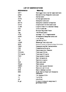
LIST OF ABBREVIATIONS Abbreviation Meaning CATT Card agglutination test for trypanosomiasis cDNA Complementary Deoxyribonucleic acid D.W. Distilled water DEPC Di ethyl pyro carbonate DNA Deoxyribonucleic acid dNTPs Deoxyribonucleotide triphosphate EDTA Ethylene diamine tetra acetic acid ELISA Enzyme-linked Immunosorbent Assay EtBr Ethidium bromide GRP Glucose Regulated Protein Hsp Heat Shock protein IPTG Isopropyl – β- D- thiogalactoside kDNA Kinetoplastid Deoxyribonucleic acid LB Luria- Bertani LMP Low Melting Point mRNA Messenger ribonucleic acid NCBI National Center for Biotechnology Information PAGE Polyacrylamide Gel Electrophoresis PBS Phosphate buffer saline PCR Polymerase chain Reaction PK Buffer Proteinase K Buffer PK Enzyme Proteinase K Enzyme PVDF Polyvinylidene fluoride R. T. Room temperature RE Restriction enzyme RNA Ribonucleic acid RNase Ribonuclease RMSD Root-mean-square Deviation RPM Revolution per minute SDS Sodium dodecyl sulphate SOC Super Optimal Broath with Catabolite Repression TAE Tris- Acetate EDTA TE Tris EDTA UV Ultra Violet X- gal 5 -bromo-4-chloro-3-indolyl-beta-D- galactopyranoside Units of Measurement % Percentage µg Microgram µl Microlitre 0C Degree Celcius A Absorbance bp Base pair cm Centimeter Da Dalton g Gram (s) h Hour (s) IU International unit Kbp Kilo base pair KDa Kilo Dalton Kg Kilogram M Molar ma Milli ampere mg Milli gram min Minute (s) Min Minute ml Milli litre mm Mili meter mM Milli molar ng Nano gram OD Optical density pmol Picomole sec Second (s) U Unit V Volt V / cm Volt per centimeter V / V Volume / volume W / V Weight / volume APPENDICES APPENDIX – I 1. Agarose Gel Electrophoresis buffer 1.1. TAE buffer (50x) Stock solution: Tris base 121 gm Glacial acetic acid 28.55 gm 0.5 M EDTA acid (pH 8.0) 50 ml Water upto 500 ml Working concentration of TAE buffer (1x) TAE buffer 10 ml Water 490 ml 1.2. TE (Tris/EDTA) Buffer Tris-HCl (pH 7.5) 10 mM EDTA 1 mM Make from 1M stock of Tris-HCl (pH 7.5) and 500 mM stock of EDTA (pH 8.0). Working solution: 1M Tris-HCl (pH 7.5) 1.0 ml 500mM EDTA (pH 8.0) 0.2 ml Water to 100 ml 2. Phosphate Buffered Saline buffer (1X), pH 7.4 NaCl 8.0 gm Na HPO .2H O 1.44 gm 2 4 2 i KH PO 0.24 gm 2 4 KCl 0.20 gm Distilled water to make 1000 ml 3. Trypanosome separation buffer (PSG buffer, pH 8.0) 3.1. Solution A Na HPO (anhydrous) 8.000 gm 2 4 NaH PO .2H O 0.780 gm 2 4 2 NaCl 4.250 gm Distilled water to make 1000 ml 3.2. Solution B Glucose solution dextrose 10 gm Distilled water to make 400 ml Just before use 6 parts of solution A was mixed with 4 part of solution B 4. Proteinase K buffer Tris base (ph 8) 100mM EDTA 10mM NaCl 50mM SDS 2% β Mercapto ethanol 20mM ii APPENDIX – II 1. Giemsa stain Giemsa stain 1 ml Distilled water 9 ml Slides were stained for 45 minutes. 2. 6X loading dye Bromophenol blue 0.25% Xylene cyanol 0.25% Glycerol 50% EDTA 2 mM 3. Ethidium bromide solution (10 mg/ ml) Ethidium bromide 0.2gm Sterile Water 20 ml 4. Agarose (0.8%) Agarose 800 mg TAE buffer 100 ml 5. Agarose (1.2%) Agarose 1.2 gm TAE buffer 100 ml iii APPENDIX – III 1. Phenol: Chloroform: Iso amyl alcohol; 25:24:1 Phenol 250 ml Chloroform 240 ml Iso amyl alcohol 100 ml 2. Alcohol (70%) Alcohol 70 ml Water 30 ml 3. Luria Bertani (LB) Medium ( 500 ml) Tryptone 1.0% (5 gm) Yeast extract 0.5% (2.5 gm) NaCl 0.5% (2.5 gm) Adjust the pH to 7.0 with NaOH For LB plates, add 1.5% (7.5 gm) agar to the LB broth and autoclave. 4. Luria Bertani (LB) Agar (500 ml) Tryptone 1.0% (5 gm) Agar 1.5% (7.5 gm) Yeast extract 0.5% (2.5 gm) NaCl 0.5% (2.5 gm) Adjust the pH to 7.0 with NaOH iv 5. LB plates with ampicillin/ IPTG/ X- Gal LB plates with ampicillin were made by adding ampicillin to a final concentration of 50µg/ml after cooling of LB agar to 500C. Then 100 µl of 100mM IPTG and 20 µl of 20mg/ml X- Gal was spreaded over the surface of the LB- ampicillin plate and allowed to absorb for 30 minutes at 37ºC prior to use. 6. SOC medium (100ml) Tryptone 2 gm Yeast extract 0.5 gm NaCl (1M) 1 ml KCl (1M) 0.25 ml Mg+2 stock (2M) 1ml Glucose (2M) 1ml 2M Mg+2 stock (100ml) MgCl 6H O 20.33 gm 2. 2 MgSO . 7H O 24.65 gm 4 2 7. X-GAL solution X-GAL 20mg/ml Dissolved in 100% N,N dimethyl formamide. 20 µl X-GAL was used for 25-30ml LB agar medium in LB plate. v 8. IPTG solution IPTG 100mM Dissolved in distilled water. 100µl IPTG was used for 25-30ml LB agar medium in LB plate. 9. Ampicillin solution Ampicillin 50µg/ml Dissolved in distilled water as 10mg/ml stock. To make 25ml of LB broth medium containing 50µg/ml Ampicillin, 125 µl of 10mg/ml ampicillin stock was used. For 500 ml LB agar 2.5 ml of 10 mg/ml ampicillin stock was added, ensure that LB agar cooled to 50oC before adding ampicillin. vi 1. INTRODUCTION Trypanosomosis caused by Tryponosona evansi is an important haemoprotozoan disease of domesticated animals, pets and wild animals. It is commonly termed as Surra in all animal species and “Tibersa” in camels. Trypanosoma evansi has the widest geographical range of all the pathogenic trypanosome species and the host species principally affected are camel, horses and dogs. Among the several species of trypanosomes, Trypanosoma evansi is the most commonly occuring species in India causing the disease. For non-prevalence of other species, non-availability of the vector is the main cause. The disease results in anaemia, lowering down of milk yield and working capacity. Hence, besides causing economic losses, it also causes high mortality in valuable animals. Animal trypanosomosis is now a days considered as a permanent constraint for livestock productivity in Africa, Asia and Latin America with their geographical distribution still evolving (Desquesnes et al., 2013). Surra is a five component system including hosts, protozoa, environment, vector and reservoir. The causative agent, Trypanosoma evansi, an intercellular haemoflagellate protozoan is transmitted mechanically by haematophagous flies like Tabanus, Lyperosia and Stomoxys genera and is endemic in Africa, Asia and South America The haematophagous flies, including Tabanids and Stomoxys, are implicated in transferring infection from host to host, acting as mechanical vectors. In Brazil, vampire bats are also implicated in a unique type of biological transmission. The clinical signs of surra are indicative but are not sufficiently pathognomonic, thus, diagnosis must be confirmed by laboratory methods (Dia et al., 1997). The disease in susceptible animals, including camels (dromedary and bactrian), horses, buffalo, cattle and pigs is manifested by pyrexia, directly associated with parasitaemia, together with a progressive anaemia, loss of condition and lassitude. 1 Such recurrent episodes lead to intermittent fever (as high as 44°C in horses (Manohar et al., 1984; Gill, 1977) and parasitaemia during the course of the disease. Oedema, particularly of the lower parts of the body, rough coat in camels, urticarial plaques and petechial haemorrhages of the serous membranes are sometimes observed. In advanced cases, parasites invade the central nervous system (CNS), which can lead to nervous signs (progressive paralysis of the hind quarters and, exceptionally, paraplegia), especially in horses, but also in other host species before complete recumbency and death. Abortions have been reported in buffaloes and camels (Gutierrez et al., 2005; Lohr et al., 1986) and there are indications that the disease causes immunodeficiency (Dargantes et al., 2005., Onah et al., 1998). There is considerable variation in the pathogenicity of different strains and the susceptibility of different host species. The disease may manifest as an acute or chronic form, and in the latter case may persist for several months, possibly years. The disease is often rapidly fatal in camels and horses, but may also be fatal in buffalo, cattle, llamas and dogs, however these host species may develop mild or subclinical infections. Wild animals such as deer, capybara and coati can become infected and ill (including death), but they may also constitute a reservoir. Animals subjected to stress – malnutrition, pregnancy, work – are more susceptible to disease. Biologically T. evansi is very similar to T. equiperdum, the causative agent of dourine (Brun et al., 1998, Claes et al., 2005), and morphologically resembles the slender forms of the tsetse-transmitted species, T. brucei brucei, T. b. gambiense and T. b. rhodesiense. Most of the molecular characterisations indicate that various strains of T. evansi isolated from Asia, Africa and South America are very homogeneous and may have a single origin (Ventura et al., 2002), but other works suggest that T. evansi could have emerged from T. brucei in several instances (Jensen et al., 2008; Lai et al., 2008). Molecular characterisation using random amplified polymorphic DNA 2
Description: