lectura Rx simple de abdomen PDF
Preview lectura Rx simple de abdomen
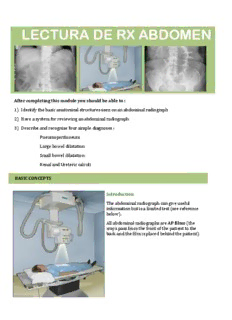
LECTURA DE RX ABDOMEN After completing this module you should be able to : 1) Identify the basic anatomical structures seen on an abdominal radiograph 2) Have a system for reviewing an abdominal radiograph 3) Describe and recognise four simple diagnoses : Pneumoperitoneum Large bowel dilatation Small bowel dilatation Renal and Ureteric calculi BASIC CONCEPTS Introduction The abdominal radiograph can give useful information but is a limited test (see reference below). All abdominal radiographs are AP 9ilms (the xrays pass from the front of the patient to the back and the Jilm is placed behind the patient). Most adults are too big to ' 9it ' on to one radiograph and so will often have two 9ilms to cover the whole abdomen. Make sure you look at both Jilms. For a tall patient occaisionally three Jilms are necessary in order to include the diaphragm and pubic symphysis (for example the three radiographs displayed are for a single patient). DENSITY OF TISSUES Four densities are seen on plain radiographs : Soft tissues Gas Bone Fat Soft tissues appear grey. For example the solid organs such as the kidneys - annotated on this Gas appears black or dark grey. For example in the various parts of the bowel visible on a plain DENSITY OF TISSUES Four densities are seen on plain radiographs : Soft tissues Gas Bone Fat Fat appears dark grey but ligher than soft tissues. Fat is seen throughout the abdomen but there are speci9ic areas to look for fat, for example, the properitoneal fat line (annotated on this 9ilm). Fluid will have the same density as soft tissue. For example the bladder may contain 9luid and will look like a soft tissue mass in the pelvis. DENSITY OF TISSUES Bone appears white or light grey Calci9ication is seen in a number of different structures. It willl have the same density as bone. For example : Costal cartilage calci9ication Mesenteric lymph nodes Vascular calci9ication - arterial or venous (phleboliths) REVIEW THE FILM. SYSTEMATIC APPROACH Use a systematic approach - look at : bowel gas pattern : dilated bowel an absence of bowel gas thickened bowel wall position of bowel gas abdominal organs : are they enlarged? are they clearly outlined? is there abnormal gas or calcification overlying the organs? bones : increased density metastases fractures etc. free gas : an erect chest radiograph is often more useful for looking for free gas abnormal calci9ication : gallstones renal calculi ! appendicolith calcification in the wall of an aneurysm etc. unusual gas collections : abscess gas in gallbladder gas in kidneys or bladder wall Deciding if an abdominal film is technically adequate is less problematic than when reviewing a chest radiograph. 1. Ensure it is the correct patient : This has become less of a problem with digital films but it is still vital to ensure the correct film is being reviewed. 2. Adequate coverage : The film should include the diaphragms, the pubic symphysis and the edges of the bowel at the lateral aspects of the film. Image 1: is inadequate because the pubic symphysis and diaphragms are not included. Image 2 : is inadequate because the edge of the bowel is not visible laterally and the diaphragms and pubic symphysis are not visible. Deciding if an abdominal film is technically adequate is less problematic than when reviewing a chest radiograph. 3. Exposure : In theory if a film is correctly exposed it should be possible to see the outline of the psoas muscle on the film. In practice overlying bowel gas and soft tissue such as fat may make it difficult to see the psoas shadow. 4. Rotation : This is not such a significant problem as encountered with chest radiographs because the film is taken with the patient lying supine. Image 3 : is inadequate because it does not include the diaphragms and pubic symphysis. ANATOMY It is often possible to see an outline of the kidneys due to the fat surrounding them which appears darker than the soft tissue of the kidneys. The spleen is harder to see on plain film and is usually not visible. Normally no bowel is visible in the right upper quadrant due to the large soft tissue mass of the liver. Occaisionally the liver can extend inferiorly - this is known as a Riedel's lobe. ! Señala en las radiogra9ías previas el hígado, el bazo y los riñones. Señala el lóbulo de Riedel Look for the psoas muscles. The radiograph shown is from a young man and therefore the psoas muscles are particularly visible. The absence of a psoas shadow may indicate retroperitoneal pathology. ! Señala el músculo psoas. ANATOMY Although the bladder is full of fluid it is often visible on the the plain radiograph as the fluid will have the same density as soft tissue. Look for a soft tissue mass in the pelvis. ! Señala en las radiogra9ías previas la vejiga. The stomach is seen beneath the left hemidiaphragm and often extends across the midline. ! Señala el estómago
Description: