Hymenolepis nana - University of Massachusetts Amherst PDF
Preview Hymenolepis nana - University of Massachusetts Amherst
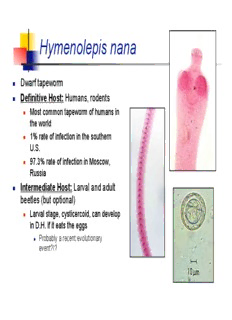
Hymenolepis nana Dwarf tapeworm Definitive Host: Humans, rodents Most common tapeworm of humans in the world 1% rate of infection in the southern U.S. 97.3% rate of infection in Moscow, Russia Intermediate Host: Larval and adult beetles (but optional) Larval stage, cysticercoid, can develop in D.H. if it eats the eggs Probably a recent evolutionary event?!? Hymenolepis nana Geographic distribution: Cosmopolitan. Mode of Transmission: Ingestion of infected beetle Ingestion of food contaminated with feces (human or rodent) Fecal/oral contact Control: remove rodents from house Pathology and Symptoms: Generally none because worm is so small (about 40 mm). Cysticercoid Hymenolepis nana life cycle Hymenolepis diminuta Rat tapeworm Definitive Host: Humans and rats Human infections are uncommon Intermediate Host: grain beetles (Tribolium) Required Geographic Distribution: Cosmopolitan Mode of Transmission to D.H.: Ingestion of infected beetle. Hymenolpeis diminuta Pathology: Usually asymptomatic because worms are relatively small (90 cm maximum). Heavy infections are rare. No fecal/oral infection Diagnosis: Eggs in feces. Eggs do not have polar filaments. Treatment: Praziquantel Prevention: Remove rats from home. Notes: Easily maintained in laboratories so has been used as the “model” tapeworm to study metabolism, reproduction, genetics, physiology, etc. H. diminuta human infections are rare Echinococcus granulosis A.K.A – Sheep Tapeworm Definitive Host: Carnivores including dogs, wolves, and coyotes Intermediate Host: Herbivores including sheep and mice. Geographic Distribution: Most common in sheep raising countries New Zealand and Australia highest incidence Echinococcus Life Cycle Hydatidosis Caused by the larval stage. After egg hatches, oncosphere leaves intestines and goes to another location Divides to create more worms Forms a hydatid cyst. Single chamber filled with fluid and larvae Tough, outer wall Grows very slowly. May take 20 years for symptoms to start The Hydatid Cyst The cyst is lined by a multilayer parasite tissue with the innermost layer being the germinal layer This layer is a undifferentiated “stem cell” layer that can spawn the formation of “brood capsules” which are themselves lined by GL The daughter cysts (the encircled body) "bud" into the center of the fluid-filled cyst. Thousands of protoscolices can fill the hydatid (hydatide sand) Protoscolices are the infective stage for dogs (each one will grow into an adult worm) Hydatides usually grow slowly but steadily (1-5 cm per year) They are usually well tolerated until their size becomes a problem or they rupture Cyst rupture or leakage can result in allergic reactions and metastasis
Description: