Histological investigations on Dendrodoris nigra (Stimpson, 1855) (Gastropoda, Nudibranchia, Dendrodorididae) PDF
Preview Histological investigations on Dendrodoris nigra (Stimpson, 1855) (Gastropoda, Nudibranchia, Dendrodorididae)
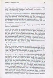
Molluscan Research 20(1): 79-94 (1999) Histological investigations on Dendrodoris nigra (Stimpson, 1855) (Gastropoda, Nudibranchia, Dendrodorididae) Heike Wiigele , Gilianne D. Brodie 2) and Annette Klussmann-Kolb Lehrstuhl fiir Spezielle Zoologie, Ruhr-Universitat Bochum, 44780 Bochum, Germany (2) Department of Marine Biology & CRC Reef Research Centre, James Cook University of North Queensland, Townsville, Australia Abstract The histology of the major organ systems (digestive, reproductive, nervous, circulatory, excretory and respiratory, as well as epidermis) of the nudibranch Dendrodoris nigra (Stimpson, 1855) are described for the first time and the results are compared with those derived from other members of the Doridoidea. It is shown that some characters which have been used to differentiate the genus Dendrodoris Ehrenberg, 1831 from other doridoideans (i.e., retractability of gills, lack of hard structures in the anterior digestive system, presence of pericardial glands) are problematic when used for phylogenetic analysis. This is especially true when taking into consideration that little is known about details of these structures in the Doridoidea as a whole. Key words: Nudibranchia, Doridoidea, Dendrodoris nigra, histology, phylogeny Introduction Two recent papers dealing with the taxonomy of species belonging to the genus Dendrodoris Ehrenberg, 1831 have clarified the taxonomy of species from the Indo-Pacific (Brodie et al. 1997) and the Atlantic (Valdés et al. 1996), and demonstrated complexes of highly variable species in both instances. Although the external features may be very variable in some species e.g. Dendrodoris migra (Stimpson, 1855) and Dendrodoris grandiflora (Rapp, 1827), the internal organ systems seem to be rather more conservative with relatively little variation (Brodie et al. 1997). Furthermore, a comparison of the organ systems of different Dendrodoris species shows that Dendrodoris is a well marked genus within the family Dendrodorididae O(cid:8217)Donoghue, 1924. The synonymy and validity of genera within the Dendrodorididae have been reviewed recently (Valdés et al. 1996; Valdés & Ortea 1997). : ; ; The family Dendrodorididae is unusual within the Doridoidea in that it lacks cuticular structures within the buccal mass. This character 1s shared by the Phyllidiidae Rafinesque, 1815, a family with secondary gills on the ventral side between the notum and the foot. The absence of jaws and radula is the single character which, according to Bergh (1876), unites these two families, and for which he created the taxon Porostomata. Although many authors have followed his classification (Pruvot-Fol 1954, Odhner in Franc 1968, Schmekel & Portmann 1982, Thollesson 1998), some mentioned doubts and considered the taxon an artificial grouping (Brunckhorst 1993, Rudman 1998). Since only the lack of characters unites these two rather different families, there is a demand for further characters to support or to falsify the relationship. Brunckhorst (1993: 84) 80 H. Wagele, G.D. Brodie and A. Klussmann-Kolb came to the conclusion, after analysing the phylogenetic relationship within the Phyllidiidae, using Dendrodoris and Chromodoris as outgroups, that Dendrodoris shared more derived characters with the Cryptobranchia than with the Phyllidiidae. He concluded (cid:8220)the grouping of dendrodorids and phyllidiids together as the Porostomata is untenable.... The Dendrodorididae clearly belong with other doridoids ... but their phylogenetic position is yet to be clarified.(cid:8221) This view was diametrically opposed by Valdés (1996) who stated that (cid:8220)the superfamily Porodoridoidea is a monophyletic group(cid:8221) and that (cid:8220)the loss of the radula has only occurred once in the evolution of the dorids. (cid:8220) There is no doubt that this situation requires further clarification. In this paper we present characters of Dendrodoris nigra which are difficult to find by macroscopic investigation and which will help to elucidate the phylogenetic relationship of the Dendrodorididae. Although we present several characters in a new light, it is evident that only after investigating many more members of the Doridoidea with the same thoroughness that the relationship of the Dendrodorididae can be determined. Material and Methods Three specimens of Dendrodoris nigra (length of living specimens 23, 43, 53 mm) from Dingo Beach ,(Queensland, Australia, collected by the authors in August 1995 and July 1997) were embedded in hydroxyethlymethacrylate for serial sectioning (2-3 (m). Sections were stained with toluidine blue. Comparisons were made with sections of other doridoideans belonging to the Cryptobranchia and Phanerobranchia. For anatomical drawings and general description of the organ systems of D. nigra the reader is referred to Brodie et al. (1997). Results Epithelia Epidermis: The dorsal notal epithelium consists mainly of high columnar to flask- shaped cells with basally lying nuclei (Fig.1A). The cytoplasm stains homogeneously light blue to transparent without further differentiation. Cells of similar size with several smaller, light blue staining grana and cells with a dark violet staining network (cells producing acid mucopolysaccharids) are interspersed. Very few ciliated cells can be observed. Black pigment grana, which obviously are not confined to a certain cell type, are located in the connective tissue of the notum beneath the basal lamina of the epidermis. The epithelium on the ventral side of the notum shows similar cell types, but the cells are less tall. In the dorsal area, the epithelium very often forms grooves or invaginations, which do not differ in their cellular appearance from the rest of the epithelium (Fig. 1B). Subepithelial glandular follicles composed of one or several cells are present in the lateral part of the notum. These glandular cells are similar in appearance to the acid mucoploysaccharide producing cells in the epidermis, but staining slightly darker. Rhinophores: The epithelium consists of small, prismatic cells with basally lying nucleus and no visible differentiation of cytoplasm. Some glandular cells are interspersed, containing a large light blue staining vacuole. The margins of the Histology of Dendrodoris nigra Hl Figure 1. (cid:8217) ; : Dendrodoris nigra. Epithelia. A, dorsal notum epithelium with crnleiyatg Ts Be granules; B, dorsal notum epithelium of an invagination; C, basal part Sie eae bes glands (arrows); D, glandular follicles around the mouth; arrow indicates the ct the mouth. Scale bars: A 50 zm, B-D 100 tm. 82 H. Wagele, G.D. Brodie and A. Klussmann-Kolb * Figure 2. Dendrodoris nigra. Digestive tract. A, transverse section of labial disc with pharynx on the right and oral tube (asterisk) on the left side; B, oral gland with cross section of collecting ducts, annexed glandular area staining dark (arrow) and glandular area staining light; C, semi cross section of pharynx; cuticular lining (arrow) barely visible; D, cross section of right salivary gland. Abbreviations: ph - pharynx; Scale bars: A-C 100 um, D 50 um. Histology of Dendrodoris nigra 83 Figure 3. (cid:8217) Dendrodoris nigra. A, cross section showing transition from pharynx into orsgphagus (asterisk) with proximal part of oesophagus above the asterisk and on bottom on rig : side, juvenile nidamental glands and oral gland on the left side above pharynx (arrows); B, part of distal oesophagus; C, cross section of digestive glandular lobes, D, cross pa of vas deferens (arrow) and prostatic duct with subepithelial lying glandular follicles o prostate gland. Abbreviations: ngl - nidamental glands, oe - oesophagus, ph - pharynx, pr - prostate gland; Scale bars: A-D 100 fm. 84 H. Wagele, G.D. Brodie and A. Klussmann-Kolb Figure 4. Dendrodoris nigra. A, vestibular gland of immature specimen; asterisk indicates the opening into the vestibulum; arrow indicates the distal oviduct; B, detail of vestibular gland of mature specimen; epithelium showing a high microvilli border (arrows); C, blood vessel situated between the fused retractor muscles (arrow).; D, eye with optic ganglion beneath and cerebro-pleural complex on right side. Abbreviations: fo - foot, dgl - digestive gland intermingled with excretory system. Scale bars: A, C-D 100 tm, B 50 um. Histology of Dendrodoris nigra 85 5 ae AfAy T | Figure 5. : Dendrodoris nigra. A, cross section of anterior part of body, showing body cavity eee by a thick muscle layer (arrows), labial disc with pharynx, pedal ganglia on both sides 0 the pharynx and the pleural (asterisk) only on the left side; B, cross section of syrinx. Scale bars: A-B 100 fim. . thinophoral lamellae of one animal shows few larger cells containing non- staining vacuoles, similar to the special vacuole cells of other Doridoidea (see Wagele 1998). Foot epithelium: The cells are tall and ciliated, with nuclei lying basally to medially. Few epithelial glandular cells with violet staining grana are present Subepithelial glandular follicles with two to four glandular cells containing blue to violet grana are very common. Gill (Fig. 1C): The epithelium consists of cuboidal cells with large nuclei. Glandular cells, which stain homogeneously light blue or have violet grana, are interspersed. Ciliated cells are also present. Gill glands (Fig. 1C, arrows) art present at the bases of the gills. They are composed of many cells with bluis contents and large nuclei. The gills are underlaid by thick muscle layers, concentrating anteromedian first in one U-shaped muscle, later separating into two strings running ventrally to the head region. 86 H. Wagele, G.D. Brodie and A. Klussmann-Kolb Digestive tract Oral tube: The epithelium consists of high columnar cells interspersed with some glandular cells with bluish contents and a few glandular cells containing violet grana. A thick glandular layer, composed of pyriform glandular follicles filled with grana of rather uniform size, staining bluish violet, surrounds the mouth area (Fig. 1D). Labial disc (Fig. 2A): The labial disc is not covered by a cuticle. Scattered glandular cells containing vacuoles uniformly staining violet can be observed. Two strong muscles insert at the labial disc (Fig. 5A). Oral (ptyaline) gland (Fig. 2B): the oral gland is formed by a thick layer of glandular cells resulting in a spongy appearance. The large vacuoles in these cells, which have pycnotic nuclei, do not stain. Along the outgoing ducts glandular cells with bluish contents can be observed. The two main ducts are characterized by a thick ring muscle layers. After fusion of these ducts, the common duct leads ventrally along the pharynx within the labial disc and opens into the lumen of the oral tube. Pharynx (Fig. 2C, 3A, 5A): The pharynx is tube-like and long and is composed of several layers. From outer to inner side following layers can be observed: exterior circular muscle layer with areas of transverse muscles; layer of cells with large vacuoles giving the appearance of a network; layer of circular muscles; pharyngeal epithelium without any glandular cells; thin cuticle. Salivary glands (Fig. 2D): The salivary glands form a rather compact circle around the transition between pharynx and oesophagus. Only one microscopic duct leading into the pharynx is observable. No distinct lumen within the glandular tissues is visible, and no distinct arrangement of glandular cells into an epithelium is discernible. The glandular cells exhibit a medium sized functional nucleus, less commonly a pycnotic nucleus. They are completely filled with densely packed, very light blue staining grana, showing a superficial similarity to the oral glandular tissue. Oesophagus (Fig. 3A, B): The oesophagus is surrounded by a layer of circular and longitudinal muscle fibres of varying thickness. No lumen is observable throughout the oesophagus except for the most anterior part, which is filled with pink secretions. It is difficult to describe the number of different glandular cell types, since all different types may represent different functional phases of one and the same type. Following phases are discernible: cells with blue to violet staining large vacuoles; cells with reddish to violet staining large vacuoles; cells with different coloured (from pink to light violet) but rather homogeneously staining cytoplasm.The nuclei always lie basally. A further glandular cell type with filiform contents, staining dark violet, is present in the anterior part of the oesophagus (Fig. 3A). In the distal part of the oesophagus, the cells with homogeneously staining contents dominate (Fig. 3B). The whole oesophagus is underlaid by an extremely thick muscular layer. Histology of Dendrodoris nigra 87 Stomach: Especially at the entrances into the digestive gland, the stomach wall is folded. The epithelium is composed of small, elongate and ciliated cells with small, elongate, basally lying nuclei. Digestive gland: Many entrances from the stomach area into the digestive gland are visible. Probably only one type of glandular cells is present in the digestive gland, since transitional stages can(cid:8217)be observed: early stage cells contain small bluish grana or vacuoles, increasing in size in the following phases and forming a grape-shaped (clustered) structure, now staining more greenish to light blue (Fig. 3C). When contents of vacuoles is emptied, small spaces or areas with microscopic grana staining violet remain. Caecum: No caecum (transformed right digestive gland) opening into the stomach is distinguishable. Intestine: The inner wall of the intestine is folded throughout its length. After its origin from the stomach the intestine is especially highly convoluted. No typhlosole is discernible. The epithelium consists of elongate, non-glandular cells, with long cilia and basally to medially lying nuclei. Some violet staining glandular cells are interspersed in the proximal part of the intestine that follows after bending and running posteriorly. The intestine is surrounded by a layer of circular muscles followed (inwards) by a ring of longitudinal muscles. No specialization of cells is discernible in the area where the intestine bends and then runs posteriorly, called caecum or pyloric gland by different authors respectively (Brodie et al. 1997; Valdas et al. 1996). Reproductive system at eS Gonad and ampulla: The gonad forms an incomplete layer over the digestive gland, with parts extending deep into the digestive glandular tissue. In cross section the digestive glandular tissue appears intermingled with gonad and kidney. The ampulla is sausage-shaped and filled with autosperm. The epithelium of the ampulla is composed of cuboidal to flat cells. Distal genital tract: The vas deferens shows a thick tubular prostate gland. The glandular cells lining the duct are elongate, filled with light blue staining grana and basal nuclei. A dense ciliation is present within duct. Subepithelial glandular cells are enormous, of similar appearance as the epithelial glandular cells, but with large nuclei, arranged into follicles (Fig. 3D). The proximal and distal sections of the vas deferens (non-prostatic parts) are lined by an epithelium with cuboidal, ciliated cells only. A layer of circular muscles surrounds the non-glandular parts. No cuticularized penial structures (i.e. cuticular lining and/or spines) are visible in any of the specimens investigated. In the smaller specimen the female mass is not differentiated into distinct glands (Fig. 3A), but in the two larger ones itis composed of three distinct parts, namely the capsule gland, proximal and distal mucous glands. The capsule gland consists of numerous narrow coils. The cells of the epithelium are broad and columnar containing numerous, large, light-blue staining granules. 88 H. Wagele, G.D. Brodie and A. Klussmann-Kolb Spherical to elliptic nuclei are lying at the base of the glandular cells. Supporting cells with short cilia are alternating with the glandular cells. The proximal mucous gland is connected to the capsule gland by a long coiled duct lined by densely ciliated cells. The epithelium of the proximal mucous gland is folded in narrow coils similar as in the capsule gland. The glandular cells are prismatic and contain heterogeneous mucous secretions staining purple or dark red. Supporting cells bear very long cilia. The distal mucous gland is composed of various parts with different staining properties. These may represent various functional stages and are therefore difficult to interpret. All glandular cells of the distal mucous gland contain purple to red staining mucous secretions. The basally lying nuclei of the glandular cells are pycnotic. The supporting cells have long cilia. The opening of the oviduct lies more caudally than the openings of the vagina and the vas deferens. The vaginal duct is narrow and opens proximally into the vestibulum next to the vas deferens. The epithelium of the bursa copulatrix is composed of apocrin secreting cells. Only a few muscle cells surround the bursa. The receptaculum seminis is filled with allosperm, these being orientated but not attached to the folded wall. Contrary to the bursa, a distinct layer of muscles surrounds the receptaculum. In the smaller specimen, the vestibular gland lies completely within the notal tissue and therefore is not visible anatomically in this animal (Fig. 4A). In the larger specimens, it intrudes into the visceral cavity as a compact mass. The cells are small, elongate, with basally (in smaller) to medially lying nuclei (in larger specimens) and apically lying, microscopic, violet-staining grana. The cells in the larger specimen exhibit a brush-like appearance (Fig. 4B). Nervous system The circumoesophageal central nervous system, with fused cerebral and pleural ganglia, surrounds the pharynx (Fig. 5A). All ganglia are surrounded by a thin layer of connective tissue. Nerve cells are concentrated in the periphery of the ganglia. The statocyst has many elongate otoconia (length about 5 um). The eye is connected to a small optic ganglion by a very short optic nerve (Fig. 4D). Circulatory system In the anterior dorsal wall of the pericardium folds ((cid:8220)pericardial glands(cid:8221)) are present, which are not glandular, but are composed of tiny cells and small nuclei being similar in appearance to those of the connective tissue. A single, well formed blood vessel is present, which runs anteriorly in the ventral side of the muscle layer surrounding the visceral cavity, and posteriorly within the two fused gill retractors (Fig. 4C); sending ducts into the foot in two directions. The blood gland is characterized by minute cells and nuclei, forming a rather compact mass on the right side of body next to the cerebropleural complex, with lacunae interspersed.
