Table Of ContentHigh-Precision Measurement of Rydberg State Hyperfine Splitting in a
Room-Temperature Vapour Cell
Atreju Tauschinsky,∗ Richard Newell, H. B. van Linden van den Heuvell, and R. J. C. Spreeuw†
Van der Waals-Zeeman Institute, Institute of Physics, University of Amsterdam,
PO Box 94485, 1090 GL Amsterdam, The Netherlands
(Dated: January 30, 2013)
WepresentdirectmeasurementsofthehyperfinesplittingofRydbergstatesinrubidium87using
Electromagnetically Induced Transparency (EIT) spectroscopy in a room-temperature vapour cell.
With this method, and in spite of Doppler-broadening, line-widths of 3.7MHz FWHM, i.e. signifi-
cantly below the intermediate state natural linewidth are reached. This allows resolving hyperfine
splittingsforRydbergs-stateswithn=20...24. WiththismethodweareabletodetermineRyd-
3
1 berg state hyperfine splittings with an accuracy of approximately 100kHz. Ultimately our method
0 allows accuracies of order 5kHz to be reached. Furthermore we present a direct measurement of
2 hyperfine-resolved Rydberg state Stark-shifts. These results will be of great value for future exper-
iments relying on excellent knowledge of Rydberg-state energies and polarizabilities.
n
a
PACSnumbers: 32.10.Fn,32.60.+i,32.80.Ee,42.50.Gy
J
9
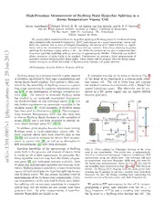
2 Rydberg atoms have recently received a great amount A schematic drawing of the setup is shown in Fig. 1.
ofattention,motivatedbytheirlargepolarizabilitiesand At the heart of the experiment is a custom-made rubid-
]
h strongdipole-dipolecoupling. Thisinterestisoftenstim- ium vapour cell. The cell is 10cm long and contains
p ulated by the suitability of Rydberg atoms to engineer twointernalstainlesssteelelectrodesof95×20mm2 size
-
long-rangeinteractionsforquantuminformationprocess- spaced 5.35(3)mm apart. The electrodes can be con-
m
ing [1–3] or the investigation of strongly correlated sys- nected to a DC power supply and an Agilent 33250A
o
tems [4–6]. The research in ultra-cold Rydberg atoms function generator.
t
a has resulted in two landmark experiments demonstrat-
.
s ing dipole-blockade for two individual atoms [7, 8], but
c
also further experiments on mesoscopic ensembles in the
i
s blockade regime [9]. Cold ensembles of Rydberg atoms
y
have been used for electrometry [10–12]. Electromagnet-
h
p ically Induced Transparency (EIT) has also been used
[ to observe Rydberg dipole blockade in cold ensembles of
atoms [13–15] and it has been proposed to directly ob-
1
serve dipole blockade using EIT [16, 17].
v
7
Inaddition,greatprogresshasalsobeenmadeexciting
0
Rydberg atoms in room-temperature vapour cells. In-
9
deed, coherent effects have been observed here as well
6
. [18, 19] and sensitive methods for electric field measure-
1
ments in vapour cells [20], as well as an alternative to
0
3 EIT measurements [21] have been developed.
1
Excellent knowledge of the spectroscopy of Rydberg FIG. 1: (Color online) (a) Schematic drawing of the setup
:
v states both in the presence and absence of electric fields used in the experiments. The probe laser is independently
Xi is crucial for all of these experiments. In particular, Ry- locked to a saturated absorption frequency-modulation (FM
dberg hyperfine structure may limit the fidelity of quan- spectroscopy setup not shown here. The reference cell used
r
to compensate long-term frequency drifts was only used for
a tum gates [22] and undermine coherent evolution. Here
the Stark-map measurements shown in Fig. 4, but not for
weshowthathigh-precisionhyperfinespectroscopyofru-
the hyperfine data presented in Figs. 2 and 3. DM: Dichroic
bidiumRydbergstatesispossibleinaroom-temperature
Mirror. (b) Energy level diagram with the weak probe laser
vapourcellandinvestigatethehyperfinesplittingforvar- coupling the 5s ground to the 5p excited state with Rabi
3/2
ious Rydberg states. We also present hyperfine-resolved frequency Ω and the strong coupling laser connecting the
p
measurementsoftheRydbergstatepolarizability. Previ- excited state to a Rydberg state with Rabi frequency Ω .
c
ous measurements of the zero-field Rydberg state hyper-
fine splitting rely on mm-wave transitions in a magneto- We perform EIT spectroscopy in this cell by counter-
optical trap, but the results are less precise than those propagating a probe laser resonant with the 5s →
1/2
presented here. No prior measurements of hyperfine- 5p transition of 87Rb and a coupling laser coupling
3/2
resolved Stark-shifts are known to us. the 5p state to a Rydberg state through the cell. The
2
probe laser is derived from a Toptica DL-100 external frequency Ω →0, where a model of the form
p
cavity diode laser at 780.24nm frequency-stabilized by
(cid:90) ∞ i
saturated-absorption frequency-modulation (FM) spec- χ∝ N(v)dv (1)
troscopy in a separate vapour cell to the F=2 to F(cid:48)=2 −∞ γp−i∆p+ γc−i(Ω∆2c/p4+∆c)
hyperfinetransition. Thecouplinglaserisderivedfroma
for the susceptibility χ of the probe transition is valid
frequency-doubled amplified diode laser system (Toptica
[23]. Here Ω is the coupling Rabi frequency, the probe
TA-SHG Pro) at ≈480nm and scanned across a Ryd- c
and coupling detunings depend on the velocity of the
berg resonance. Both lasers propagate through the cell
atoms through Doppler shifts:
parallel to the long axis of the field plates and are over-
lapped over the entire length of the cell. The gaussian ∆ =∆0−k v
p p p
beamwaistsareapproximately0.4mmfortheprobeand
∆ =∆0+k v
1.0mm for the coupling lasers, with peak intensities of c c c
0.4mW/cm2 and 4.3mW/cm2 respectively. and γp and γc are decay rates given by γp =1/2Γ5p with
Thecouplinglaserismodulatedbyachopperwheelat Γ5p the natural decay rate of the excited state and γc =
approximately4kHzforlock-indetectionoftheEITsig- 1/2Γr withΓr thenaturaldecayrateoftheRydbergstate.
nal. A reference vapour cell without electric field plates Additional broadening effects can be included in γc (see
is used to measure and compensate for drifts of the cou- below). N(v) is a one-dimensional Maxwell-Boltzmann
pling laser frequency during scans. velocity distribution describing the velocity of the atoms
inthevapourcell. Theintegralovervisequivalenttothe
We measure Rydberg state hyperfine splittings by
averaging over all velocity classes that occurs in a room-
scanning the coupling laser across a Rydberg resonance
temperature vapour cell and can be solved analytically
byturningthegratingoftheECDLheadoftheSHGsys-
for a Maxwell-Boltzmann velocity distribution [23].
tem with the built-in piezo-element. The resulting spec-
The imaginary part of χ determines the absorption of
trum is shown in Fig. 2 for the 20s-state. The frequency
the probe laser. We fit the data to an incoherent sum of
axis is calibrated by applying a 7MHz sinusoidally vary-
six peaks of the form (1) after analytic integration as in
ing voltage to the field plates of the vapour cell, thereby
[23]. Eachofthetwohyperfinepeaksisfittedwithanin-
creatingsidebandsofthestateatawell-definedfrequency
dependent coupling Rabi frequency, but sidebands share
spacing.
the coupling Rabi frequency of the main peaks. The in-
termediatestatelinewidthisfixedtotheliteraturevalue,
γ =2π×3.03MHz [24]. The excited state linewidth γ
p c
is fitted to a common value for all peaks. The fixed sep-
aration of the sidebands allows us to precisely calibrate
the frequency axis and thus extract accurate values for
the hyperfine splitting from our data.
Fig. 2 shows a spectrum for a Rydberg state 20s and
the corresponding fit. The data points are an average of
860 individual traces, aligned by fitting two gaussians to
themainhyperfinepeaksandcenteringthemidpointbe-
tweenthepeaksbeforeaveraging. Dataandfitarevirtu-
allyindistinguishable,confirmingthequalityofourmea-
surements. The linewidth of our features is particularly
remarkable: The fitted γ is typically 2π×2MHz, sig-
c
nificantly smaller than the intermediate state linewidth,
even though all measurements are done in a vapour cell
at room temperature, and with a free-running coupling
laser. The observed width of a single hyperfine peak of
about 2π×3.7MHz FWHM is however still somewhat
FIG. 2: (Color online) Spectrum of 20s hyperfine structure larger than the limit of 2π×1.7MHz for vanishing γ
c
including positive and negative 2nd order sidebands used for and Ω that can be observed in rubidium at room tem-
c
frequencycalibration. Bluedots: averageof860traces;Light-
perature. This is due to both the finite linewidth of the
blue line: fit based on equation (1). The lower part of the
free-runningcouplinglasersystemaswellastransit-time
figure shows the residual of this fit. The field was modulated
broadening due to atoms mving in and out of the beam
at 7MHz. First-order sidebands are not visible as their exci-
tation is dipole-forbidden. radially [25], which we estimate at approximately 1MHz
for our beam width.
WeperformsimilarmeasurementsforRydbergsstates
We assume the weak-probe limit, i.e. the probe Rabi with principal quantum numbers n = 20...24. At n >
3
n ν σ σ ∆ bands for the frequency calibration. We then find a dif-
hfs fit piezo scaling
20 7 782 4 +57/−17 -43 ference of the measured hyperfine splittings for the two
21 6 497 3 +40/−20 14 cases of between 50kHz and 300kHz, with the two high-
22 5 442 5 +22/−61 88 est n showing the largest errors, and the three lower n
23 4 780 7 +45/−106 -44 showingerrorsoflessthan80kHz. Theerrorbarsshown
24 4 229 9 +47/−281 -142 inthefigurearebasedontheseresults. Thenonlinearity
in the piezo scan is the biggest uncertainty identified in
TABLE I: Table of measured hyperfine splitting in the range the frequency calibration.
n=20...24,aswellasfittingerror,errorderivedfrompiezo Ourmeasurementsareinexcellentagreementwiththe
nonlinearitiesanddistancetoscalinglawfit,allgiveninkHz. expected(n−δ)−3 scaling. Hereδ isthequantumdefect
of the state, depending on both n and (cid:96) and taken from
[27], leading to an effective principal quantum number
24theDoppler-broadenedlinewidthoftheEITresonance
n∗ = n − δ. The inset of Fig. 3 shows our data to-
is too large to observe individual peaks. At n < 20 the
getherwithearlierresultsusingmicrowavetransitionsto
spectral tuning range of our laser system is limited.
other Rydberg states from reference [27] as well as low-
n data from reference [26] after removing the expected
n∗-scaling. We see excellent agreement with the low-n
data but an offset of approximately 10% compared to
the results of [27]. The excellent agreement of our mea-
surementswithlow-ndatamightindicatethatthisoffset
isduetosystematicsinthedataof[27]. Thedataof[27]
isinagreementwithourmeasurementsassuminganerror
of 1 in the principal quantum number of their data. An
equation for the scaling of the hyperfine splitting based
on our data is given in (2). From this we expect a hy-
perfine splitting of approximately 80kHz at n = 80. A
best fit with variable exponent yields a scaling law with
a power of −2.95(11) for our data.
FIG. 3: (Color online) Scaling of the hyperfine splitting with ν =37.1(2)GHz(n−δ)−3 (2)
effective principal quantum number n∗ = (n−δ), extracted hfs
from measurements such as presented in Fig. 2. The solid Finallywepresenthyperfine-resolvedmeasurementsof
lineisbasedona(n−δ)−3-scalingwithonlythepre-factoras
Stark shifts in fields of up to 130V/cm for 20s. The up-
fit parameter. The shaded area signifies the 95% confidence
per part of Fig. 4 shows the overall Stark shift of state
regionofthisfit. Theinsetshowsthesamedataafterremov-
ing the (n−δ)−3 scaling in comparison to low-n data from 20s. The three independent lines are due to different
reference [26] and slightly higher-lying states from reference 5p3/2 hyperfine states; while the probe laser is locked to
[27]. Error bars indicated are estimated on the basis of piezo theF(cid:48) =2transition,otherlinescanbeshiftedintoreso-
scan nonlinearity, see text. nancebyDopplershiftinthevapourcell. Duetothedif-
ferentwavelengthsoftheprobe-andcouplinglaserthese
The resulting hyperfine splittings are shown in Fig. shifts are only partially compensated by the counter-
3, with our results also listed in table I. The error bars propagating beams; the remaining shifts are expected to
listed in the table are standard errors obtained from the be reduced by a factor λc/λp = 480/780 compared to the
fit. By separately analysing 300 individual traces of the 5p hyperfine splittings, in good agreement with our
3/2
20s measurement we find a mean hyperfine splitting of observations. In the topmost line no hyperfine splitting
7.801MHz with a standard error of the mean of 7.2kHz, is visible, as the excitation of the F(cid:48)(cid:48) = 1 component of
i.e. 19kHz larger than the results quoted above. As the the Rydberg state is dipole forbidden from 5p F(cid:48) =3.
3/2
fitting of the sidebands can be difficult without averag- InboththeF(cid:48) =1andF(cid:48) =2linesthehyperfinesplitting
ing we consider the values quoted in table I to be more oftheRydbergstateisinprinciplevisibleintheindivid-
reliable. ualtraces. However,thesignalinF(cid:48) =2ismuchstronger
In addition to this it is worth noting here that nonlin- than in F(cid:48) =1, making the splitting almost indiscernible
earitiesintheresponseofthepiezo-elementusedtotune for F(cid:48) =1 in this plot.
the coupling laser frequency can in principle also skew The overall shift of the Rydberg state is in excel-
our results, although this can be minimized by making lent agreement with calculations based on wavefunc-
surethattheobservedpeaksarenotnearaturningpoint tions obtained with the Numerov method [28], as can
ofthefrequencyscan. Weestimatethemagnitudeofthis be seen in the dashed line overlaid with the F(cid:48) = 3
effect by using only either the lower or the upper side- state which has no free parameters. Fitting a parabola
4
can be seen. This is compatible with a misalignment of
the field plates by approximately 1mrad, equivalent to
100µmdifferenceinplateseparationattheedges, which
wehaveobservedinearliermeasurementsofhigher-lying
states in which the hyperfine splitting is entirely negligi-
ble.
In conclusion we have presented high-precision mea-
surements of the hyperfine splitting of Rydberg states
in 87Rb, achieving kHzaccuracy even in a room-
temperature vapour. These measurements obey the ex-
pected (n−δ)−3-scaling very well and are in excellent
agreement with low-n data such as presented in [26].
However,ourmeasurementsshowasmallsystematicshift
comparedtomeasurementsathighernpresentedin[27].
We furthermore present hyperfine-resolved measure-
ments of Rydberg state Stark shifts. To our knowledge
no prior measurements of this kind have been demon-
strated. These show no change in the hyperfine splitting
as the electric field is increased, and no further splitting
of m levels, in agreement with our calculations.
F
The measurements presented above show how a reso-
FIG. 4: (Color online) Hyperfine-resolved Stark shifts of 20s lutionfarbelowtheDopplerlimitispossibleforRydberg
measuredbyapplyinganelectricfieldtotheelectrodesinthe
statespectroscopyinroom-temperaturevapourcells. Us-
vapour cell. The three lines in the upper part of the figure
ing a vapour cell with internal electrodes as described in
correspond to three different hyperfine states F(cid:48) = 1,2,3 in
this paper this makes high-accuracy Stark spectroscopy
the intermediate 5p state. The dashed line overlaid with
3/2
the F(cid:48) =3 state shows the excellent agreement of the overall extremely simple. This can be of great value for future
Stark shifts with calculations based on the Numerov method experiments relying on excellent knowledge of Rydberg-
[28]. Thebottompartofthefigureshowstherelativeshiftof state energies and polarizabilities.
thetwohyperfinestatesafterremovingtheoverallStark-shift,
WewouldliketothankN.J.vanDrutenforhelpwith
clearly indicating that the hyperfine splitting remains con-
the manuscript. This work is part of the research pro-
stant in an electric field, and no splitting of m -components
F
grammeoftheFoundationforFundamentalResearchon
is observed.
Matter (FOM), which is part of the Netherlands Organ-
isation for Scientific Research (NWO). We acknowledge
to the Stark shift in Fig. 4 we extract a value of support from the EU Marie Curie ITN COHERENCE
α=0.0720(8)MHz/(V/cm)2 from this data, in excel- Network.
lent agreement with the theoretically expected value of
α=0.0722MHz/(V/cm)2. The uncertainty in this de-
termination of α is dominated by the accuracy with
which the average separation of the electric field plates
∗ Electronic address: [email protected]
is known; the uncertainty from the frequency calibration
† Electronic address: [email protected]
is lower by one order of magnitude.
[1] D.Jakschetal.,PhysicalReviewLetters85,2208(2000).
We attribute the faint line visible above F(cid:48) = 3 to [2] M. D. Lukin, M. Fleischhauer, and R. Cote, Physical
inhomogeneous electric fields at the edges of the cell, in Review Letters 87, 037901 (2001).
particular in the gap between the electrodes and the cell [3] M. Mu¨ller et al., Physical Review Letters 102, 170502
(2009).
walls.
[4] N.Henkel,R.Nath,andT.Pohl,PhysicalReviewLetters
The lower part of Fig. 4 shows the hyperfine splitting
104, 195302 (2010).
of the F(cid:48) = 2 line after removing the overall quadratic [5] T. Pohl, E. Demler, and M. D. Lukin, Physical Review
Stark shift of the state. This has been done by fitting Letters 104, 043002 (2010).
the model of eq. 1 to each individual trace and aligning [6] P. Schauß et al., Nature 490, 87 (2012).
the center point between the two peaks across all traces. [7] E. Urban et al., Nature Physics 5, 110 (2009).
[8] A. Ga¨etan et al., Nature Physics 5, 115 (2009).
As can clearly be seen no further splitting into m sub-
F [9] Y. O. Dudin and A. Kuzmich, Science 336, 887 (2012).
levels occurs for these hyperfine states, and the splitting
[10] A. Tauschinsky et al., Physical Review A 81, 063411
remainsconstantacrosstherangeoffieldspresentedhere.
(2010).
Thisisinagreementwithnumericalcalculationswehave [11] R. Abel, C. Carr, U. Krohn, and C. Adams, Physical
performed. Athighfieldsaslightbroadeningofthepeaks Review A 84, 023408 (2011).
5
[12] J. Carter and J. Martin, Physical Review A 83, 032902 [21] D. Barredo et al., arXiv:1209.6550 (2012).
(2011). [22] T. G. Walker and M. Saffman, arXiv:1202.5328 (2012).
[13] J. Pritchard et al., Physical Review Letters 105, 193603 [23] J. Gea-Banacloche, Y.-Q. Li, S.-Z. Jin, and M. Xiao,
(2010). Physical Review A 51, 576 (1995).
[14] C. S. Hofmann et al., arXiv:1211.7265 (2012). [24] D. A. Steck, http://steck.us/alkalidata 2, (2010).
[15] T. Peyronel et al., Nature 488, 57 (2012). [25] J.ThomasandW.Quivers,PhysicalReviewA22,2115
[16] G. Gu¨nter et al., Physical Review Letters 108, 013002 (1980).
(2012). [26] A. Corney, Atomic and Laser Spectroscopy (Oxford Uni-
[17] B. Olmos, W. Li, S. Hofferberth, and I. Lesanovsky, versity Press, Oxford, 1977).
Physical Review A 84, 041607 (2011). [27] W. Li, I. Mourachko, M. W. Noel, and T. F. Gallagher,
[18] B. Huber et al., Physical Review Letters 107, 243001 Physical Review A 67, 052502 (2003).
(2011). [28] M.Zimmerman,M.Littman,M.Kash,andD.Kleppner,
[19] H. Ku¨bler et al., Nature Photonics 4, 112 (2010). Physical Review A 20, 2251 (1979).
[20] M. G. Bason et al., New Journal of Physics 12, 065015
(2010).

