Glaucoma PDF
Preview Glaucoma
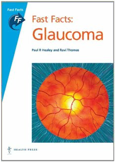
‘a great overview of glaucoma, certainly for those that need to Fast Facts F get acquainted with the subject, but no less for experienced a s ophthalmologists’ t Fast Facts: F Peter J. Ringens, Department of Ophthalmology a c VU Medical Centre, Amsterdam t s Glaucoma G l a u Fast Facts: c o Glaucoma m a Paul R Healey and Ravi Thomas 9 Definitions and epidemiology 20 Pathophysiology, natural history and prognosis 30 Diagnosis and clinical features 68 Principles of management 78 Medical treatment 92 Laser and surgical treatment 97 Monitoring 118 Prevention ISBN 978-1-905832-40-8 70 titles by 150 world experts at www.fastfacts.com 9 781905 832408 > FF Glaucoma1e pages.qxd 17/2/10 13:56 Page 1 Fast Facts Fast Facts: Glaucoma Paul R HealeyMBBS(Hons) BMedSc(Cell Biol) MMed(Clin Epidemiol) PhD(Med) FRANZCO Clinical Senior Lecturer University of Sydney Department of Ophthalmology Centre for Vision Research Westmead Millennium Institute & Save Sight Institute New South Wales, Australia Glaucoma Specialist Eye Associates Sydney, Australia Ravi ThomasMBBS MD FRANZCO Director of Glaucoma Services Queensland Eye Institute and Professor, University of Queensland Brisbane, Australia Previously: Professor & Head, Department of Ophthalmology Christian Medical College, Vellore, India Director, LV Prasad Eye Institute Banjara Hills, Hyderabad, Andhra Pradesh, India Declaration of Independence This book is as balanced and as practical as we can make it. Ideas for improvement are always welcome: [email protected] © 2010 Health Press Ltd. www.fastfacts.com FF Glaucoma1e pages.qxd 17/2/10 13:56 Page 2 Fast Facts: Glaucoma First published March2010 Text © 2010 Paul R Healey, Ravi Thomas © 2010 in this edition Health Press Limited Health Press Limited, Elizabeth House, Queen Street, Abingdon, Oxford OX14 3LN, UK Tel: +44 (0)1235 523233 Fax: +44 (0)1235 523238 Book orders can be placed by telephone or via the website. For regional distributors or to order via the website, please go to: www.fastfacts.com For telephone orders, please call +44 (0)1752 202301 (UK and Europe), 1 800 247 6553 (USA, toll free), +1 419 281 1802 (Americas) or +61 (0)2 9698 7755 (Asia–Pacific). Fast Facts is a trademark of Health Press Limited. All rights reserved. No part of this publication may be reproduced, stored in a retrieval system, or transmitted in any form or by any means, electronic, mechanical, photocopying, recording or otherwise, without the express permission of the publisher. The rights of Paul R Healey and Ravi Thomas to be identified as the authors of this work have been asserted in accordance with the Copyright, Designs & Patents Act 1988 Sections 77 and 78. The publisher and the authors have made every effort to ensure the accuracy of this book, but cannot accept responsibility for any errors or omissions. For all drugs, please consult the product labeling approved in your country for prescribing information. Registered names, trademarks, etc. used in this book, even when not marked as such, are not to be considered unprotected by law. A CIP record for this title is available from the British Library. ISBN 978-1-905832-40-8 Healey PR (Paul) Fast Facts: Glaucoma/ Paul R Healey, Ravi Thomas Cover: Glaucoma is an increased pressure in the eyeball due to an excessive amount of aqueous humour (the fluid that fills the eyeball). Seen here are the blood vessels (red) and the optic disc (yellow), which is raised with a central bulge or 'cup' (white). David Mack/Science Photo Library Medical illustrations by Dee McLean, London, UK. Typesetting and page layout by Zed, Oxford, UK. Printed by Latimer Trend and Company Limited, Plymouth, UK. Text printed on biodegradable and recyclable paper manufactured using elemental chlorine free (ECF) wood pulp from well-managed forests. © 2010 Health Press Ltd. www.fastfacts.com FF Glaucoma1e pages.qxd 17/2/10 13:56 Page 3 Glossary 5 Introduction 7 Definitions and epidemiology 9 Pathophysiology, natural history and prognosis 20 Diagnosis and clinical features 30 Principles of management 68 Medical treatment 78 Laser and surgical treatment 92 Monitoring 97 Prevention 118 Useful resources 128 Index 130 © 2010 Health Press Ltd. www.fastfacts.com FF Glaucoma1e pages.qxd 17/2/10 13:56 Page 4 © 2010 Health Press Ltd. www.fastfacts.com FF Glaucoma1e pages.qxd 17/2/10 13:56 Page 5 Glossary Amblyopia: a decrease in vision for Iridocorneal angle:the angle created by which no cause can be found on the junction of the cornea and iris; the examination; in appropriate cases, outflow channels for aqueous drainage amblyopia is correctable by therapeutic (trabecular meshwork) are located here measures Lamina cribrosa: a sieve-like structure in (Primary) angle closure: obstruction of the optic nerve head through which the trabecular meshwork by the iris in nerve fibers from the retina pass; blood the absence of any detectable preceding vessels enter and leave the eye through causative disease this structure Angle-closure glaucoma: glaucomatous Myopia: condition of the eye where, optic neuropathy due to raised IOP with the accommodation at rest, parallel caused by obstruction of the rays of light focus at a point in front of iridocorneal angle by the iris the retina, usually because the axial length of the eyeball is larger than CDR:(optic) cup-to-disc ratio normal Cyclophotocoagulation:procedure to Neuroretinal rim: the area of the optic destroy the ciliary processes, usually disc occupied by the axons of the optic performed with a diode laser nerve (the ‘left over’ space is the cup) Cycloplegic agent: topical drops used to Neuroretinal rim notch: a focal area of paralyse the ciliary muscle; they also loss of the neuroretinal rim; notches are dilate the pupil often seen in glaucoma Fundus:the inner part of the eye Open-angle glaucoma: glaucoma in the visualized on ophthalmoscopy presence of anatomically normal (fundoscopy): optic disc, retina, macula, iridocorneal angle structures, as seen on blood vessels, etc. gonioscopy Hypermetropia:condition of the eye Optic cup:the space left over in the where, with the accommodation at rest, optic disc after accommodating the parallel rays of light focus at a point retinal axons that form the optic nerve behind the retina, usually because the axial length of the eyeball is smaller than Optic disc: the region of the fundus normal where the axons aggregate to form the optic nerve and exit the eye Hypotony:a syndrome of reduced vision and/or retinal swelling or folds caused PAS: peripheral anterior synechiae; an by a very low eye pressure adhesion of the iris to the iridocorneal angle IOP: intraocular pressure PGA:prostaglandin analog 5 © 2010 Health Press Ltd. www.fastfacts.com FF Glaucoma1e pages.qxd 17/2/10 13:56 Page 6 Fast Facts: Glaucoma Primary angle-closure suspect:a patient whose angles are at risk of closure but who has no structural or functional signs of the disease Primary glaucoma: glaucoma with no detectable preceding causative disease Relative afferent pupillary defect (RAPD):an abnormal response in which one pupil dilates, rather than constricts, when a light is shone alternately on it and the other eye Scotoma:a visual-field defect Secondary exotropia: an outward deviation of the eye due to loss of vision Secondary glaucoma:glaucoma with an identifiable cause of angle damage Tonometry: measurement of the pressure within the eye Trabecular meshwork: mesh-like structure at the iridoscleral angle, which allows aqueous humor to flow from the eye Vascular dysregulation: a condition in which blood flow is not properly distributed to meet the demands of different tissues (includes Raynaud’s phenomenon) 6 © 2010 Health Press Ltd. www.fastfacts.com FF Glaucoma1e pages.qxd 17/2/10 13:56 Page 7 Introduction Glaucoma is a chronic neurodegenerative disease of the optic nerve (the second cranial nerve). It is the most common neurodegenerative disease, affecting about 70 million people worldwide. Glaucoma is the second most common disease causing blindness after cataract, although the speed and degree of vision loss vary. Glaucoma blindness is irreversible but preventable. However, most people with glaucoma are not diagnosed or treated. As yet, we have no tests that show the pathogenic mechanism at work in glaucoma. So, clinically, the disease is diagnosed and treated as a syndrome. Unfortunately, symptoms do not usually become noticeable until the late stages of glaucoma, because of neural compensation mechanisms. Nevertheless, quality of life can still be impaired relatively early in the course of the disease. Clinical diagnosis consists of identifying the signs of structural damage in the eye (spatially localized loss of ganglion cells on the retina and at the optic disc) with matching loss of function (reduction in differential light sensitivity or amplitude of visual evoked potentials in the corresponding part of the visual field). A number of risk factors for the onset and progression of glaucoma have been identified, of which raised intraocular pressure (IOP) is the most important. Corticosteroid use and contact between the iris and trabecular meshwork (angle closure) are modifiable risk factors for glaucoma, which act via raised IOP. Cardiovascular disease (including high and low blood pressure) is also a risk factor, acting via both raised IOP and possibly reduced perfusion of the optic nerve. Treatment of glaucoma is based on reducing risk factors (almost always IOP) and improving quality of life. Lowering the IOP is a generic strategy for protecting the optic nerve, even when the initial IOP is not particularly high. The aim is to keep the IOP at a level at which disease progression is anticipated to be at an acceptably low rate. Medicines that may protect the visual pathways at a cellular level are being researched and developed, but no treatment can regenerate the optic nerve. 7 © 2010 Health Press Ltd. www.fastfacts.com FF Glaucoma1e pages.qxd 17/2/10 13:56 Page 8 Fast Facts: Glaucoma The management of glaucoma requires life-long monitoring for risk factors and checking optic nerve structure and function to determine whether the risk or disease state has changed. Results can be fed back into a management plan and the desired (target) IOP revised where necessary. The diagnosis and treatment of glaucoma is made difficult by a number of factors. • Visual disability often does not become apparent until the patient is almost blind. • The ‘normal’ appearance of the optic disc varies enormously, making early diagnosis of structural damage difficult. • Accurate measurement of vision loss from glaucoma requires expensive visual field analyzers and training for the clinician and their staff. False positive visual field test results are common in inexperienced patients. • Risk factors for glaucoma in the absence of glaucomatous optic neuropathy are usually not sufficient to warrant prophylactic treatment (with the exception of a very high IOP or angle closure). • The degree of IOP lowering required to stabilize glaucoma is different for each patient. • Worsening of glaucoma usually occurs over years, making change difficult to recognize. • Lowering the eye pressure surgically is challenging. • We do not fully understand how glaucoma occurs, and have no treatments to directly prevent or cure it. The aim of Fast Facts: Glaucoma is to provide a clear understanding of glaucoma: what it is, how to detect it and how to treat it. We hope this book will serve as a ready reference for all medical and eyecare practitioners, an aid for students and scientists involved in the study of eye disease and a sound overview for anyone interested in this challenging disease. 8 © 2010 Health Press Ltd. www.fastfacts.com FF Glaucoma1e pages.qxd 17/2/10 13:56 Page 9 1 Definitions and epidemiology Several well-conducted population-based studies have investigated the prevalence and incidence of glaucoma and the risk factors associated with the disease. Additional information about risk factors and the natural history of glaucoma has been gleaned from well-conducted randomized controlled trials of treatment. Definitions Because it is diagnosed as a syndrome, the frequency (prevalence) of glaucoma varies depending on how it is defined. As the condition develops slowly, most definitions come from cross-sectional studies. Loose definitions are based on either the appearance of the optic nerve head or a visual field defect that is typical in glaucoma. The perceived influence of intraocular pressure (IOP) has been so strong that some (mostly older) studies defined glaucoma as any eye with a pressure above the normal range (8–21 mmHg for most populations), even in the absence of any detectable nerve damage. Stricter (more modern) definitions of glaucoma require correlation between structural damage at the optic disc and functional abnormalities of the visual field typical of glaucoma, or an amount of nerve tissue in the optic nerve head that is less than the extreme end of the normal population distribution (i.e. 97.5th or 99th percentile). This is based on the assumption that the smaller the area of nerve tissue in the optic nerve head, the more likely it is to have been destroyed by glaucoma. In longitudinal studies glaucoma is defined as either the loss of tissue from the neuroretinal rim of the optic nerve head or the new development of a reproducible visual field defect that is typical of glaucoma. The ‘typical’ findings of glaucoma are summarized in Table1.1. Subtypes of glaucoma Because IOP is such an important modifiable risk factor for glaucoma, subtypes of glaucoma are classified according to the cause or mechanism 9 © 2010 Health Press Ltd. www.fastfacts.com
