Erethistoides sicula, a new catfish (Teleostei: Erethistidae) from India PDF
Preview Erethistoides sicula, a new catfish (Teleostei: Erethistidae) from India
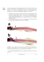
Zootaxa 1021: 1–12 (2005) ISSN 1175-5326 (print edition) www.mapress.com/zootaxa/ ZOOTAXA 1021 Copyright © 2005 Magnolia Press ISSN1175-5334(online edition) Erethistoides sicula, a new catfish (Teleostei: Erethistidae) from India HEOK HEE NG Fish Division, Museum of Zoology, University of Michigan, 1109 Geddes Avenue, Ann Arbor, Michigan 48109- 1079, USA. Email: [email protected] Abstract A new species of erethistid catfish, Erethistoides sicula, is described from the Brahmaputra River drainage in northeast India. Erethistoides sicula differs from both E. montana and E. pipri in having a longer caudal peduncle (19.6–22.3% SL vs. 14.4–18.4) and shorter pectoral spine (14.6–28.0% SL vs. 30.7–32.1). It further differs from E. montana in having a dorsally projecting bony splint on the opercle immediately posterior to its articular facet with the hyomandibula (vs. splint absent) and from E. pipri in having a more slender head (13.4–15.1% SL vs. 16.4). The diagnostic characters of Erethistoides are discussed and four new synapomorphies are proposed to diagnose the genus. Key words:Erethistoides, Erethistidae, Nepal, Ganges River drainage, South Asia Introduction Members of the genus Erethistoides are small erethistid catfishes traditionally diagnosed by a strongly depressed head and body and the presence of diverging serrations (antrorse on the distal half and retrorse on the proximal half) on the anterior edge of the pectoral spine. The genus, known from the sub-Himalayan region of the Indian subcontinent, currently includes two nominal species: E. montana Hora, 1950 and E. pipri (Hora, 1950). During a recent ichthyological survey of the northern Bengal region in India, specimens of Erethistoides were obtained, which upon further study, proved to be an undescribed species. The species is described here as Erethistoides sicula, new species. It was also found that the traditional diagnosis of Erethistoides is inadequate due to variation in the morphology of the pectoral-spine serrations. Here, four new synapomorphies diagnosing Erethistoides are identified and briefly discussed. Accepted by L. Page: 17 Jun. 2005; published: 22 Jul. 2005 1 ZOOTAXA Material and methods 1021 Measurements were made point to point with dial calipers and data recorded to tenths of a millimeter. Counts and measurements were made on the left side of specimens whenever possible. Subunits of the head are presented as proportions of head length (HL). Head length and measurements of body parts are given as proportions of standard length (SL). Measurements follow those of Ng & Dodson (1999). An asterisk following a meristic count (if present) indicates the value for the holotype. Material examined in this study is deposited in the following institutions: University of Michigan Museum of Zoology, Ann Arbor (UMMZ); and the Zoological Survey of India, Kolkata (ZSI). Erethistoides sicula sp. nov. (Fig. 1) Type material. Holotype: UMMZ 243718, 39.0 mm SL; India: West Bengal, Schutunga River (tributary of the Mansai River) at Ansole, 26°22'24"N 89°11'17"E; H. H. Ng et al., 12 April 2004. Paratypes: UMMZ 243647 (12), 15.7–37.2 mm SL; data as for holotype. Diagnosis. Erethistoides sicula differs from both E. montana and E. pipri in having a longer caudal peduncle (19.6–22.3% SL vs. 14.4–18.4) and shorter pectoral spine (14.6– 28.0% SL vs. 30.7–32.1). It further differs from E. montana in having a dorsally projecting bony splint on the opercle immediately posterior to its articular facet with the hyomandibula (vs. splint absent; Fig. 2) and from E. pipri in having a more slender head (13.4–15.1% SL vs. 16.4). The key biometric differences separating the three species are given in Table 1. TABLE 1. Diagnostic traits separating Erethistoides sicula, E. montana and E. pipri. Values are percentages of SL. E. sicula E. montana E. pipri Pectoral-spine length 14.6–28.0 30.7–31.6 32.1 Length of caudal peduncle 19.6–22.3 17.0–18.4 14.4 Head width 20.9–26.1 22.4–24.7 16.4 Description. Biometric data given in Table 2. Head broad and strongly depressed; dorsal profile slightly convex posteriorly and ventral profile almost straight. Neurocranium covered by thin skin bearing numerous elongate and flattened plaque-like tubercles; neurocranium extremely rugose and ornamented with numerous ridges and bumps. Supraoccipital spine not reaching nuchal shield. Posterior projection of the 2 © 2005 Magnolia Press NG posttemporo-supracleithrum (=Weberian lamina of de Pinna, 1996) well developed, ZOOTAXA 1021 approximately same length as supraoccipital spine and extending parallel to either side of spine. Eye ovoid, horizontal axis longest; located entirely in dorsal half of head and with bony ridge on frontal dorsal to it. Orbit with free margin. Gill openings narrow, extending from posttemporal to longitudinal line through mouth corner. TABLE 2. Biometric data for Erethistoides sicula (n=12). Holotype Range Mean±SD %SL Head length 30.3 28.2–31.1 29.8±1.12 Head width 24.9 20.9–26.1 23.3±1.79 Head depth 14.1 13.4–15.1 14.0±0.55 Predorsal length 45.6 40.0–46.5 43.0±2.22 Preanal length 69.2 64.7–69.2 67.1±1.71 Prepelvic length 50.0 45.1–50.3 48.3±1.70 Prepectoral length 25.6 22.8–27.2 24.9±1.52 Body depth at anus 10.3 8.4–10.7 9.5±0.76 Length of caudal peduncle 19.6 19.5–22.3 20.7±0.82 Depth of caudal peduncle 4.6 4.1–5.1 4.6±0.36 Pectoral-spine length 25.6 14.6–28.0 24.0±4.55 Pectoral-fin length 32.6 27.3–34.5 31.3±2.40 Dorsal-spine length 18.2 17.5–21.4 18.8±1.34 Length of dorsal-fin base 13.1 11.0–14.6 13.2±1.04 Pelvic-fin length 11.8 11.8–17.5 16.8±1.96 Length of anal-fin base 17.7 13.4–20.3 14.7±2.17 Caudal-fin length 21.8 20.1–26.4 23.1±2.19 Length of adipose-fin base 12.3 10.4–13.6 12.3±1.16 Dorsal to adipose distance 12.8 12.8–16.9 14.9±1.65 Post-adipose distance 16.7 15.9–20.0 17.8±1.26 %HL Snout length 50.0 43.5–53.8 48.8±3.92 Interorbital distance 28.0 22.8–29.3 26.3±2.33 Eye diameter 13.6 13.6–19.1 16.8±1.92 Nasal barbel length 21.2 10.6–21.2 13.3±3.49 Maxillary barbel length 105.9 87.9–116.5 102.0±8.32 Inner mandibular barbel length 29.7 27.7–41.8 33.3±5.37 Outer mandibular barbel length 38.1 38.1–51.7 46.0±3.93 A NEW ERETHISTOIDES © 2005 Magnolia Press 3 ZOOTAXA 1021 FIGURE 1. Erethistoides sicula, UMMZ 234718, holotype, 39.0 mm SL; dorsal, lateral and ventral views. 4 © 2005 Magnolia Press NG Mouth small, inferior and with papillate lips; upper jaw projecting beyond lower jaw. ZOOTAXA 1021 Oral teeth small and in irregular rows on all tooth-bearing surfaces. Premaxillary tooth band consisting of two quadratic patches on either side of midline; with conical teeth and exposed when mouth is closed. Dentary tooth band narrow, with conical teeth. Barbels in four pairs. Nasal barbel very short and slender, extending just beyond posterior margin of posterior nares. Maxillary barbel slender, extending to middle of pectoral-fin base. Outer mandibular barbel extending just beyond base of posteriormost pectoral-fin ray; inner mandibular barbel shorter, almost reaching to base of pectoral spine. Body broad and depressed, becoming more compressed towards caudal peduncle. Dorsal profile rising evenly but not steeply from tip of snout to origin of dorsal fin and sloping gently ventrally from origin of dorsal fin to end of caudal peduncle. Ventral profile horizontal to pelvic-fin base, then sloping gently dorsally from there to end of caudal peduncle. Skin with elongate and flattened plaque-like tubercles arranged in longitudinal rows along flanks. Lateral line complete and midlateral in position. Vertebrae 14+15=29 (6), 13+17=30 (1), 14+16=30 (3) or 14+17=31* (1). Abdomen smooth and flat, without adhesive apparatus. Dorsal fin located about two-fifths along body; with 5 (12) rays and straight margin. Dorsal-fin spine compressed, straight and robust; depressed spine extending to a vertical line through tips of extended pelvic fins. Anterior margin of spine smooth, posterior margin with 24 small serrations. Pectoral fin with stout, blade-like spine, sharply pointed at tip, and with 5 (4) or 6* (8) rays. Anterior spine margin with 8–25 strong serrations along entire length; proximal 4–13 serrations retrorse, distal 4–12 serrations antrorse; point at which serrations diverge at distal quarter of spine. Posterior spine margin with 4–10 strong serrations along entire length. Pectoral-fin margin straight anteriorly, slightly convex posteriorly. Coracoid with well developed posterior process, extending to two-thirds of distance between base of posteriormost pectoral-fin ray and pelvic-fin origin. Pelvic-fin origin at vertical through middle of dorsal-fin base. Pelvic fin with i,5 (12) rays and slightly convex margin; tip of adpressed fin reaching anal-fin origin. Anus located at two-thirds of distance between pelvic- and anal-fin origins. Adipose fin small, posterior end deeply incised. Fin located above posterior third of anal-fin base. Anal fin with iv,6* (11) or iv,7 (1) rays and slightly curved margin. Caudal peduncle slender. Caudal fin forked, with i,5,5,i* (2) or i,5,6,i (10) principal rays; upper and lower lobes narrow and pointed, with lower lobe slightly longer than upper. Procurrent rays symmetrical, with 9–10 rays on each surface extending only slightly anterior to fin base. Coloration. In 70% alcohol: dorsal and lateral surfaces of head and body light chocolate-brown, color somewhat unevenly distributed; tubercles along lateral line cream, forming faint thin midaxial stripe. A series of cream markings on head and body: first consisting of paired spots on snout immediately in front of anterior nares and on either side A NEW ERETHISTOIDES © 2005 Magnolia Press 5 ZOOTAXA of ethmoidal region; second consisting of paired spots on cheek region ventrolateral with 1021 respect to orbit, with cream coloration extending to maxillary barbels; third consisting of faint spot on supraoccipital process; fourth consisting of spot on nuchal plates; fifth consisting of transverse band spanning distance between dorsal and adipose fins and final consisting of transverse band on anterior half of caudal peduncle. Ventral surfaces of head and body cream. Dorsal fin hyaline, with faint transverse brown bands towards base and subdistally. Pectoral, pelvic and anal fins hyaline, sometimes with scattered melanophores. Caudal fin hyaline, with subdistal brown band. Maxillary barbels cream, annulated with brown rings; all other barbels cream. FIGURE 2. Outlines (mesial view) of right opercles of: a. Erethistoides sicula, paratype, UMMZ 243647, 36.1 mm SL; b. E. montana, UMMZ 243715, 37.5 mm SL. The bony splint posterior to the articular facet with the hyommandibula in E. sicula is indicated with an arrow. Scale bar represents 1 mm. Distribution. Known from the Mansai River drainage, itself a tributary of the Brahmaputra River in northern West Bengal state in India (Fig. 3). Habitat and biology. Erethistoides sicula was collected from a large, shallow, fast- flowing stream with a sandy bottom. The fish were usually found hiding in clumps of aquatic vegetation. Other fish species associated with this locality and habitat are: Cyprinidae - Barilius shacra, Barilius vagra, Chagunius chagunio; Psilorhynchidae - Psilorhynchus balitora, Psilorhynchus sucatio; Cobitidae - Canthophrys gongota, Lepidocephalichthys guntea; Balitoridae - Acanthocobitis botia, Schistura savona; Amblycipitidae - Amblyceps mangois; and Erethistidae - Pseudolaguvia spp. Etymology. From the Latin sicula, meaning dagger, in reference to the short pectoral spines of this species. Used as a noun. 6 © 2005 Magnolia Press NG ZOOTAXA 1021 FIGURE 3. Map showing distributions of Erethistoides sicula,E. montana and E. pipri. Discussion Erethistoides was originally diagnosed from other erethistid genera in having a strongly depressed head and body, and the presence of diverging serrations (retrorse, or proximally directed, on the proximal half and antrorse, or distally directed in the distal half) on the anterior edge of the pectoral spine (Hora, 1950). This was opposed to only antrorse serrations on the anterior edge of the pectoral spine in Hara and bifurcate serrations in Erethistes. I have examined individuals of Hara filamentosa in which some of the proximalmost serrations on the anterior edge of the pectoral spine are retrorse (as in A NEW ERETHISTOIDES © 2005 Magnolia Press 7 ZOOTAXA Erethistoides) and not antrorse as typically reported for Hara. Therefore, the utility of the 1021 pectoral spine morphology as a useful diagnostic character for distinguishing the genera is doubtful. In my study on the phylogenetic relationships of the Erethistidae, I have identified two new unambiguous synapomorphies diagnostic for Erethistoides: 1. Neural spines of the first eight post-Weberian vertebrae strongly depressed, inflected posteriorly, and positioned in a groove formed by the prezygapophyses of the vertebrae immediately posterior (Fig. 4). The neural spines of the corresponding vertebrae in other erethistid genera are not as strongly depressed, and are not strongly deflected posteriorly. 2. An enlarged maxilla that is almost as long as the palatine (Fig. 5). The maxilla is half or less than half the length of the palatine in other erethistids. FIGURE 4. Vertebral column of: a. Erethistoides sicula, UMMZ 243647, 36.1 mm SL, and b. Caelatoglanis zonatus, UMMZ 243668, 34.3 mm SL, showing conditions of first eight post- Weberian vertebrae (indicated by arrows) for Erethistoides and other erethistids. Two additional synapomorphies that are not unique to Erethistoides have also been identified: 8 © 2005 Magnolia Press NG 1. A fan-shaped mesethmoid lacking distinct cornua (vs. a Y-shaped mesethmoid in other ZOOTAXA 1021 erethistids; Fig. 5). This condition is shared only with Ayarnangra. 2. A strongly overhanging snout with the premaxillary tooth plates completely exposed when the mouth is closed (Fig. 1, ventral view). This condition is shared only with Ayarnangra. In other erethistid genera, the snout is not strongly overhanging and the premaxillary tooth plates are either not exposed, or only partially exposed when the mouth is closed. FIGURE 5. Dorsal views of skulls of: a. Erethistoides montana, UMMZ 243715, 37.5 mm SL, and b. Caelatoglanis zonatus, UMMZ 243668, 34.3 mm SL, showing conditions of mesethmoid and maxilla (indicated by arrows) for Erethistoides and other erethistids. The differences in biometrics between E. sicula and E. montana are unlikely to be due to ontogeny alone. Biplots of pectoral-spine length (Fig. 6) and caudal-peduncle length (Fig. 7) against SL show that the slopes of the regression lines are significantly different (ANCOVA; P<0.001 in both cases). It should be noted that the plots of both E. montana and E. pipri for pectoral-spine and caudal-peduncle lengths all fall outside of the 95% confidence interval of the regression lines for E. sicula (Figs. 6a and 7a). The corresponding plots for E. sicula fall inside the 95% confidence interval for E. montana (Figs. 6b and 7b), but this is most likely the result of the extremely small sample size obtained (only a single specimen of E. pipri could be examined and hence regression lines could not be calculated). In any case, the presence of the bony splint on the posterodorsal edge of the opercle additionally diagnoses E. sicula from E. montana. This character is found useful for diagnosing the two species (i.e., it does not vary much intraspecifically). The condition of the posterodorsal edge of the opercle (i.e., whether the bony splint is A NEW ERETHISTOIDES © 2005 Magnolia Press 9 ZOOTAXA present) can be observed in non-osteologically prepared specimens by careful dissection 1021 of the skin surrounding the area. FIGURE 6. Scatterplots of pectoral-spine length (PSL) plotted against standard length for Erethistoides sicula and E. montana: a. with 95% confidence intervals of regression line for E. sicula (dotted lines); b. with 95% confidence intervals of regression line for E. montana (dotted lines). 10 © 2005 Magnolia Press NG
