DTIC ADA532349: Emergency Decompressive Craniotomy with Banked Skull Flap in Subcutaneous Pocket PDF
Preview DTIC ADA532349: Emergency Decompressive Craniotomy with Banked Skull Flap in Subcutaneous Pocket
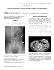
Emergency Decompressive Craniotomy with Banked Skull Flap in Subcutaneous Pocket Radiology Corner Emergency Decompressive Craniotomy with Banked Skull Flap in Subcutaneous Pocket Guarantor: COL Les Folio, USAF, MC, SFS Contributors: COL Les Folio, USAF, MC, SFS; 1, 2 2LT Steven Craig, USA; 1 LCDR Brian Singleton, USN;1, 2 2LT Brian Adams, USA1 Note: This is the full text version of the radiology corner Summary of Imaging Findings question published in the May 2006 issue, with the abbreviated answer in the June 2006 issue. 1 The abdominal KUB (Kidneys, Ureters, and Bladder, also sometimes referred to as “Flat Plate”) demonstrates a smooth, Introduction large, calcific opacity in the left lower quadrant (see figure 1). An enteric tube is also noted with its tip in stomach; surgical The following is a more detailed description of the case staples are noted peripherally to skull cap. No other introduced in the May and June issues of Military Medicine’s abnormalities are present on the KUB. Radiology Corner. The axial CT of the abdomen localizes the skull cap to the anterior abdominal wall subcutaneous tissue (see figure 2). CT also shows 10 x 6 x 1.5 cm non-enhancing homogeneous low attenuating fluid collection posterior to the skull cap. For providers that have not heard of banking the skull cap in the abdomen, this type of finding can be perplexing. This procedure became common in recent military combat operations; however, the trend may change due to unique infection potential in other continents. Figure 2. Note the skull cap (arrows) with the posterior homogeneous attenuating fluid collection (curved arrow) in left anterior abdominal wall. Figure 1 Plain computed radiology of abdomen demonstrate the skull flap overlying the left lower quadrant (arrows). Surgical clips and an enteric tube Axial head CT (figure 3) demonstrates the craniectomy site within stomach are seen. No other abnormalities. (arrows). Additionally, there is left parieto-occiptial encephalomalacia. There are metallic fragments/shrapnel 1 Department of Radiology and Radiological Sciences; Uniformed Services (bone windows not included) causing beam hardening artifact University of the Health Sciences, Bethesda, Maryland 20814-4799 in occipital bone (curved arrow). 2 Department of Radiology National Naval Medical Center, Bethesda, Maryland 20814-4799 Reprint & Copyright © by Association of Military Surgeons of U.S., 2006 Military Medicine Radiology Corner, 2006 Report Documentation Page Form Approved OMB No. 0704-0188 Public reporting burden for the collection of information is estimated to average 1 hour per response, including the time for reviewing instructions, searching existing data sources, gathering and maintaining the data needed, and completing and reviewing the collection of information. Send comments regarding this burden estimate or any other aspect of this collection of information, including suggestions for reducing this burden, to Washington Headquarters Services, Directorate for Information Operations and Reports, 1215 Jefferson Davis Highway, Suite 1204, Arlington VA 22202-4302. Respondents should be aware that notwithstanding any other provision of law, no person shall be subject to a penalty for failing to comply with a collection of information if it does not display a currently valid OMB control number. 1. REPORT DATE 3. DATES COVERED 2006 2. REPORT TYPE 00-00-2006 to 00-00-2006 4. TITLE AND SUBTITLE 5a. CONTRACT NUMBER Emergency Decompressive Craniotomy with Banked Skull Flap in 5b. GRANT NUMBER Subcutaneous Pocket 5c. PROGRAM ELEMENT NUMBER 6. AUTHOR(S) 5d. PROJECT NUMBER 5e. TASK NUMBER 5f. WORK UNIT NUMBER 7. PERFORMING ORGANIZATION NAME(S) AND ADDRESS(ES) 8. PERFORMING ORGANIZATION Uniformed Services University of the Health Sciences,Department of REPORT NUMBER Radiology and Radiological Sciences,4301 Jones Bridge Road,Bethesda,MD,20814 9. SPONSORING/MONITORING AGENCY NAME(S) AND ADDRESS(ES) 10. SPONSOR/MONITOR’S ACRONYM(S) 11. SPONSOR/MONITOR’S REPORT NUMBER(S) 12. DISTRIBUTION/AVAILABILITY STATEMENT Approved for public release; distribution unlimited 13. SUPPLEMENTARY NOTES 14. ABSTRACT 15. SUBJECT TERMS 16. SECURITY CLASSIFICATION OF: 17. LIMITATION OF 18. NUMBER 19a. NAME OF ABSTRACT OF PAGES RESPONSIBLE PERSON a. REPORT b. ABSTRACT c. THIS PAGE Same as 3 unclassified unclassified unclassified Report (SAR) Standard Form 298 (Rev. 8-98) Prescribed by ANSI Std Z39-18 Emergency Decompressive Craniotomy with Banked Skull Flap in Subcutaneous Pocket Patient Discussion is the patient’s own bone flap (removed segment of skull). This option is cost-effective, strong, immunologically compatible with the host, and cosmetically pleasing. Several This patient had an emergency craniectomy in a deployed field techniques for preserving the bone flap exist to include hospital for intracranial hypertension secondary to head freezing, placement in storage solutions, and in this case, trauma during military combat in Iraq. The removed skull cap placement in the subcutaneous tissue of the patient’s was placed subcutaneously in his abdominal wall for abdominal wall. preservation during aeromedical evacuation. After successful transfer to a referral medical center in the Discussion U.S. the patient developed persistent GI symptoms. Tissue culture of the fluid posterior to the skull flap was negative for Decompressive craniectomy is a neurosurgical procedure bacteria. aimed at relieving elevated intra-cranial pressure (ICP) by removing the patient’s rigid skull.2 Decompressive craniectomy is a surgical procedure for the treatment of elevated ICP in cases where medical management fails and in acute severe traumatic brain injury. The surgery alone has been shown to reduce ICP by 15% and up to 70% if the surgeon opens the dura.3 More than 40,000 cranial surgeries are performed in the United States each year. The most frequent principle diagnosis in patients receiving such surgeries is subdural hemorrhage.4 Recent studies demonstrate efficacy of banked bone grafts in the abdomen.5 In austere environments and special situations (like combat injuries), the skull cap was often placed into the abdominal wall for preservation to allow for potential replacement after transport to a referral medical center. This trend may change due to recent literature on increasing infection potential and improved prosthetic technology. Aeromedical evacuation from combat to US military medical centers can occur within 24-48 hours. Figure 3: Axial head CT demonstrates the craniectomy site (arrows). Encephalomalic changes are noted in the left parieto-occipital lobes. Beam Note: Follow this link for Category 1 CME or CNE in the case hardening artifact is noted from metallic shrapnel (curved arrow). of the week in the MedPix™ digital teaching file. This case demonstrates a skull cap in anterior abdominal wall http://rad.usuhs.mil/medpix/medpix.html?mode=single&recnu with low attenuation area consistent with the following m=6969&th=-1#top differential diagnosis: liquified hematoma, seroma, abscess, or proteinaceous fluid collection. Since the cultures were negative for bacteria, liquified hematoma was the working diagnosis. After surgical removal of skull cap from abdomen, it was decided to use prosthetic cranioplasty material instead. Developing literature on (MDR) Acinetobacter species1 is one of several factors weighing into using synthetic material. As the patient was recovering and before closing the gap in the skull, the patient ambulated with a hockey helmet for protection of the area. Decompressive craniectomy is a common surgical procedure used to relieve intra-cranial hypertension. Upon resolution of the intra-cranial hypertension, a cranioplasty is performed to close the hole in the skull. There are a number of suitable materials that can be used for this purpose. One such material Military Medicine Radiology Corner, 2006 Emergency Decompressive Craniotomy with Banked Skull Flap in Subcutaneous Pocket References 1 Davis KA, Moran A, McAllister K, Gray P. Multidrug-Resistant Acinetobacter Extremity Infections in Soldiers. Emerging Infectious Diseases. www.cdc.gov/eid. 2005 Aug;11(8):1218-24. 2 Flanndry T; McConnell RS; Cranioplasty: why throw the bone flap out? British Journal of Neurosurgery, 2001; 15(6): 518- 520. 3 Jourdan C; Convert J; Mottolese C; Bachour E; Gharbi S; Artru F.; Evaluation of the clinical benefit of decompression hemicraniectomy in intracranial hypertension not controlled by medical treatment, Neurochirurgie 1993;39(5):304-10. 4 Buczko W.; Cranial surgery among Medicare beneficiaries. The Journal of Trauma. 2005 Jan;58(1):40-6. 5 Movassaghi K, Ver Halen J, Ganchi P, Amin-Hanjani S, Mesa J, Yaremchuk MJ. "Cranioplasty with subcutaneously preserved autologous bone grafts." Plast Reconstr Surg. 2006 Jan;117(1):202-6. Military Medicine Radiology Corner, 2006
