DTIC ADA453368: Gene Expression Analysis of Circulating Hormone Refractory Prostate Cancer PDF
Preview DTIC ADA453368: Gene Expression Analysis of Circulating Hormone Refractory Prostate Cancer
AD_________________ Award Number: W81XWH-05-1-0175 TITLE: Gene Expression Analysis of Circulating Hormone Refractory Prostate Cancer Micrometastases PRINCIPAL INVESTIGATOR: Jonathan Rosenberg, M.D. CONTRACTING ORGANIZATION: University of California, San Francisco San Francisco, CA 94143-0962 REPORT DATE: January 2006 TYPE OF REPORT: Annual PREPARED FOR: U.S. Army Medical Research and Materiel Command Fort Detrick, Maryland 21702-5012 DISTRIBUTION STATEMENT: Approved for Public Release; Distribution Unlimited The views, opinions and/or findings contained in this report are those of the author(s) and should not be construed as an official Department of the Army position, policy or decision unless so designated by other documentation. Form Approved REPORT DOCUMENTATION PAGE OMB No. 0704-0188 Public reporting burden for this collection of information is estimated to average 1 hour per response, including the time for reviewing instructions, searching existing data sources, gathering and maintaining the data needed, and completing and reviewing this collection of information. Send comments regarding this burden estimate or any other aspect of this collection of information, including suggestions for reducing this burden to Department of Defense, Washington Headquarters Services, Directorate for Information Operations and Reports (0704-0188), 1215 Jefferson Davis Highway, Suite 1204, Arlington, VA 22202- 4302. Respondents should be aware that notwithstanding any other provision of law, no person shall be subject to any penalty for failing to comply with a collection of information if it does not display a currently valid OMB control number. PLEASE DO NOT RETURN YOUR FORM TO THE ABOVE ADDRESS. 1. REPORT DATE 2. REPORT TYPE 3. DATES COVERED 01-01-2006 Annual 1 Jan 2005 – 31Dec2005 4. TITLE AND SUBTITLE 5a. CONTRACT NUMBER Gene Expression Analysis of Circulating Hormone Refractory Prostate Cancer 5b. GRANT NUMBER Micrometastases W81XWH-05-1-0175 5c. PROGRAM ELEMENT NUMBER 6. AUTHOR(S) 5d. PROJECT NUMBER Jonathan Rosenberg, M.D. 5e. TASK NUMBER 5f. WORK UNIT NUMBER 7. PERFORMING ORGANIZATION NAME(S) AND ADDRESS(ES) 8. PERFORMING ORGANIZATION REPORT NUMBER University of California, San Francisco San Francisco, CA 94143-0962 9. SPONSORING / MONITORING AGENCY NAME(S) AND ADDRESS(ES) 10. SPONSOR/MONITOR’S ACRONYM(S) U.S. Army Medical Research and Materiel Command Fort Detrick, Maryland 21702-5012 11. SPONSOR/MONITOR’S REPORT NUMBER(S) 12. DISTRIBUTION / AVAILABILITY STATEMENT Approved for Public Release; Distribution Unlimited 13. SUPPLEMENTARY NOTES 14. ABSTRACT This annual report for the Physician Research Training Award focuses progress and challenges in the analysis of circulating hormone refractory prostate cancer micrometastases. Metastatic tissue for research is difficult to obtain in hormone refractory prostate cancer (HRPC), as most metastatic sites are not conducive to biopsy. Circulating tumor cells (CTC’s) have been found in high numbers in patients with metastatic HRPC and are easily accessible in the peripheral blood. Detecting genetic alterations that occur during the development of chemotherapy resistance will give insight into the mechanisms behind this resistance and determine potential therapeutic strategies to combat this resistance. Array CGH of CTC’s will demonstrate the genomic changes that accompany the hormone refractory state, in addition to shedding light on genetic changes that occur with chemotherapy resistance. In addition to the genomic studies, CTC enumeration was used to determine the effect of CTC number on survival in patients receiving chemotherapy for HRPC. Work performed under this grant in 41 patients with metastatic HRPC has shown that patients with > 1.8 CTC/mL have a worse prognosis than patients with ≤1.8 CTC/mL. The median survival of patients with metastatic HRPC with > 1.8 CEC’s/mL was 13 months. Median survival for patients with <1.8 CTC’s/mL has not been reached. CTC’s from chemotherapy refractory and chemotherapy naïve patients are being collected for genetic analysis. 15. SUBJECT TERMS circulating tumor cells, prostate cancer, chemotherapy resistance, genomic analysis 16. SECURITY CLASSIFICATION OF: 17. LIMITATION 18. NUMBER 19a. NAME OF RESPONSIBLE PERSON OF ABSTRACT OF PAGES USAMRMC a. REPORT b. ABSTRACT c. THIS PAGE 19b. TELEPHONE NUMBER (include area U U U UU 11 code) Standard Form 298 (Rev. 8-98) Prescribed by ANSI Std. Z39.18 Table of Contents Cover……………………………………………………………………………………1 SF 298……………………………………………………………………………..…….2 Table of Contents……………………………………………………………….…….3 Introduction…………………………………………………………….………..….....4 Body……………………………………………………………………………………..5 Key Research Accomplishments………………………………………….……….11 Reportable Outcomes………………………………………………………………..11 Conclusions…………………………………………………………………………...11 References……………………………………………………………………………..11 Introduction: This project focuses on the genetic analysis of circulating hormone refractory prostate cancer micrometastases focusing on the mechanisms of chemotherapy resistance. Metastatic tissue for research is difficult to obtain in hormone refractory prostate cancer (HRPC), as most metastatic sites are not conducive to biopsy. However, circulating tumor cells (CTC’s) have been found in high numbers in patients with metastatic HRPC. CTC’s represent an untapped resource for studying metastatic prostate cancer. These cells are easily accessible in the peripheral blood. We have previously demonstrated that circulating epithelial tumor cells are found in peripheral blood of metastatic HRPC patients at a mean concentration of 110 cells/mL. The detection of these cells and their isolation will allow new research to be conducted into the nature of advanced hormone refractory prostate cancer. The purpose of this research is to detect genetic alterations that occur during the development of chemotherapy resistance, to give insight into the mechanisms behind this resistance, and determine potential therapeutic strategies to combat it. In addition, we have examined the prognostic implication of CTC number upon survival in patients receiving chemotherapy for HRPC. This report will summarize the progress on this grant and the challenges and obstacles that have arisen and how they will be overcome. 4 Body: Tasks 1, 2: Isolation and characterization of circulating micrometastases of chemotherapy naïve and chemotherapy resistant HRPC. The departure of a key collaborator has provided a challenge to the conduct of this grant. Christopher Haqq, the collaborator who had been identified to perform the microarray analysis, left UCSF for a position in the pharmaceutical industry in early 2005. As a result, his laboratory and expertise were unavailable. As the genetic analysis of these tumors was essential to carrying out this grant, alternative collaborators have been identified. Pamela Paris, PhD, a senior postdoctoral fellow, and Colin Collins, one of the principal investigators on the UCSF Prostate Cancer Spore and an expert in oncogenomics, have developed techniques to amplify small amounts of DNA extracted from circulating tumor cells. Dr. Paris has developed a collaboration with Dr. Wen-Tien Chen at the State University of New York, Stonybrook, and Vitatex, Inc., to isolate the circulating tumor cells using a novel method based on the adhesion of these cells to a cell-adhesion matrix (CAM). Dr. Chen has developed a novel cell separation technology based on preferential adhesion of tumor cells to cell adhesion matrix (CAM) coated surfaces. Viable tumor cells attach to CAM with great avidity, but normal cells (including more than 99.9% of white cells and 99.9999% of red cells) and dead or dying tumor cells do not. When the CAM is prepared using fluorescently labeled collagen, the invasive tumor cells become labeled, since they exhibit a propensity to bind and ingest collagen. In contrast, normal cells leave the CAM undisturbed. Immunocytochemistry and real-time RT-PCR using multiple epithelial/tumor associated cell lineage markers have been used to validate the CAM-recovered cells from blood as circulating tumor cells. CAM recovered cells are viable based on Molecular Probes LIVE/DEAD Viability testing. In preliminary work done at SUNY Stonybrook, blood from 125 patients with metastatic cancer was collected and analyzed using the CAM technique. In these specimens, approximately 1x10⁶-fold enrichment of viable human tumor cells can be achieved in a single step. Tumor cells were identified with multiple criteria as viable circulating tumor cells, including by epithelial/tumor markers, uptake of CAM fragments, and nucleic acid dyes. Current assays have successfully detected viable tumor cells in blood samples from patients with prostate cancer. The CAM technique is highly specific, as no healthy donors and patients with inflammatory lung diseases have yielded a (false) positive result. Cell separation based on preferential adhesion of tumor cells to CAM coated surfaces will be used to detect circulating HRPC cells from chemotherapy naïve and chemotherapy refractory patients. Once a specimen has been processed for CTC isolation using the CAM technology, DNA will be extracted from the CTC’s that is suitable for use with array comparative genomic hybridization (aCGH) technology. Blood will be drawn at UCSF from 72 patients with chemotherapy naïve hormone refractory prostate cancer who are being enrolled on UCSF 04557, “A Phase I/II Study of Docetaxel/Prednisone and PTK787/ZK222584 in Previously Untreated Metastatic Hormone Refractory Prostate Cancer,” and 35 patients with chemotherapy-refractory hormone refractory prostate cancer enrolled on UCSF 055513, “Phase I/II Trial of Epothilone Analog BMS-247550 (Ixabepilone), 5 Mitoxantrone, and Prednisone in Hormone Refractory Prostate Cancer Patients Previously Treated with Chemotherapy.” For each patient, 20 ml of anti-coagulated blood will be routinely collected and then 4 ml aliquoted into ~ 5 Vitatex Vita-Cap tubes. The Vita-Cap tubes will be rotated at 10 rpm for 1-3 hrs at 37C, and then shipped overnight in a thermos (to prevent freezing) to VitaTex. VitaTex will perform the cell isolation and ship the cells to Dr. Paris. She will use the Promega Wizard Kit to extract the DNA from the cells. This kit is routinely used in her lab to isolate DNA from cultured cells for use with array comparative genomic hybridization (aCGH). The CTC DNA quality will be visualized by gel chromatography and quantitated by a Nano-drop uv-vis. If DNA of suitable quality (high molecular weight) and quantity (1 ug) are obtained, aCGH will be performed. If the yield is less than a microgram, linear amplification will be employed. Dr. Paris is currently optimizing amplification methods for aCGH as part of the UCSF prostate SPORE program. As the collaborators for this grant have changed, collection of CTC’s for genetic analysis has been delayed. However, at this time, samples from two patients have undergone the Vitatex isolation protocol and the DNA is being prepared for aCGH, and we are anticipating further patients be accrued in the near future. Quantitating CTCs in the blood of patients with prostate cancer has not previously proven prognostic. As part of the work on this grant evaluating the importance of CTC’s in HRPC, Dr. Rosenberg has been working with Dr. John Park and Dr. Jorge Garcia to determine whether the numbers of circulating tumor cells carries prognostic significance in patients with metastatic HRPC. This research has been developed into a manuscript that is in its final stages of preparation for journal submission. In this manuscript, we report the results of a retrospective pilot analysis that evaluated HRPC patients undergoing cytotoxic therapy to determine the utility and feasibility of CTC analysis in the detection of CTC’s, their change over time, and to assess whether or not the presence and number of CTC’s identified by FACS analysis was a predictor of outcome in men with HRPC. This was a retrospective study of 41 consecutively treated metastatic HRPC patients initiating systemic chemotherapy at the University of California, San Francisco. All patients had peripheral blood collections prior to the initiation of systemic cytotoxic chemotherapy with a taxane-based regimen. Descriptive statistics were utilized to characterize the entire patient sample. Comparisons of subsets were carried out using Fisher’s exact test for categorical variables, analysis of variance methods for continuous variables and the non-parametric Mann-Whitney test to compare distributions. The association between continuous variables was estimated by the Spearman rank correlation coefficient. The Kaplan-Meier product limit method was used to estimate the probability of survival with the log rank test performed to compare distributions of subsets. Survival was measured from the start of chemotherapy until either death or date of last contact. Forty-nine percent of patients had between 0.1 and 5.0 CTC’s/mL, 24% of patients between >5 – 15 CTC’s/mL, 15% of patients between >15 – 30 CTC’s/mL, and only 2% of patients were found to have more than 30 CTC’s/mL. The overall median (1.8 CTC’s/mL) was used to dichotomize the patient sample. HRPC patients with <1.8 CTC’s/mL had a significantly longer survival than those with higher counts of CTC’s/ml (p = 0.02) (Figure). The median survival of patients with metastatic HRPC with > 1.8 CEC’s/mL was 13 months. Median OS for patients with <1.8 CTC’s/mL has not been reached. After a median follow-up of approximately 15 months, due to the number of 6 early deaths, the median survival for the entire cohort is 18.4 months. Nineteen of the 41 patients enrolled in the study have died. All deaths occurred within 20 months of diagnosis and 10 patients survived beyond that time point up to 65 months from diagnosis. We have demonstrated that the quantification of CTC’s in patients with HRPC can be used as a prognostic tool to predict outcome in this cohort of patients. Higher number of CTC’s/mL appears to correlate with shorter survival in patients with metastatic disease. Our findings, combined with the results from other groups, suggest that CTC’s may have relevance and can be used to predict outcome in patients with HRPC. Figure. Overall Survival and CTC’s/mL in HRPC Patients Vertical axis indicates proportion of patients alive after the start of first-line chemotherapy according to CTC number/mL. Patients with ≤1.8 cells/mL have improved survival compared with patients who have greater numbers of CTC’s. In addition to the work described above, Dr. Rosenberg has been collaborating with Dr. Lydia Sohn, of the University of California, Berkeley, who is developing a new approach to flow cytometry, termed “Nanocytometry”. Nanocytometry will be applied to circulating prostate tumor cells and be used in the future as a novel way to isolate the cells if the technology is validated. This novel technology employs an artificial pore, an integrated microfluidic chip, and nano-based electronics, which together she will adapt to sort cancer cells based on their cell-surface markers. The artificial pore and its associated nano- or molecular-sensing techniques will allow significant improvements over 7 conventional FACS, because the system permits: 1) electronic detection, allowing compaction, ease of use, and low cost; 2) unmanipulated (e.g. label-free) cells to be passed directly through the device, as the detection label is incorporated directly into the device itself; 3) a virtually limitless number of simultaneous detections of cell surface markers (in contrast to FACS which is often limited in the number of simultaneous labels); 4) small sample volumes (microliters or less), with the capacity to run continuously, thereby allowing the analysis of milliliter volumes of sample at a time; 5) cell separations using very few cells, and thus improved sensitivity; 6) little physical stress on the cells during interrogation and isolation; and 7) the opportunity for cell propagation after cell separation (Table). Additionally, these advantages hold true when comparing the Nanocytometer to magnetic bead/column selection, which is used most often for cell separations based on a single parameter. Dr. Sohn envisions constructing the Nanocytometer with numerous pores in parallel in order to achieve a rate of cell separation comparable to that of FACS (Table). Because of the use of unmanipulated cells and the slower flow speed, cells that are separated by the Nanocytometer pass through the device virtually without damage. The isolation of unmanipulated, nondamaged cells increases the number of potential applications of the recovered cells including further propagation for additional analysis. NanoCytometer Conventional FACS •Electronic detection and therefore •Optical detection and therefore large, compact, easy to use, and inexpensive complex, and costly •Cells are label-free •Cells require fluorescent labeling or immunostaining •Limitless number of simultaneous •Limited in the number of simultaneous interrogation of cell labels •Small sample volumes (µL or less), with •Large sample volumes (mL or more) the capacity to run continuously •Large populations of cells are needed for •Separations using very few cells & cell separations improved sensitivity •Little physical stress on cells during •Large physical stress imparted on cells interrogation and isolation due to sheer flow •Analyzes hundreds of cells/sec, and with •Analyzes thousands of cells/sec N x N arrays of pores, thousands of cells/sec Nanotechnology brings significant potential advantages over current technologies in terms of being able to perform manipulations of cells using very small volumes and very small numbers of cells. By developing a more sensitive technique to perform cell separations, Nanocytometry could provide an important new technology applicable to cancer. Ultimately, Nanocytometry could increase physicians' ability to detect minimal residual disease and remission states and could improve upon scientists' ability to study cell populations that occur in very small numbers (e.g. rare circulating malignant cells and endothelial cells from patients with solid tumors). 8 The basis of Dr. Chen’s proposed Nanocytometer is an on-chip artificial pore that is integrated with microfluidics and employs nanoelectronic-sensing techniques. She has recently demonstrated the ability to fabricate such a pore and in addition, has demonstrated that this pore is effective in molecular sensing on length scales from micrometers to nanometers to single molecules. The on-chip artificial pore is based on the resistive-pulse technique of particle sizing: a particle passing through a pore displaces conducting fluid, which causes a transient increase, or pulse, in the pore’s electrical resistance, that, in turn, is measured as a decrease in current. This technique has been used in the past to measure the size and concentration of a variety of particles, such as cells, viruses, and colloids. Because the magnitude of the pulse is directly related to the diameter of the particle that produced it, Dr. Sohn has used the resistive-pulse technique and her artificial pore to detect the nanometer increase in diameter of a submicron latex colloid upon binding to an unlabeled specific antibody. This success has permitted her to perform two important types of immunoassays: an inhibition assay and a sandwich assay. In addition, she has successfully used her on-chip artificial pore to sense single molecules of unlabeled lambda (l) phage DNA. Dr. Chen envisions producing a fully-integrated, high-throughput device that could be immediately clinically applicable, consisting of parallel N x N arrays of Nanocytometers that can perform sensitive cell separations simultaneously. This high-throughput device will be used in an exploratory fashion to isolate circulating HRPC cells for CTC analysis. Tasks 3,4,5,6 Analyze and compare gene signatures of circulating tumor cells to biomarkers previously identified, identify markers of chemotherapy resistance and response in CTC’s in HRPC. These tasks will be carried out once adequate numbers of samples have been collected and subjected to aCGH. Dr. Paris has identified a set of DNA based biomarkers as part of the UCSF prostate SPORE program that associate with aggressive disease.(1) The work will evaluate the prognostic capacity of these markers using CTC DNA from patients with advanced prostate cancer, as well as looking for markers of chemotherapy resistance in comparing aCGH profiles from chemotherapy-naïve and -refractory patients. In addition, we will be examining the differences in lymph node–only metastases, bone-only metastases, and mixed metastases in HRPC. Task 7 Due to increased clinical responsibilities resulting from the departure of a faculty member in the UCSF Urologic Oncology group, Dr. Rosenberg was not able to enroll in the ATCR coursework during year 1. However, he plans to begin to take coursework in year 2, as a new faculty member has been hired to lighten the clinical load. Coursework that is being considered includes: Introduction to Statistical Analysis (S. Glantz, Director) Instruction in descriptive statistics; techniques for testing hypotheses (ANOVA, t-tests, linear regression, non-parametric methods); and sampling. Introduction to Statistical Computing in Clinical Research (M. Pletcher, Director) 9 Instruction in use of computer software for managing and analyzing clinical research data; roles of spreadsheet and relational database programs; use of STATA for managing, cleaning, describing, and analyzing data. Biostatistical Methods for Clinical Research II (D. Glidden, Director) Instruction in multiple predictor analyses as a tool for control of confounding and for constructing predictive models. Topics will include linear regression and logistic regression. The STATA statistical package will be used throughout. Systematic Reviews (Meta-Analysis) EPI 214 (D. Grady, Director) Instruction in the methods of systematic and unbiased identification of primary research studies; abstraction of data; determination of summary estimates and evaluation of heterogeneity. Publishing and Presenting Clinical Research EPI 212 (S. Hulley, Director) Instruction in preparing abstracts, posters, all aspects of manuscripts, and oral presentations; instruction in oral presentations includes videotaping and critique of trainees’ presentations. Biostatistical Methods for Clinical Research III BIOSTAT 209 (D. Glidden, Director) A continuation of the Winter Quarter course in multivariable statistical analysis that includes instruction in survival analysis and analysis of repeated measures and clustered data. The course culminates with student presentations of statistical analyses of their own research projects. Grant Writing Workshop (E. Holly and T. Mitchell) Instruction in grant writing principles, with specific guidelines for preparing successful grant applications using PHS 398 forms, will be presented with emphasis on federal guidelines and regulations for research with human study participants. Outcomes Research EPI 211 (A. Bindman, Director) Instruction in types of questions that can be addressed with large administrative and clinical databases; gaining access to these databases; determining validity of information; risk adjustment; linking datasets; and building registries. Dr. Rosenberg has met weekly with Dr. Eric Small as part of the mentoring plan. In addition he has met regularly with Dr. Paris regarding the genomic analysis. 10
The list of books you might like

Better Than the Movies

Credence

The Strength In Our Scars

What Happened to You?

West Knits Book Three

Remèdes de nos grand-mères en 150 recettes maison

Les tribulations d'un chinois en Chine by Jules Verne
Greek Government Gazette: Part 2, 2006 no. 1611
Greek Government Gazette: Part 2, 2006 no. 1885

Cada hora muere una persona por culpa del tabaco en Andalucía
Beyond Discrete E-Services
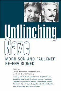
Unflinching Gaze: Morrison and Faulkner Re-Envisioned

Farzana the woman who saved an empire
Greek Government Gazette: Part 1, 2006 no. 48
Health Policy 1993 - 1994: Vol 26 Table of Contents

La sangre y el ámbar

bozner hochzeitsmesse 5° bolzano sposi

Benedicam Dominum, Op.62
BAB III AKUAKULTUR UDANG HARIMAU 3.1 PENGENALAN Menurut sejarah aktiviti akuakultur

Vade mecum da jurisprudência: dizer o direito 2020
beam-column connection flexural behavior and seismic collapse performance of concentrically

