DTIC ADA349836: Identification of Mammary Specific Transcription Factors. PDF
Preview DTIC ADA349836: Identification of Mammary Specific Transcription Factors.
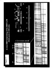
AD_ GRANT NUMBER: DAMD17-94-J-4497 TITLE: Identification of Mammary Specific Transcription Factors PRINCIPAL INVESTIGATOR: Julia M. Michelotti, Ph.D. CONTRACTING ORGANIZATION: National institutes of Health Bethesda, Maryland 20892 REPORT DATE: January 1998 TYPE OF REPORT: Final PREPARED FOR: Commander U.S. Army Medical Research and Materiel Command Fort Detrick, Frederick, Maryland 21702-5012 DISTRIBUTION STATEMENT: Approved for public release- distribution unlimited The views, opinions and/or findings contained in this report are those of the author(s) and should not be construed as an official Department of the Army position, policy or decision unless so designated by other documentation. DHCWLnrniBPCCTEB j REPORT DOCUMENTATION PAGE Form Approved OMB No. 0704-0188 Public reporting burden for this collection of information is estimated to average 1 hour per response, including the time for reviewing instructions, searching existing data sources, gathering and maintaining the data needed, and completing and reviewing the collection of information. Send comments regarding this burden estimate or any other aspect of this collection of information, including suggestions for reducing this burden, to Washington Headquarters Services, Directorate for Information Operations and Reports, 1215 Jefferson Davis Highway, Suite 1204, Arlington, VA 22202-4302, and to the Office of Management and Budget, Paperwork Reduction Project (0704-0188), Washington, DC 20503 1. AGENCY USE ONLY {Leave blank) 2. REPORT DATE 3. REPORT TYPE AND DATES COVERED January 1998 Final (30 Sep 94 - 30 Sep 97) 4. TITLE AND SUBTITLE 5. FUNDING NUMBERS Identification of Mammary Specific Transcription Factors DAMD17-94-J-4497 6. AUTHOR(S) Julia M. Michelotti, Ph.D. 7. PERFORMING ORGANIZATION NAME(S) AND ADDRESS(ES) 8. PERFORMING ORGANIZATION National Institutes of Health REPORT NUMBER Bethesda, Maryland 20892 9. SPONSORING / MONITORING AGENCY NAME(S) AND ADDRESS(ES) 10. SPONSORING/MONITORING U.S. Army Medical Research and Materiel Command AGENCY REPORT NUMBER Fort Detrick, Maryland 21702-5012 J980728 025 It 11. SUPPLEMENTARY NOTES 12a. DISTRIBUTION / AVAILABILITY STATEMENT 12b. DISTRIBUTION CODE Approved for public release; distribution unlimited 13. ABSTRACT (Maximum 200 words) The Mouse Mammary Tumor Virus (MMTV) achieves its highest levels of expression in the mammary glands of lactating mice. Previous work showed that the MMTV Long Terminal Repeat (LTR) has a modest level of activity in non-mammary cells but is expressed most efficiently in mammary epithelial cells. It has been shown that optimal expression of several milk proteins including beta-casein in the mammary epithelial cell line (CID-9) are dependent upon the presence of extracellular matrix (ECM) and lactogenic hormones. To see whether the MMTV LTR regulation is similar to beta-casein the PI analyzed the transcriptional regulation of full length MMTV LTR in CID9 cells. Using an LTR deletion series the PI determined that the MMTV minimal promoter (-200) is sufficient to provide the maximal ECM response. In addition, transient transfection analysis reveals that the ECM-induced response occurs only when LTR DNA is stably integrated into CID9 cells. This finding suggests that chromatin structure may play an important role in ECM induced transcription. To test whether ECM induction is via changes in histone acetylation the PI treated the cells with deacetylase inhibitors that have been shown to affect gene transcription presumably at the level of histone/DNA interaction. Sodium butyrate inhibited overall MMTV LTR gene transcription by 60% and trichostatin A by 80%. However, these inhibitors did not suppress the fold induction by ECM. The inhibition of transcription is also independent of hydrocortisone and stable integration. This surprising finding suggests that these inhibitors effect other factors besides DNA/histone interactions. Recent studies have shown the activity of cis-acting factors is affected by their state of acetylation which may explain why transcriptional inhibition occurs on the transient template. However, analysis of the chromatin template by in vivo DMS footprinting analysis identified a differentiation-specific cleavage in the minimal promoter (-151) of the LTR in CID9 cells cultured in the presence of ECM. This is the first evidence that ECM-induced activation of gene transcription may induce strucructural changes in the DNA of differentiated cells. 14. SUBJECT TERMS 15. NUMBER OF PAGES Breast Cancer 16 16. PRICE CODE 17. SECURITY CLASSIFICATION 18. SECURITY CLASSIFICATION OF THIS 19. SECURITY CLASSIFICATION 20. LIMITATION OF ABSTRACT OF REPORT PAGE OF ABSTRACT Unclassified Unclassified Unclassified Unlimited NSN 7540-01-280-5500 Standard Form 298 (Rev. 2-89) Prescribed by ANSI Std. Z39-18 298-102 FOREWORD Opinions, interpretations, conclusions and recommendations are those of the author and are not necessarily endorsed bv the ü S Army. J ' ' X_ Where copyrighted material is quoted, permission has been obtained to use such material. _K_ Where material from documents designated for limited distribution is quoted, permission has been obtained to use the material. —K_ Citations of commercial organizations and trade names in this report do not constitute an official Department of Army endorsement or approval of the products or services of these organizations. X In conducting research using animals, the investigator(s) ad hered to the "Gu— ide for the — Cagr e — —a n——d— —U s— ef ow.f. w L a**b4 oW rV» aW tW o*k rW yWt < Animals," prepared by the Committee on Care and use of Laboratory Animals of the Institute of Laboratory Resources, national Research Council (NIH Publication No. 86-23, Revised 1985). For the protection of human subjects, the investigator(s) adhered to policies of applicable Federal Law 45 CFR 46. X In conducting research utilizing recombinant DNA technology, the investigator(s) adhered to current guidelines promulgated by the National Institutes of Health. __2S_ In the conduct of research utilizing recombinant DNA, the investigator(s) adhered to the NIH Guidelines for Research Involving Recombinant DNA Molecules. __><_ In the conduct of research involving hazardous organisms, the investigator(s) adhered to the CDC-NIH Guide for Biosafety in Microbiological and Biomedical Laboratories. Bjf - Signature Date Table of Contents Cover SF 298 Foreword Table of Contents Introduction 5 Results -j Conclusion g References 9 Figures 1-3 Introduction The Mouse Mammary Tumor Virus (MMTV) which causes mammary carcinomas in mice has been used a model system to study the influence of chromatin structure on gene regulation and the mechanism for mammary tissue specific gene expression. MMTV is expressed most highly in the mammary gland during pregnancy and lactation and infection of the pups occurs when viral particles are secreted in the milk. Therefore, it has been suggested that the MMTV LTR may be regulated in a similar manner to other milk protein genes. During pregnancy the alveolar epithelial cells form alveoli which secrete milk proteins directionally into a central lumen in response to prolactin, growth hormones and glucocorticoids. The transcription process by which the milk proteins are regulated has been characterized for several of the genes including Whey acidic protein (WAP) and ß -casein. One tissue culture system that retains many characteristics of the primary mammary gland is the cell line CID-9 that was developed by Dr. Bissell and colleagues to study transcriptional regulation of the ß-casein gene promoter (1). CID-9 cells are readily transfectable and secrete ß-casein into a central lumen when placed in serum-free media in the presence of lactogenic hormones and extra-cellular matrix (ECM). The ECM used in this system is a solublized basement membrane extracted from Engelbreth- Holm-Swarm (EHS) mouse sarcoma cells. Its major components are laminin, followed by collagen, proteoglycans and several growth factors such as TGF-ß and FGF that are secreted by the tumor cells. When cultured in differentiation media (without serum in the presence of ECM and lactogenic hormones) CID-9 cells form glandular structures that are functionally identical to differentiated primary mouse mammary epithelial cells (PMME's). It was shown previously that the activation of ß-casein and the MMTV LTR by ECM requires that the DNA be stably integrated since transiently transfected DNA is not activated by ECM (2). only at higher doses that the LTR is repressed. They propose a model where a moderate level of histone acetylation enhances LTR transcription and that only when a heavy state of acetylation occurs is the LTR repressed. We have been unable to show activation of the full length LTR with either drug in CID9 cells which are a more relevant model for MMTV LTR gene expression. However, we have preliminary results in C127 derived cells that the LTRLUC construct that we use is also upregulated at low doses of Trichostatin A confirming the work published by the Beato laboratory. In this study we show a DMS cleavage that is specific for CID9 cells stably transfected with the MMTV LTR occuring only when the cells are grown in differentiating conditions. This cleavage maps to a sequence that has been implicated by several independent laboratories as a negative regulatory element for MMTV (7,8,9). It occurs directly adjacent to a putative ETS binding site that has been shown to bind recombinant ETS-1 in mobility shift assays (data not shown). This is the first time that a structural change in DNA in vivo has been shown for mammary epithelial cells differentiated in tissue culture. Further studies are necessary to determine which sequences in the MMTV minimal promoter are necessary for the ECM response and may be critical for optimal MMTV expression in the mammary gland of lactating mice. Results In this study we analyzed the MMTV LTR and determined which DNA sequence elements mediate the response to differentiation on extracellular matrix. We show (Figure 1A and B) that the 5' 125 bp of the LTR is required for optimal expression of the LTR in both undifferentiated (growth media on plastic) and differentiated cells (differentiation media plus ECM). The proximal 200 bp of the LTR which includes the glucocorticoid response elements (GRE's), NF1 and OTF1 binding sites is sufficient to mediate the ECM response (Figure 1C). We show in vivo DMS footprinting using LMPCR (Figure 2) which implicates a sequence at -151 which is cleaved when the cells are plated on Polyhema, an agent which causes differentiation analogous to ECM, and not on plastic. This study implicates DNA structure or a transacting factor altering the DMS sensitivity at this site in differentiated epithelial cells. In addition, we tested whether the ECM-response mechanism is altered by the presence of chemicals that modify histone structure by altering their acetylation state. It has been shown previously that sodium butyrate, an inhibitor of histone deacetylase, inhibits transcription from the MMTV LTR (3). We tested the effect of sodium butyrate and a more specific inhibitor Trichostatin A on the transcription of the LTR in both the presence and absence of hydrocortisone and ECM (Figure 3 A and ref. 2). We show that the LTR is inhibited by both sodium butyrate and Traichostatin A and that the effect is independent of hydrocortisone and ECM. CONCLUSION The MMTV LTR is upregulated by ECM only when the DNA is stably integrated into the CID-9 cells. We propose that a combination of chromatin organization, three-dimensional nuclear structure and cooperative transcription factor binding determine the activity of the MMTV LTR. The regulation of mammary epithelial cells by their extracellular environment most likely plays an important role in the high expression of MMTV in the lactating mammary gland. This study addresses the question of how tissue specific expression of the MMTV LTR is established. Since all of the factors that bind to the LTR are expressed in many cell types, we designed a study to determine additional mechanisms by which the LTR expression is enhanced or restricted. This study elucidates two interesting mechanisms by which the LTR is regulated. The first is an upregulation of the MMTV LTR by the presence of factors that are present in the lactating mammary gland (eg. ECM). The second is a downregulation of the LTR by agents that cause accumulated histone acetylation that presumably alters chromatin structure. There are several ways in which ECM could modulate MMTV gene expression. First the ECM could induce the levels or binding activity of NF1 or OTF1. However, in vitro EMS A indicates that these required factors are present and able to bind DNA independent of the ECM (data not shown). This is in contrast to the ECM dependent induction of albumin expression in hepatocytes where liver specific gene transcription depends on the presence of liver-enriched transcription factors which are upregulated in hepatocytes cultured on ECM (4). The fact that MMTV is repressed in the presence or absence of ECM upon treatment with inhibitors of histone deacetylase suggests that histone modifications do play a role in specific gene regulation. However, the fact that the beta-casein BCE-1 enhancer (another ECM responsive gene) is activated by the same treatments suggests this is not a generalized phenomenon (2). One potential explanation is that MMTV requires an ordered histone/DNA interaction for proposed structural transitions that permit subsequent loading of necessary factors and activation of transcription. In a recent paper Beato and colleagues analyzed the effect of sodium butyrate and Trichostatin A on the MMTV LTR stably integrated into C127 (mammary epithelial derived) cells (5). They show that at low doses of acetylase inhibitor there is a slight activation of the LTR and it is References 1) Schmidhauser, C, G.F. Casperson, C.A. Myers, K.T. Sanzo, S. Solton, and M.J. Bissell. (1992) A novel transcriptional enhancer is involved in the prolactin and extracellular matrix-dependent regulation of beta-casein gene expression. Mol Biol Cell. 3:699-709. 2) Myers, C, C. Schmidhauser, J. Mellentin-Michelotti, G. Fragoso, G.L.Hager and M.J. Bissell. The BCE-1 enhancer from the bovine beta-casein enhancer requires integration in a complex chromatin structure for functional activation. In preparation. 3) Bresnick, E.H., S. John, D.S. Berard, P. LeFebvre, and G.L. Hager. (1990) Glucocorticoid receptor-dependent disruption of a specific nucleosome on the mouse mammary tumor virus promoter is prevented by sodium butyrate. PNAS USA 87:3977-3981. 4) DiPersio, CM., D.A. Jackson, and K.S. Zaret. (1991) The extracellular matrix coordinately modulates liver transcription factors and hepatocyte morphology Mol Cell. Biol. 11:4405-4414. 5) Bartsch, J., M. Truss, J. Bode, and M. Beato. (1996) Moderate increase in histone acetylation activates the mouse mammary tumor virus promoter and remodels its nucleosome structure. PNAS, USA 93:10741-10746. 6) Mellentin-Michelotti, J., S. John, W.D. Pennie, T. Williams and G.L. Hager. (1994) The 5' enhancer of the mouse mammary tumor virus long terminal repeat contains a functional AP-2 element. J. Biol. Chem. 269:31983-31990. 7) Hartig, E., B. Nierlich, M. Sigrun, G. Nebl, and A.C.B. Cato. (1993) Regulation of expression of mouse mammary tumor virus through sequences located in the hormone response element: involvement of cell-cell contact and a negative regulatory factor. J. of Virol. 67(2)813-821. 8) Tanaka, H, Y. Dong, Q. Li, S. Okret, and J. A. Gustafsson. (1991) Identification and characterization of a cis-acting element that interferes with glucocorticoid-inducible activation of the mouse mammary tumor virus promoter. PNAS, USA 88:5393-5397. 9) Langer, S.J. and M.C. Ostrowski. (1988) Negative regulation of transcription in vitro by a glucocorticoid response element is mediated by a trans-acting factor. Mol. Cell Biol. 8:3872-3881. 10) LeFebvre, P., D.S. Berard, M.G. Cordingley, and G.L. Hager. (1991) Two regions of the mouse mammary tumor virus long terminal repeat regulate the activity of its promoter in mammary cell lines. Mol. Cell. Biol. 11:2529-2537. Figure 1 Analysis of MMTV deletion constructs in CID9 cells (stable pools) shows that the 5' end is required for optimal expression and that the minimal promoter (-200) contains an ECM-response element. A and B) The relative Luciferase activity of full length LTR LUC (LTR) and several 5' deletion constructs (-1070, -870, and -200) which are deleted from the 5' end at -1320 bp up to the sequence indicated (10). Ban2-LUC contains the Ban2 element (-1075 to -978) cloned in front of-200 LUC (6). These plasmids were stably transfected into CID9 cells and maintained as stable pools in culture before testing the activity of each transfectant in the presence or absence of ECM. In each case, the cells were cultured in Differentiation media (DMEM/F12, glutamine,5ug/ml insulin, 3ug/ml prolactin, no serum, plus of minus lug/ml hydrocortisone) in the absence (A) or presence (B) of ECM for several days before harvest and analysis. The numbers are the average of several experiments and are corrected for total protein. C) A ratio of the previous two graphs showing the ECM-response for each transfectant in the presence (ihp) or absence (ip) of Hydrocortisone. The legend at the right shows which plasmid is represented. D) A ratio of the Luciferase activity for each construct plotted +Hydrocortisone / -Hydrocortisone illustrating the fact that the ECM response is independent of the hormone response of the LTR. Figure 2 In vivo DMS footprinting of MMTV LTR showing a DMS-specific cleavage at -151, a putative ETS-1 binding site. CID-9 cells stably transfected with LTR-LUC were analyzed in both the undifferentiated and differentiated state using DMS and Potassium Permanganate cleavage followed by LMPCR with a nested primer set ending at -165. Cells were plated and grown for two days in serum-free media on plastic or Polyhema (prohibits cells from adhering that has been shown to cause ECM-like differentiation in CID-9 cells) coated plates (2). Lanes 1-5 are DMS and Lanes 6-10 are Potassium Permanganate treated. Lanes 1 and 6 are control DNA from a murine cell line 1040.2 that contains the MMTV LTR. Lanes 2,4,7 and 9 are undifferentiated cells and lanes 3,5,8 and 10 are differentiated on Polyhema. There is a differentiation specific DMS-cleavage at -151 in differentiated cells (arrow: lanes 3 and 5) that correlates with a putative ETS-1 binding site as shown in the diagram. Figure 3 The MMTV LTR is transcriptionally down-regulated by inhibitors of histone deacetylase when it is integrated into chromatin in stable transfectants or transiently introduced. A) MMTV LTR stable transfectants were plated in plastic in differnetiation media (DMEM/F12, 5ug/ml insulin, 3ug/ml prolactin, with or without lug/ml hydrocortisone (+). Sodium butyrate and Trichostatin A were prepared as a 100X stock in water and 100X stock in ethanol respectively. The cells were treated 48 hours after plating and harvested 24 hours after treatment. Sodium Butyrate
