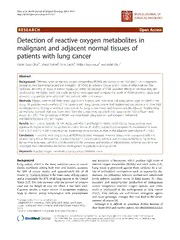
Detection of reactive oxygen metabolites in malignant and adjacent normal tissues of patients with lung cancer. PDF
Preview Detection of reactive oxygen metabolites in malignant and adjacent normal tissues of patients with lung cancer.
Okuretal.WorldJournalofSurgicalOncology2013,11:9 http://www.wjso.com/content/11/1/9 WORLD JOURNAL OF SURGICAL ONCOLOGY RESEARCH Open Access Detection of reactive oxygen metabolites in malignant and adjacent normal tissues of patients with lung cancer Hacer Kuzu Okur1*, Meral Yuksel2, Tunc Lacin3, Volkan Baysungur3 and Erdal Okur4 Abstract Background: Different types of reactive oxygen metabolites (ROMs)are knownto be involved incarcinogenesis. Several studies have emphasized the formation of ROMs in ischemic tissues and in cases of inflammation. The increased amountsof ROMs intumor tissues can either be because of their causative effects or because they are produced bythe tumor itself. Our study aimed to investigateand compare the levels of ROMs intumor tissue and adjacent lung parenchyma obtained from patients withlung cancer. Methods: Fifteenpatients(all male, mean age 63.6 ± 9years) with non-small cell lung cancer were enrolled inthe study. Allpatients were smokers.Of the patients with lung cancer, twelve had epidermoid carcinoma and threehad adenocarcinoma.During anatomical resection ofthelung, tumor tissue and macroscopically adjacent healthy lung parenchyma (control) that was 5cm away from thetumor were obtained. The tissues were freshly frozen and stored at−20°C. The generation of ROMs was monitored using luminol- and lucigenin-enhanced chemiluminescence (CL) techniques. Results: Bothluminol(specificfor.OH,H O ,andHOCl-)andlucigenin(selectiveforO.-)CLmeasurementswere 2 2 2 significantlyhigherintumortissuesthanincontroltissues(P<0.001).LuminolandlucigeninCLmeasurementswere 1.93±0.71and2.5±0.84timesbrighter,respectively,intumortissuesthanintheadjacentparenchyma(P=0.07). Conclusion:Inpatientswithlungcancer,allROMlevelswereincreasedintumortissueswhencomparedwiththe adjacentlungtissue.Becausetheincreaseinlucigeninconcentration,whichisduetotissueischemia,ishigherthan theincreaseinluminol,whichisdirectlyrelatedtothepresenceandseverityofinflammation,ischemiamaybemore importantthaninflammationfortumordevelopmentinpatientswithlungcancer. Keywords:Reactiveoxygenmetabolites,Lungcancer,Oxidativestress Background and activation of leucocytes, which produce high levels of Lung cancer, which is the most common cause of cancer reactive oxygen metabolites (ROMs) and nitric oxide [3,4]. deaths worldwide among both men and women, accounts Lung tissue is protected against these oxidants by a var- for 28% of cancer deaths and approximately 6% of all iety of antioxidant mechanisms such as superoxide dis- deaths[1].Tobaccosmokeisthemaincauseoflungcancer mutases. An imbalance between oxidant and antioxidant andisresponsiblefor87%ofalllungcancersintheUnited levels induces oxidative stress in lung tissues [5]; thus, States[1,2]. Therisk increases withthe amountof tobacco excessive and inappropriate production of endogenous used and the length of time for which it has been used. and/or exogenous reactive oxygen species and nitric Cigarette smoke, a major source of exogenous oxidants, oxide is implicated in the pathogenesis of lung cancer leads to chronic airway inflammation with accumulation [1,6,7]. Free radicals are well-known mutagenic agents and cause genotypic changes that may lead to the devel- *Correspondence:[email protected] opmentofcancer[8]. 1DepartmentofPulmonology,SureyyapasaChestDiseaseandThoracic Although it is difficult to quantitate ROMs because of SurgeryTrainingandResearchHospital,Basibuyuk-Maltepe,Istanbul34726, Turkey theirshort-livedandreactivenature,thechemiluminescence Fulllistofauthorinformationisavailableattheendofthearticle ©2013Okuretal.;licenseeBioMedCentralLtd.ThisisanOpenAccessarticledistributedunderthetermsoftheCreative CommonsAttributionLicense(http://creativecommons.org/licenses/by/2.0),whichpermitsunrestricteduse,distribution,and reproductioninanymedium,providedtheoriginalworkisproperlycited. Okuretal.WorldJournalofSurgicalOncology2013,11:9 Page2of5 http://www.wjso.com/content/11/1/9 (CL)methodusedinthepresentstudyisasimpleandre- awiderangeofapplications,especiallythosethatinvolve producible technique. The two CL probes, luminol and monitoringofreactiveoxygenspecies[10]. lucigenin, differ in their selectivity. Lucigenin is particu- In our experiment, tissues were thawed and washed larly sensitive to the superoxide radical (O.-), whereas with saline. Luminescence of the tissue samples was 2 luminol detects hydrogenperoxide(H O ),hydroxyl radi- recorded at room temperature using a Junior LB 9509 2 2 cals (.OH), hypochlorite (ClO-), peroxynitrite (ONOO-), luminometer(EG&GBerthold,BadWildbad,Germany)in andlipidperoxylradicals[9]. thepresenceofenhancers.Tissuespecimenswerecutinto Detection of high levels of ROMs, which are known to two pieces and placed into tubes containing PBS-HEPES be increased by ischemia or inflammation, in tumor tis- buffer (0.5 mol/L phosphate buffered saline containing 20 sues may be attributable either to their role in the eti- mmol/L HEPES, pH 7.2). ROMs were quantitated after ology of cancer or to the presence of the tumor itself. addition of the enhancer (lucigenin or luminol) to a final The present study was designed to explore the intensity concentration of 0.2 mmol/L. After the measurements of oxidative stress in patients with lung cancer by asses- weremade,thetissueswereremovedfromthetubes,dried sing the generation of ROMs in tumor and normal lung on filter papers, and weighed. All chemiluminometric parenchyma and comparing the levels of metabolites in countswereobtainedat1-minintervalsfor5min,andthe both tissues. resultswereexpressedasareasunderthe curve(AUCs)of relativelightunits(rlu)for5minpermgoftissue[9].The Methods calculationwasbasedontheintegrationofthecurveusing Patients thetrapezoidalrule(alinearapproximation). Fifteen consecutive patients with non-small cell lung carcinoma who were operated on in our clinic were Statisticalanalysis includedinthestudy.Allpatientswere smokers andhad Thevariousdegreesofoxidativestressindifferenttypesof no preoperative neoadjuvant treatment and no previous lung tumors, including epidermoid carcinomaand adeno- history of carcinoma of any type. Patients with tumors carcinoma,andadjacentlungtissueswerestatisticallyana- less than 2 cm in size or endobronchial tumors were not lyzed. All data are expressed as means ± SEM. Groups of included because obtaining a 1-cm sample from such a datawerecomparedusingapairedttest.Theresultswere small tumor might disturb the pathological examination. consideredsignificantwhenthePvaluewaslessthan0.05. After routine preoperative evaluation and staging, the Calculations were performed using GraphPad Prism 3.0 patientsunderwentanatomicallungresection(eitherlobec- (GraphPadSoftware,SanDiego,CA,USA). tomyorpneumonectomy)togetherwithmediastinallymph node dissection. Informed consent of the patients and Results and discussion approval of the ethics committee of Marmara University We examined a total of 15 patients, all of whom were MedicalSchoolwereobtained. male with a mean age of 63.6 ± 9 years (range 48 to 74 years).Patients’characteristicsaresummarizedinTable1. Experimentaldesign In all patients, the lucigenin and luminol CLvalues were Immediately after completion of lung resection and re- higher in cancerous lung tissues than in adjacent lung tis- moval of the surgical specimen, two tissue samples were sues. The luminol CL level in tumor tissues was signi- obtained from each patient: the first sample was obtained ficantlyhigherthanthatincontroltissues(4.32±0.38rlu/ from the lung tumor and the second was obtained from mg tissue vs. 2.26 ± 0.22 rlu/mg tissue, P <0.001) adjacent lung parenchyma (for the control group) at least (Figure 1a). Similarly, the lucigenin CL level showed a 5 cm away from the gross margin of the lung tumor. The marked increase in the tumor tissues as compared to con- tissues were then washed with saline (0.9% NaCl) to re- troltissues(4.49±0.44rlu/mgtissuevs.1.91±0.12rlu/mg moveblood,freshlyfrozen,andstoredat−20°Cuntilthey tissue,P<0.001)(Figure1b). wereexamined. In this study, we investigated the role of oxidative stress in lung cancer. We demonstrated that ROM Chemiluminescencemeasurement levels, as measured by the CL method, were significantly To assess the role of ROMs in this study, CL derived higher in the cancerous tissues of lung cancer patients from luminol and lucigenin was measured as an indica- than incontroltissues obtainedfrom thesame patients. tor of radical formation. CL measurements are based on Theantitumordefensemechanismsofthebodyresultin light emission with specific enhancers such as luminol, continuous oxidative stress and inflammatory responses, lucigenin, luciferase, and horseradish peroxidase. The which contribute to the various types of oxidative damage benefits of this method include rapidity, ultrasensitive observed in lung cancer [11]. It may be hypothesized that detection limits, and broad applicability. In clinical re- chronic injury to the epithelium caused by the production search, the sensitivity of this method has led to its use in ofROMsmayleadtolungcancer.Freeradicalsthatplaya Okuretal.WorldJournalofSurgicalOncology2013,11:9 Page3of5 http://www.wjso.com/content/11/1/9 Table1Characteristicsofpatients Age(years) Locations TNM Histologicaltype 1 59 RUL TNM Adenoca 2 0 0 2 73 RUL TNM Epidca 1 1 0 3 71 LLL TNM Epidca 1 1 0 4 65 RUL TNM Epidca 1 0 0 5 58 RUL TNM Epidca 1 1 0 6 65 RUL TNM Adenoca 2 1 0 7 60 RLL TNM Epidca 1 1 0 8 74 RML TNM Epidca 2 0 0 9 49 LLL TNM Adenoca 2 1 0 10 72 LLL TNM Epidca 1 1 0 11 78 LUL TNM Epidca 2 0 0 12 53 LLL TNM Epidca 2 0 0 13 67 LUL TNM Epidca 2 1 0 14 62 RUL TNM Epidca 2 0 0 15 48 LLL TNM Epidca 1 1 0 Adenoca,adenocarcinoma;Epidca,epidermoidcarcinoma;LLL,leftlower lobe;LUL,leftupperlobe;RLL,rightlowerlobe;RML,rightmediallobe; RUL,rightupperlobe;TNM,tumor,node,metastasis(classificationsystem). role in carcinogenesis may originate from cigarettesmoke, air pollution, or activated phagocytes, the latter of which areaconsequenceofchronicinflammatorydiseases[12]. In a report that examined lipid peroxidation in whole blood samples obtained from patients with lung cancer, the authors reported that the level of malondialdehyde (MDA, a lipid peroxidation product) in patients with early-stage (stage I and II) lung cancer was not signifi- cantly altered compared to the control group [7]. How- Figure1Lucigenin(a)andluminol(b)chemiluminescence(CL) ever, in patients with advanced-stage lung cancer, the levelsinthecontrolandtumorgroups.Rlu,relativelightunits. MDA level was significantly higher. Additionally, the level of whole blood glutathione (an intracellular antioxidant) was decreased in patients with both early- and advanced- are released from lung tumor specimens. In an animal stage lung cancer. In another study, the ROM levels in study, rats were exposed to cigarette smoke, and the serum samples from patients with small cell carcinoma, luminol-enhanced CL technique was used to measure adenocarcinoma, and epidermoid carcinoma were higher ROMs in lung and laryngeal tissues [14]. The results indi- thanthelevelsdetectedincontrolsubjects[13].Ziebaetal. cated that cigarette smoking increased luminol-enhanced reported that lipid peroxidation was higher in cancerous CL measurements, and vitamin E decreased ROM release tissue samples than in normal lung parenchyma and significantly.H O canbeconvertedto.OHinthepresence 2 2 demonstrated that thiobarbituric acid-reactive substances, ofmetalionsviatheFentonreactionorbyneutrophilinfil- which are markers of lipid peroxidation, were positively tration; alternatively, it can be converted to HOCl- in the correlatedwithbothclinicalstageandspontaneousgener- presence of chloride ions via the myeloperoxidase enzyme. ationofH O intumortissue[4]. In our study, luminol-enhanced CL amplification can also 2 2 In our study, luminol-enhanced CL measurements were beusedtomeasure.OHandHOCl-.HOCl-isusuallypro- significantly higher in tumor specimens than in adjacent duced by polymorphonuclear leukocytes at an inflamma- pulmonary parenchyma tissues. Our method, which is tion site. In a group of patients with non-small cell lung based on the measurement of spontaneously released cancer,myeloperoxidaseactivitywaslowerandglutathione ROMs,isalsocapableofmeasuringH O andanothersub- levels were higher in patients with malignant disease [15]. 2 2 stances such as hydroxyl radicals (.OH), hypochlorite ions Reports of increased inflammatory markers and C-reactive (ClO-), peroxynitrite ions (ONOO-), and lipid peroxyl ra- protein [16] as well as an acute phase response in cancer dicals. Our results show that H O and other compounds patientsindicatethatbothsystemicandlocal(thatis,atthe 2 2 Okuretal.WorldJournalofSurgicalOncology2013,11:9 Page4of5 http://www.wjso.com/content/11/1/9 tumor site) inflammatory reactions are closely associated Authors’contributions withthepresenceofatumor[4].Inaddition,somestudies HKOandMYdesignedtheresearch,TL,VBandEOperformedtheoperations andcollectedthetissuesamples.MYperfomedthelaboratory indicate that the tumor tissue itself could be the source of measurementsandanalyses.HKOandEOanalysedthedata.HKO,MYand ROMsandlipidperoxidationproducts[17]. EOwrotethepaper.Allauthorsreadandapprovedthefinalmanuscript. Ischemia is the major cause of superoxide generation in Authordetails cancertissues.Ischemialeadstoglucoseandadenosinetri- 1DepartmentofPulmonology,SureyyapasaChestDiseaseandThoracic phosphate (ATP) depletion, which in turn leads to a de- SurgeryTrainingandResearchHospital,Basibuyuk-Maltepe,Istanbul34726, crease in sodium-potassium ATPase (Na-K ATPase) Turkey.2VocationalSchoolofHealthRelatedProfessions,Departmentof MedicalLaboratory,MarmaraUniversity,HaydarpasaCampus,Istanbul34668, activity and causes depolarization of the cells. In these Turkey.3DepartmentofThoracicSurgery,SureyyapasaChestDiseaseand depolarized cells, intracellular calcium increases, resulting ThoracicSurgeryTrainingandResearchHospital,Basibuyuk-Maltepe,Istanbul inactivationofthexanthine-hypoxanthinesystemandpro- 34726,Turkey.4DepartmentofThoracicSurgery,AcibademUniversity MedicalSchool,Istanbul,Gulsuyu_Maltepe,Istanbul34726,Turkey. duction of superoxide radicals. Superoxide radicals, which arehighlyreactive,damagemembranephospholipids,cellu- Received:23August2012Accepted:24December2012 lar proteins, and DNA [18]. In our study, the levels of Published:17January2013 lucigenin-enhancedCLwerehigherintumorsamplesthan in parenchymal tissues. Formation of superoxide radicals References 1. MasriF:Roleofnitricoxideanditsmetabolitesasmetabolitesas canbepreventedviasuperoxidedismutase(SOD)enzymes. potentialmarkersinlungcancer.AnnThoracMed2010,5:123–127. In an immunohistochemical study, the expression levels of 2. BilelloKS,MurinS,MatthayRA:Epidemology,etiologyandpreventionof both Mn-SOD (mitochondrial form) and Cu/Zn-SOD lungcancer.ClinChestMed2002,23:1–15. 3. ChowCK:Cigarettesmokingandoxidativedamageinthelung.AnnNY (cytoplasmic or nuclear form) in the lungs of patientswith AcadSci1993,28:289–298. squamous cell carcinoma were significantly lower than the 4. ZiebaM,SuwalskiM,KwiatkowskaS,PiaseckaG,Grzelewska-RzymowskaI, expressionlevelsoftheseenzymesinluminalcellsandun- StolarekR,NowakD:Comparisonofhydrogenperoxidegenerationand thecontentoflipidperoxidationproductsinlungcancertissueand involvedepitheliumofthesamepatients[19].Theseresults pulmonaryparenchyma.RespMed2000,94:800–805. demonstrate that intracellular and intramitochondrial 5. YooDG,SongYJ,ChoEJ,LeeSK,ParkJB,YuJH,LimSP,KimJM,JeonBH: SODsarenotcapableofreducingsuperoxideradicalgener- AlterationofAPE1/ref-1expressioninnon-smallcelllungcancer:The implicationsofimpairedextracellularsuperoxidedismutaseandcatalase ation,andourstudyalsoprovidedevidencetosupportthis antioxidantsystems.LungCancer2008,60:277–284. findingbasedonlucigenin-enhancedCLmeasurements. 6. WisemanH,HalliwellB:DamagetoDNAbyreactiveoxygenandnitrogen ThemonitoringofROMgenerationinfreshlyfrozentis- species:Roleininflammatorydiseaseandprogressiontocancer. BiochemJ1996,313:17–19. suesamplesusingtheCLtechniquehasbeendescribedin 7. EsmeH,CemekM,SezerM,SaglamH,DemirA,MelekH,UnluM:High twostudiesbyOhoietal.[20,21].Cicenasetal.andToklu levelsofoxidativestressinpatientswithadvancedlungcancer. et al. also used this method for freshly frozen tissue sam- Respirology2008,13:112–116. 8. ChungFL,ChenHJ,GuttenplanJB,NishikawaA,HardGC:2,3-epoxy-4- ples of breast and colon cancers as well as for urogenital hydroxynonanalasapotentialtumor-indicatingagentoflipid tissues [9,22,23]. Our study is the first to measure ROM peroxidation.Carcinogenesis1993,14:2073–2077. levelsinlungcancertissuesamples. 9. HaklarG,OzveriES,YukselM,AktanAO,YalcinAS:Differentkindsof reactiveoxygenandnitrogenspeciesweredetectedincolonandbreast A limitation of our study is the small sample size (15 tumors.CancerLett2001,165:219–224. patients).Thiswasasmallpilotstudy.Similarstudieswith 10. KrickaLJ:Clinicalapplicationsofchemiluminescence.AnalChimActa largersamplesize,atleast100patients,areneededtocon- 2003,500:279–286. 11. MooneyLA,PereraFP,VanBennekumAM,BlanerWS,KarkoszkaJ,CoveyL, firmourresults. HsuY,CooperTB,FrenkelK:Genderdifferencesinautoantibodiesto oxidativeDNAbasedamageincigarettesmokers.CancerEpidemBiomar 2001,10:641–648. Conclusion 12. FeigDI,ReidTM,LoebLA:Reactiveoxygenspeciesintumorigenesis. In general, evidence suggests that lucigenin-enhanced CancerRes1994,54:1890–1894. superoxide radical generation is increased by ischemia in 13. GencerM,CeylanE,AksoyN,UzunK:Associationofserumreactive oxygenmetabolitelevelswithdifferenthistopathologicaltypesoflung tissues, whereas the luminol level is increased in the pres- cancer.Respiration2006,73:520–524. ence of inflammation and is directly related to inflamma- 14. ÜneriC,SariM,BağlamT,PolatŞ,YükselM:EffectsofvitaminEon tory severity. In our study, the mean concentrations of cigarettesmokeinducedoxidativedamageinlarynxandlung. Laryngoscope2006,116:97–100. lucigenin and luminol in cancerous lung tissue were sig- 15. IlonenIK,RasanenJV,SihvoEL,KnuuttilaA,SalmenkiviKM,AhotupaMO, nificantlyhigherthantheirconcentrationsinadjacentlung KinnulaVL,SaloJA:Oxidativestressinnon-smallcelllungcancer:roleof parenchyma. A higher increase in the concentration of nicotinamideadeninedinucleotidephosphateoxidaseandglutathione. ActaOncol2009,48:1054–1061. lucigenin compared with luminol shows that tissue ische- 16. vanderBrekelAJS,ScholsAMW,DentenerMA,tenVeldeGP,BuurmanWA, mia may be more important than inflammation for tumor WoutersEF:Metabolisminpatientswithsmallcelllungcarcinoma developmentinlungcancerpatients. comparedwithpatientswithnon-smallcelllungcarcinomaandhealthy controls.Thorax1997,52:338–341. 17. LeroyerV,WernerL,ShaughnessyS,GoddardGJ,OrrFW: Competinginterests ChemiluminescenceandoxygenradicalgenerationbyWalkercarcinoma Alltheauthorsdeclarethattheyhavenocompetinginterests. cellsfollowingchemotacticstimulation.CancerRes1987,47:4771–4775. Okuretal.WorldJournalofSurgicalOncology2013,11:9 Page5of5 http://www.wjso.com/content/11/1/9 18. SorgO:Oxidativestress:atheoreticalmodelorabiologicalreality? CRBiol2004,327:649–662. 19. PiyathilakeCJ,BellWC,OelschlagerDK,HeimburgerDC,GrizzleWE:The patternofexpressionofMnandCu-Znsuperoxidedismutasevaries amongsquamouscellcancersofthelung,larynx,andoralcavity. HeadNeck2002,24:859–867. 20. OhoiI,SoneK,TobariH,KawanoE,NakamuraK:Asimplechemiluminescence methodformeasuringoxygen-derivedfreeradicalsgeneratedin oxygenatedratmyocardium.JpnJPharmacol1993,61:101–107. 21. OhoiI,TobariH,NakamuraK:Measurementofoxygen-derivedfree radicalgenerationintheregionally-ischemicratheartbythe chemiluminescencemethod.JpnJPharmacol1993,62:415–418. 22. CicenasJ,KüngW,EppenbergerU,Eppenberger-CastoriS:Increasedlevel ofphosphorylatedShcAmeasuredbychemiluminescence-linked immunoassayisapredictorofgoodprognosisinprimarybreastcancer expressinglowlevelsofestrogenreceptor.Cancers2010,2:153–164. 23. TokluH,ŞehirliÖ,ŞahinH,ÇetinelŞ,YeğenBC,ŞenerG:Resveratrol supplementationprotectsagainstchronicnicotine-inducedoxidative damageandorgandysfunctionintheraturogenitalsystem. MarmaraPharmaceuticalJournal2010,14:29–40. doi:10.1186/1477-7819-11-9 Citethisarticleas:Okuretal.:Detectionofreactiveoxygenmetabolites inmalignantandadjacentnormaltissuesofpatientswithlungcancer. WorldJournalofSurgicalOncology201311:9. Submit your next manuscript to BioMed Central and take full advantage of: • Convenient online submission • Thorough peer review • No space constraints or color figure charges • Immediate publication on acceptance • Inclusion in PubMed, CAS, Scopus and Google Scholar • Research which is freely available for redistribution Submit your manuscript at www.biomedcentral.com/submit
