Common Problems in Family Medicine Booklet PDF
Preview Common Problems in Family Medicine Booklet
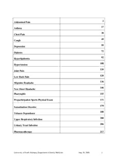
2 Abdominal Pain 17 Asthma 30 Chest Pain 49 Cough 58 Depression 72 Diabetes 92 Hyperlipidemia 108 Hypertension 120 Joint Pain 128 Low Back Pain 136 Migraine Headache 146 New Onset Headache Pharyngitis 155 Preparticipation Sports Physical Exam 171 179 Somatization Disorder 188 Tobacco Dependence 200 Upper Respiratory Infection 206 Urinary Tract Infection Pharmacotherapy 217 University of South Alabama, Department of Family Medicine June 30, 2008 1 Abdominal Pain 789.0X GENERAL CONSIDERATIONS Scope of This Article This article discusses atraumatic abdominal pain in adults and older children. Definition of Abdominal Pain Complaint of pain in the area between the ribcage and pelvis. Overview Abdominal pain is frequent symptom encountered by a large portion of the otherwise healthy population (in some studies more than 50%). Most commonly it is a benign complaint reflecting a disease process that can be treated symptomatically. However, it may be the presenting symptom to an acute life-threatening illness that requires rapid assessment and immediate triage to an acute care facility. Red Flags: Red flags are historical or physical findings that suggest a higher severity of illness, requiring further investigation. Red Flag Possible Etiology Possible Consequence Fever Infection Dehydration, sepsis Diarrhea Infection Dehydration Persistent constipation Intestinal obstruction Dehydration Hematochezia Lower or upper GI bleed Hemodynamic instability Persistent Vomiting Multiple etiologies Dehydration, metabolic acidosis Hematemesis Upper GI bleed Hemodynamic instability Severe Pain Perforation, peritonitis Sepsis, hemodynamic instability Pregnancy Ectopic pregnancy, placental Increased fetal and maternal abruption morbidity, mortality Jaundice Liver failure, biliary Hepatic encephalopathy obstruction University of South Alabama, Department of Family Medicine June 30, 2008 2 History The history should include: Duration and frequency of pain Severity and nature of pain. Location and radiation of pain. Aggravating/alleviating factors, such as food, antacids, exertion, defecation. Associated symptoms, including fever, chills, weight loss or gain, nausea, vomiting, diarrhea, constipation, hematochezia, melena, jaundice, change in the color of urine or stool, change in the diameter of stool. Family history of bowel disorders. Alcohol intake. Medication use, including over the counter medications such as aspirin and NSAIDs. Menstrual history. Pain Duration Acute: Pain of less than a few days duration, that has worsened progressively until the time of presentation. Chronic: Pain that has remained unchanged for months or years. Pain that does not clearly fit either category might be called subacute, and requires consideration of the differential diagnoses for both acute and chronic pain. Pain Type Pain Type Character Localization Cause Examples Dull and aching, Distention or Intestinal Visceral Pain but can be Poorly localized spasm of a obstruction, colicky hollow organ cholecystitis Parietal Acute Parietal Pain Sharp Well localized peritoneal appendicitis irritation University of South Alabama, Department of Family Medicine June 30, 2008 3 Pain Location Causes of Abdominal Pain by Location Location Causes Hepatitis Cholecystitis Cholangitis Right Upper Quadrant Pancreatitis Budd-Chiari Syndrome Pneumonia Splenic Abscess Splenic Infarct Left Upper Quadrant Gastritis Gastric Ulcer Pancreatitis Appendicitis Salpingitis Ectopic Pregnancy Right Lower Quadrant Inguinal Hernia Nephrolithiasis Inflammatory Bowel Disease Diverticulitis Salpingitis Ectopic Pregnancy Left Lower Quadrant Inguinal Hernia Nephrolithiasis Inflammatory Bowel Disease Peptic Ulcer Disease GERD Gastritis Epigastric Pancreatitis MI Pericarditis Aortic Aneurysm Early Appendicitis Gastroenteritis Periumbilical Bowel Obstruction Aortic Aneurysm Ventral Hernia Gastroenteritis Mesenteric Ischemia Metabolic Diffuse Irritable Bowel Syndrome Bowel Obstruction Peritonitis University of South Alabama, Department of Family Medicine June 30, 2008 4 Pain Severity The severity of the pain generally is related to the severity of the disorder, especially if acute in onset. As an example, the pain of biliary obstruction, renal colic, or mesenteric infarction is of high intensity, while the pain of gastroenteritis is less marked. Age, mental status, and general health may affect the patient’s clinical presentation. A patient taking corticosteroids may have significant masking of pain, and the elderly often present with less intense pain. Associated Symptoms Symptoms that occur in relation to abdominal pain may give important information. Nausea and vomiting occur with a number of disorders. Weight loss may occur in association with malignancy. A change in bowel habits suggests a colonic lesion. Women should be asked whether they are sexually active, the number of sexual partners, whether any sexual partners are new, and whether any sexual partners are experiencing symptoms suggestive of a sexually transmitted infection. Physical Examination General Exam General appearance. Patients with peritonitis try to remain immobile. Vital signs, including measurement of orthostatic changes in blood pressure and heart rate. Obstruction or peritonitis can cause large amounts of third spacing of fluid and intravascular volume depletion or overt shock. Jaundice. Abdominal Exam Auscultation. Hypoactive sounds may be noted in advanced peritonitis or ileus. High- pitched bowel sounds are a feature of early bowel obstruction. Gentle percussion is useful to identify acute peritonitis. Percussion is also used to identify ascites, liver span, and bladder and splenic enlargement. Tympany signifies distended bowel, while dullness may signify a mass. Palpation must be performed gently and while the patient is distracted. Muscular rigidity or ―guarding‖ is an important and early sign of peritoneal inflammation. Palpation also may detect enlarged organs, masses, or hernias. Rectal and Pelvic Exam A rectal is generally required in all patients with acute abdominal pain. Fecal impaction might be the explanation for signs and symptoms of obstruction in the elderly. Stool for occult blood should also be obtained. A pelvic exam is generally required in all women with acute lower abdominal pain, and is critical for determining whether abdominal pain is due to pelvic inflammatory disease, an adnexal mass or cyst, uterine pathology, or an ectopic pregnancy. University of South Alabama, Department of Family Medicine June 30, 2008 5 ACUTE ABDOMINAL PAIN Surgical Abdomen (AKA ―Acute Abdomen‖) The first priority in patients with acute abdominal pain is to determine who has a rapidly worsening prognosis in the absence of surgical intervention. Only after the clinician is satisfied that the abdominal presentation is not an acute surgical emergency can consideration of other diagnostic possibilities begin. Patients should not eat or drink while a diagnosis of a surgical abdomen remains under consideration. The two syndromes that cause most surgical abdomens are obstruction and peritonitis. The latter encompasses most severe abdominal pathology since intraperitoneal hemorrhage or viscus perforation typically present with common features of peritonitis. Obstruction Obstruction generally presents as pain together with anorexia, bloating, nausea, and vomiting. Physical examination may reveal distension and high-pitched or absent bowel sounds. Abdominal percussion reveals tympany from proximally dilated loops of bowel. An abdominal mass, if present, may suggest an etiology for the obstruction. Peritonitis Patients with peritonitis of any cause tend to appear ill and lie still to minimize their discomfort. Rebound tenderness, abdominal wall rigidity, and percussion tenderness are classically thought to reflect peritonitis. Other subtle signs of peritonitis include diminished bowel sounds and pain worsened when an examiner lightly bumps the stretcher. Initial Diagnostic Testing Complete blood count with differential Electrolytes, BUN, creatinine, and glucose Aminotransferases, alkaline phosphatase, and bilirubin Lipase Urinalysis Pregnancy test in women of childbearing potential While these laboratory tests are important, they are not sufficient to rule in or rule out a diagnosis of surgical abdomen, as a surgical abdomen is a clinical diagnosis. Subsequent Diagnostic Testing Patients clearly in need of urgent laparotomy may proceed directly to the operating room for diagnosis and management. In particular, patients with a painful pulsatile abdominal mass, with or without bruit, should be suspected to have a ruptured aortic aneurysm. University of South Alabama, Department of Family Medicine June 30, 2008 6 However, many patients will not have a firm diagnosis after initial assessment, and in these cases, careful observation of the patient’s course will be the most important factor in their management. In addition, the following additional investigations can also be considered: Blood and urine cultures, in the presence of fever or unstable vital signs. CT Scan. When available, CT scanning is the test of choice for detecting partial or complete bowel obstruction, hernias, diverticulitis, renal stones, and aortic aneurysms. It can also be useful in investigating appendicitis, peritonitis, ischemia due to strangulation/adhesions, and pancreatitis. CT scan may play a decision-making role when evaluating demented or obtunded patients. Plain Abdominal Films. Where CT scanning is immediately available, abdominal plain films are not necessary, as they do not provide additional information. However, in the absence of CT scanning, flat and upright or lateral decubitus radiographs are a crucial step in decision making for the suspected surgical abdomen, as proximally dilated loops of bowel are the hallmark of intestinal obstruction, and free intraperitoneal air can confirm a suspicion of hollow organ perforation. Ultrasound. Ultrasound is the preferred test in pregnancy, and evaluating biliary and pelvic pathology. If more readily available, it may also be useful for many of the conditions listed above under CT Scan. Right Upper Quadrant Pain Usually caused by involvement of the liver or biliary tree. Initial assessment The presence of fever and jaundice in a patient with right upper quadrant pain leads to a clinical diagnosis of ascending cholangitis. Acute cholecystitis can also present as a systemically unwell patient with low-grade fever. Patients with an acute rise in aminotransferases and right upper quadrant pain most likely have choledocholithiasis, particularly if there is also an acute rise in bilirubin. Initial Diagnostic Testing Complete blood count with differential. Electrolytes, BUN, creatinine, and glucose. Aminotransferases, alkaline phosphatase, and bilirubin. Lipase. Abdominal ultrasound. (Plain films of the abdomen are unlikely to yield much information.) Subsequent Diagnostic Testing (if available) Endoscopic ultrasound. Endoscopic retrograde cholangiopancreatography (ERCP). Magnetic resonance cholangiopancreatography (MRCP). University of South Alabama, Department of Family Medicine June 30, 2008 7 Epigastric Pain Epigastric pain that is relatively sudden in onset is suggestive of pancreatitis, particularly when it radiates to the back and is associated with nausea, vomiting, and anorexia. Epigastric pain that is less acute and is associated with bloating, abdominal fullness, heartburn, or nausea can be classified as dyspepsia. Most of these patients can safely undergo a therapeutic trial or watchful waiting. However red flags that suggest a need for further investigation include: Age over 50. Weight loss. Persistent vomiting. Dysphagia. Anemia. Hematemesis, melena, or heme positive stool. Palpable abdominal mass. Family history of upper gastrointestinal carcinoma. Previous gastric surgery. Refractory/recurrent symptoms. It is also important to consider nonabdominal etiologies of upper abdominal pain: Cardiac pain. Pleural or pulmonary pathology. Initial Diagnostic Testing Many patients can be managed with a therapeutic trial of antisecretory therapy without further investigation. However, those with red flags or suspicion of pancreatitis should have the following: Complete blood count with differential. Electrolytes, BUN, creatinine, and glucose. Aminotransferases, alkaline phosphatase, and bilirubin. Lipase. Elevated lipase in the presence of epigastric pain is very suggestive of pancreatitis. Amylase is a less specific alternative, if lipase is not available. Subsequent Diagnostic Testing Esophagogastroduodenoscopy (EGD), especially for those with red flags and dyspepsia. Testing for Helicobacter pylori may be considered, especially in refractory/recurrent cases. Abdominal ultrasound if pancreatitis is being considered, since biliary disease is a common etiology for pancreatitis. CT Scan is more sensitive for the diagnosis of pancreatitis than ultrasound; consider if doubts remain after an ultrasound has been obtained. University of South Alabama, Department of Family Medicine June 30, 2008 8 Lower Abdominal Pain Pain in the lower abdomen can be associated with pathology in the following: Distal intestinal tract. Upper abdominal structures with pain radiating into the lower abdomen. The pelvis. The history should include risk factors for infectious and ischemic causes, medication use (e.g., NSAIDs, laxatives), and family history of inflammatory bowel disease (IBD). Patients should be asked about urinary symptoms such as frequency, urgency, and dysuria. Left and/or right lower quadrant pain, when occurring together with diarrhea, is suggestive of colitis and/or ileitis. Diverticulitis presents more frequently as left lower quadrant pain, often with leukocytosis. In older patients, abdominal pain and a change in bowel habits can be the first sign of colon cancer. It is also important to consider nonabdominal etiologies of upper abdominal pain: Retroperitoneal pathology. Cystitis can cause suprapubic pain. Renal colic results in pain that may radiate to the lower abdomen. Lower abdominal pain (pelvic pain) in women is frequently caused by disorders of the internal female reproductive organs. (Discussed below.) Initial Diagnostic Testing Complete blood count with differential. Urinalysis (and culture if results dictate). Subsequent Diagnostic Testing Stool studies in patients with severe or persistent lower abdominal pain associated with diarrhea, and immunosuppressed patients, should include culture for enteric pathogens, microscopy for ova and parasites, and measurement of Clostridium difficile toxin. However, many patients with less severe presentations will often have self-limited illness, and can be managed expectantly. Colonoscopy in patients with illness exceeding two weeks with negative cultures, systemically unwell patients, immunosuppressed patients, and when ileal pathology is suspected. (Note that patients over age 50 are also candidates for screening colonoscopy, and their presenting symptoms provide an opportunity to discuss this.) CT scan, especially in cases suggestive of diverticulitis. University of South Alabama, Department of Family Medicine June 30, 2008 9 Lower Abdominal Pain in Women Additional history in women should include: Regularity and timing of menstrual periods. Possibility of pregnancy. Presence of vaginal discharge or bleeding. Recent history of dyspareunia or dysmenorrhea. In addition to the causes of lower abdominal pain discussed above, other common etiologies of acute lower abdominal pain in women include: Pelvic inflammatory disease (PID). Adnexal cysts or masses with bleeding. Ovarian torsion. Ectopic pregnancy. Uterine pain due to infection (endometritis) or torsion of leiomyomas. A pelvic examination is part of the physical examination whenever pelvic pathology is in the differential diagnosis. Purulent cervical discharge, cervical tenderness, uterine enlargement, or adnexal masses may be detected. PID should be considered when acute left, right, or bilateral abdominal pain is accompanied by fever and an elevated white blood count with left shift. Initial Diagnostic Testing In addition to tests discussed above, women with lower abdominal pain should have the following: Pregnancy test in women of childbearing potential, even when pregnancy is felt unlikely. Wet prep of any abnormal vaginal discharge. Tests for Chlamydia and gonococcus in women with risk factors for sexually transmitted infections, mucopurulent cervical discharge, or suspected PID. Subsequent Diagnostic Testing Pelvic ultrasound, especially if pregnancy test is positive, or pelvic exam is suggestive of PID or adnexal masses. Generalized Abdominal Pain (not meeting the criteria of ―surgical abdomen‖) Generalized abdominal pain with vomiting and/or diarrhea, alone or in association with systemic symptoms, often represents an acute self-limited illness, such as viral or bacterial enteritis or colitis, or toxin-mediated food poisoning. Multisystem symptoms, such as upper respiratory tract involvement or myalgias, may suggest a viral etiology. University of South Alabama, Department of Family Medicine June 30, 2008 10
Description: