Common Diseases of Wild and Cultured Fishes in Alaska PDF
Preview Common Diseases of Wild and Cultured Fishes in Alaska
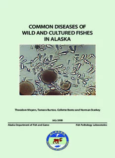
DISEASES OF WILD AND CULTURED FISHES IN ALASKA Theodore Meyers, Tamara Burton, Collette Bentz, Jayde Ferguson, Davis Stewart, and Norman Starkey July 2019 Alaska Department of Fish and Game Fish Pathology Laboratories The Alaska Department of Fish and Game printed this publication at a cost of $9.83 in Anchorage, Alaska, USA. About This Field Guide This field guide is a product of the Ichthyophonus Diagnostics, Educational and Outreach Program which was initiated and funded by the Yukon River Panel’s Restoration and Enhancement fund and facilitated by the Yukon River Drainage Fisheries Association in conjunction with the Alaska Department of Fish and Game. The original impetus driving the production of this guide was from a concern that Yukon River fishers were discarding Canadian-origin Chinook salmon believed to be infected by Ichthyophonus. It was decided to develop an educational program that included the creation of a field guide containing photographs and descriptions of frequently encountered parasites within Yukon River fish. This field guide is to serve as a brief illustrated reference that lists many of the common (and not so common) parasitic, infectious, and noninfectious diseases of wild and cultured fishes encountered in Alaska. The content is directed towards lay users, as well as fish culturists at aquaculture facilities and field biologists and is not a comprehensive treatise nor should it be considered a scientific document. Interested users of this guide are directed to the listed fish disease references for additional information. Information contained within this field guide is published from the laboratory records of the Alaska Department of Fish and Game, Fish Pathology Section that has regulatory oversight of finfish health in the State of Alaska. This third printing includes several new entries, some new photographs and updated information on previous diseases and parasites. This version may be downloaded as a PDF from the ADF&G website at the following web address: http://www.adfg.alaska.gov/static/species/disease/pdfs/fish_disease_book.pdf 3 Text written and provided by: Theodore Meyers, Tamara Burton, Collette Bentz, and Jayde Ferguson, Alaska Department of Fish and Game, Fish Pathology Laboratories, 333 Raspberry Road, Anchorage, Alaska 99518; P.O. Box 115526 (physical 3333 Glacier Highway), Juneau, Alaska 99811-5526. Manuscript Photographs: Alaska Department of Fish and Game, Fish Pathology Laboratory photo archives except where indicated. Publication design by Southfork Graphic Services. Cover Photograph: Ichthyophonus growing in laboratory culture. ©2019 Alaska Department of Fish and Game, third printing The original printing of this publication was produced with funding and support from the Yukon River Panel and its members and the Yukon River Drainage Fisheries Association. Current funding is provided by the ADF&G, Commercial Fisheries and Sport Fish Divisions. The Alaska Department of Fish and Game (ADF&G) administers all programs and activities free from discrimination based on race, color, national origin, age, sex, religion, marital status, pregnancy, parenthood, or disability. The department administers all programs and activities in compliance with Title VI of the Civil Rights Act of 1964, Section 504 of the Rehabilitation Act of 1973, Title II of the Americans with Disabilities Act of 1990, the Age Discrimination Act of 1975, and Title IX of the Education Amendments of 1972. If you believe you have been discriminated against in any program, activity, or facility please write ADF&G ADA Coordinator, P.O. Box 115526, Juneau, AK 99811-5526; U.S. Fish and Wildlife Service, 4401 N. Fairfax Drive, MS 2042, Arlington, VA 22203; or Office of Equal Opportunity, U.S. Department of the Interior, 1849 C Street NW MS 5230, Washington DC 20240. For information on alternative formats and questions on this publication, please contact the department’s ADA Coordinator at (VOICE) 907-465-6077, (Statewide Telecommunication Device for the Deaf) 1-800-478-3648, (Juneau TDD) 907-465-3646, or (FAX) 907-465-6078. Table of Contents Viruses Kudoa .................................................................................. 62 Aquareovirus .........................................................................2 Myxobolus neurotropus ...................................................... 64 Erythrocytic Inclusion Body Syndrome (EIBS) ....................4 Myxobolus squamalis ......................................................... 66 Erythrocytic Necrosis Virus (VENV) .....................................6 Tetracapsuloides bryosalmonae (PKD) .............................. 68 Infectious Hematopoietic Necrosis Virus (IHNV) ................8 Helminths (Worms) North American Viral Hemorrhagic Acanthocephalans (spiny headed worms) ........................7 0 Septicemia Virus (NA-VHSV) ..............................................1 0 Anisakid Larvae (nematode) ............................................. 72 Pacifc Salmon Paramyxovirus ...........................................12 Philometra (nematode) .......................................................74 Piscine Orthoreovirus (PRV) ...............................................14 Philonema (nematode) .......................................................76 Bacteria Black Spot Disease (trematode) .........................................78 Bacterial Coldwater Disease (BCWD) .................................16 Encysted Digenean Metacercariae (trematode) .............. 80 Bacterial Gill Disease ...........................................................18 Larval Diplostomulum of the Eye (trematode) ............... 82 Bacterial Kidney Disease (BKD) ......................................... 20 Gyrodactylus and Dactylogyrus (monogene) ................. 84 Enteric Redmouth Disease (ERM) ..................................... 22 Piscicola (annelid) .............................................................. 86 Furunculosis .........................................................................24 Diphyllobothrium (cestode) ............................................... 88 Fusobacteria-like Agent .................................................... 26 Schistocephalus (cestode) ................................................. 90 Marine Tenacibaculosis ..................................................... 28 Triaenophorus (cestode) ....................................................92 Motile Aeromonas and Pseudomonas Septicemia ........... 30 Arthropods Mycobacteriosis of Fish .......................................................32 External Parasitic Copepods ..............................................94 Vibriosis............................................................................... 34 Salmincola (copepod) ....................................................... 96 Fungi Sarcotaces (copepod) ........................................................ 98 Phaeohyphomycosis of Safron Cod ................................. 36 Non-infectious Diseases Phoma herbarum ............................................................... 38 Bloat (Water Belly) ........................................................... 100 Protozoa Blue Sac Disease of Fry ..................................................... 102 Epistylis (Heteropolaria) .................................................... 40 Coagulated Yolk Disease (White Spot Disease) .............. 104 Hexamita..............................................................................42 Drop-out Disease ............................................................. 106 Ichthyobodiasis (Costiasis) ................................................ 44 Gas Bubble Disease (GBD)................................................ 108 Ichthyophonus .................................................................... 46 Mushy Halibut Syndrome ................................................. 110 Saprolegniasis – Cotton Wool Disease ............................. 48 Neoplasia (Tumors) ...........................................................112 Trichodiniasis ...................................................................... 50 Organ and Tissue Anomalies ............................................116 Trichophrya (Capriniana) .................................................. 52 Pigment Aberrations in Fish .............................................118 White Spot Disease ............................................................ 54 Sunburn (Back-Peel) ........................................................ 120 X-Cell Tumors ..................................................................... 56 Reference Cnidaria Glossary of Terms ............................................................. 122 Ceratonova (Ceratomyxa) shasta ........................................ 58 Fish Disease References ....................................................127 Henneguya .......................................................................... 60 VIRUSES Aquareovirus I. Causative Agent and Disease associated with epizootic fish mortal- Aquareovirus is a genus in the virus ity producing severe hemorrhaging in family Reoviridae. These icosahedral fingerlings and yearlings resulting in up (60-80 nm) 11 segmented double- to 80% mortality. stranded RNA viruses (over 50) have been isolated from a variety of marine IV. Transmission and freshwater aquatic animals world- Transmission is horizontal via water wide including finfish, and bivalve mol- or from fish to fish. Isolates from bivalve luscs. Genetic analyses have identified 7 mollusks likely represent virus that has different genotypes or species (A-G) of been shed into the water column from a aquareoviruses. Most of these viruses fish host and then bioaccumulated into produce self-limiting infections of low shellfish tissues by filter feeding. pathogenicity and are not associated with extensive disease or mortality. V. Diagnosis Exceptions include isolates from 7 fish Detection of Aquareovirus is by species that have been associated with isolation of the virus in cultures of fish mortality, most notably the grass susceptible fish cell lines inoculated carp aquareovirus (G). The viral agents with infected tissue. The virus causes a are most often isolated from asymp- unique cytopathic effect (CPE) charac- tomatic adult carrier fish during routine terized by focal areas of cellular fusion screening examinations. (syncytia) and cytoplasmic destruction creating a vacuolated or foamy appear- II. Host Species ance. The exception is grass carp species In the Pacific Northwest states of G that produces a diffuse CPE. Presump- Washington, Oregon and California, tive identifications are made based on adult Chinook salmon appear to be the the typical CPE and are confirmed by most frequent species infected with serology, electron microscopy or poly- aquareovirus A or B. The virus has also merase chain reaction (PCR). been isolated from adult coho and chum salmon and steelhead. Rainbow trout VI. Prognosis for Host have been experimentally infected with The prognosis for the fish host is the virus resulting in mild hepatitis with good in the majority of cases where the no overt disease or mortality. In Alaska, virus is not a primary pathogen. There aquareoviruses have been isolated are no corrective therapies for viral from Chinook salmon (species B) and infections in fish except avoidance. geoduck clams (species A). VII. Human Health Signifcance III. Clinical Signs There are no human health concerns Fish naturally infected with aquareo- associated with aquareoviruses. viruses generally do not exhibit clinical signs of disease. Experimental infections can produce focal necrotic lesions in the livers of rainbow trout, chum salmon and bluegill fry. Other pathogenic exceptions include the grass carp species G that is 2 VIRUSES Large rounded plaques of syncytial cell CPE (arrow) of Aquareovirus in bluegill fry cells. Double capsid morphology of Aquareovirus particles (arrow) in negative stain; transmission electron microscopy, X 91,000. 3 VIRUSES Erythrocytic Inclusion Body Syndrome (EIBS) I. Causative Agent and Disease V. Diagnosis Erythrocytic inclusion body syn- Isolation and replication of the drome (EIBS) is caused by an unclassi- virus in available fish cell lines has fied icosahedral virus (70-80 nm) that been unsuccessful. Thus, diagnosis is infects erythrocytes of several salmonid by observation of the small pale blue fishes in fresh and seawater. Typically, inclusion bodies in the cytoplasm of EIBS presents with single or multiple infected erythrocytes with confirmation pale, basophilic inclusions (0.4-1.6 by transmission electron microscopy um) in the cytoplasm of erythrocytes (TEM). The virus is found free in the in stained peripheral blood smears. cytoplasm or more commonly occurs in Affected fish may be asymptomatic, membrane bound cytoplasmic inclusion but more often have varying degrees bodies within erythrocytes. of anemia and secondary bacterial and fungal infections. In severe cases of VI. Prognosis for Host uncomplicated anemia, cumulative fish Severe fish losses caused directly mortality over 20% has been reported by EIBS are rare. However, fish become with hematocrits less than 20%. weakened from the anemia and mortal- ity from other associated environmental II. Host Species stressors or secondary pathogens can be EIBS has been found in Chinook, significant. The disease generally is self- coho and Atlantic salmon in the Pa- limiting with recovery and immunity in cific Northwest, Japan, Norway and the survivors. British Isles. Other salmonid species showing variable susceptibilities by ex- VII. Human Health Signifcance perimental injection with infected blood There are no human health concerns homogenates include cutthroat trout, with the EIBS virus. masou salmon and chum salmon. NOTE: In Alaska, only one case of EIBS III. Clinical Signs has been reported in 2004 affecting Fish are lethargic, anorexic and juvenile Chinook salmon in seawa- anemic with chronic mortality often ter netpens. Subsequent studies have associated with secondary infections shown that EIBS in Japanese farmed by other pathogens. Five stages of EIBS coho salmon may be caused by a strain have been described: preinclusion, inclu- of Piscine Orthoreovirus (PRV-2). sion body formation, cell lysis with low Molecular studies have determined that hematocrits, recovery with increasing PRV is present in Alaskan coho and hematocrits and full recovery. Chinook salmon. See PRV chapter for more detail. IV. Transmission The disease can be transmitted horizontally while surviving fish gener- ally recover and develop an acquired immunity against reinfection that is transferable by passive immunization. 4 VIRUSES Erythrocytes of Chinook salmon with small basophilic cytoplasmic inclusion bodies (arrow) typical of EIBS, X 1000. EIBS inclusion body (not membrane-bound) composed of virus particles in the cytoplasm of an erythrocyte; transmission electron microscopy, X 56,400. 5 VIRUSES Erythrocytic Necrosis Virus (VENV) I. Causative Agent and Disease IV. Transmission Erythrocytic necrosis or viral Transmission of this virus is likely erythrocytic necrosis (VEN) is caused horizontal from fish to fish based on the by several similar iridoviruses having few experimental studies using water- double-stranded DNA and a hexagonal borne exposure. Adult carrier fish of shape ranging in size from 130-330 nm. susceptible species are likely reservoirs The viruses infect erythrocytes causing of the virus that is transmitted to juve- a hemolytic disease often resulting in nile fish. Anadromous fish likely become anemia and secondary infections by infected during their marine phase of other pathogens including VHSV Type life. There is some suggestion that the IVa virus is vector-borne and one instance of infection in juvenile salmonids in II. Host Species freshwater suggesting vertical transmis- There are likely several different sion from adult anadromous parent fish. strains of the virus worldwide in the marine environment infecting a large V. Diagnosis variety of more than 20 anadromous and Diagnosis is made with blood smears marine fish species. In Alaska, VENV showing characteristic eosinophilic has been detected in Pacific herring inclusion bodies (1-4 um) present in the from several locations but has not yet cytoplasm of erythrocytes when stained been observed in salmonids. Results with Giemsa or Wright stains. Impres- from experimental infections and occur- sion smears of hematopoietic head rence of epizootics in young-of-the-year kidney can be substituted for blood. The Pacific herring indicate that juveniles are virus is confirmed by the observation more susceptible than older fish. of iridovirus particles associated with inclusion bodies using electron micros- III. Clinical Signs copy (TEM). VEN viruses have not been Adult herring generally show no successfully cultured in available fish clinical signs of disease. In juvenile cell lines, however an unvalidated PCR Pacific herring, fish are anemic exhibit- is available for the virus in Pacific her- ing nearly white gills and pale visceral ring. organs. Liver color may be green due to breakdown of blood hemoglobin VI. Prognosis for Host releasing excess biliverdin. Hemotocrits The virus in Alaskan juvenile Pacific may be as low as 2 to 10%, erythrocytes herring caused one of the first natural are fragile causing hemolysis of blood epizootics reportedly associated with samples, and immature erythrocytes mass fish mortality. Chronic to subacute predominate in peripheral blood. High mortality in juvenile Pacific herring can mortality with dead fish on the shoreline also occur, especially when stressed. accompanied by extensive congregations of predator birds may occur in areas VII. Human Health Signifcance where juvenile herring are weakened by No human health concerns are as- the disease. sociated with VEN virus. 6
Description: