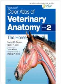
Color Atlas of Veterinary Anatomy, Volume 2, The Horse, 2e PDF
Preview Color Atlas of Veterinary Anatomy, Volume 2, The Horse, 2e
Color Atlas of Veterinary Anatomy Volume 2 The Horse Commissioning Editor: Robert Edwards Development Editor: Lynn Watt/Ailsa Laing Project Manager: Nancy Arnott Designer/Design Direction: Stewart Larking Illustration Manager: Merlyn Harvey Illustrator: Samantha Elmhurst Color Atlas of Veterinary Anatomy Volume 2 The Horse Second Edition Raymond R. Ashdown With radiographs provided by BVSc PhD MRCVS Elizabeth A. Baines Emeritus Reader in Veterinary Anatomy MA VetMB DVR DipECVDI MRCVS University of London Lecturer in Veterinary Radiology London Department of Veterinary Clinical Sciences Royal Veterinary College Stanley H. Done London BA BVetMed PhD DECPHM DECVP FRCVS FRCPath Visiting Professor of Veterinary Pathology University of Glasgow Veterinary School, Glasgow Former Lecturer in Veterinary Anatomy Royal Veterinary College London Photography by Susan A. Evans MIScT AIMI MIAS Former Chief Technician in Anatomy Department of Veterinary Basic Sciences Royal Veterinary College London Edinburgh London New York Oxford Philadelphia St Louis Sydney Toronto 2011 © 2011 Elsevier Ltd. All rights reserved. No part of this publication may be reproduced or transmitted in any form or by any means, electronic or mechanical, including photocopying, recording, or any information storage and retrieval system, without permission in writing from the publisher. Details on how to seek permission, further information about the Publisher’s permissions policies and our arrangements with organizations such as the Copyright Clearance Center and the Copyright Licensing Agency, can be found at our website: www.elsevier.com/permissions. This book and the individual contributions contained in it are protected under copyright by the Publisher (other than as may be noted herein). First edition 1987 Second edition 2011 ISBN 978-0-7234-3414-6 British Library Cataloguing in Publication Data A catalogue record for this book is available from the British Library Library of Congress Cataloging in Publication Data A catalog record for this book is available from the Library of Congress Notices Knowledge and best practice in this fi eld are constantly changing. As new research and experience broaden our understanding, changes in research methods, professional practices, or medical treatment may become necessary. Practitioners and researchers must always rely on their own experience and knowledge in evaluating and using any information, methods, compounds, or experiments described herein. In using such information or methods they should be mindful of their own safety and the safety of others, including parties for whom they have a professional responsibility. With respect to any drug or pharmaceutical products identifi ed, readers are advised to check the most current information provided (i) on procedures featured or (ii) by the manufacturer of each product to be administered, to verify the recommended dose or formula, the method and duration of administration, and contraindications. It is the responsibility of practitioners, relying on their own experience and knowledge of their patients, to make diagnoses, to determine dosages and the best treatment for each individual patient, and to take all appropriate safety precautions. To the fullest extent of the law, neither the Publisher nor the authors, contributors, or editors, assume any liability for any injury and/or damage to persons or property as a matter of products liability, negligence or otherwise, or from any use or operation of any methods, products, instructions, or ideas contained in the material herein. Working together to grow libraries in developing countries www.elsevier.com | www.bookaid.org | www.sabre.org The publisher’s policy is to use paper manufactured from sustainable forests Printed in China PREFACE This book is intended for veterinary students and practising veterinary The dissections follow the pattern of prosections that were used for surgeons. Important features of topographical anatomy are presented teaching at the Royal Veterinary College for many years. in a series of full-colour photographs of detailed dissections. The The aim of these dissections and photographs is to reveal the topog- structures are identifi ed in accompanying coloured line drawings, and raphy of the animal as it would be presented to the veterinary surgeon the nomenclature is based on that of the Nomina Anatomica Veteri- during a routine clinical examination. Therefore, lateral views pre- naria (1992). Latin terms are used for muscles, arteries, veins, lymphat- dominate and we have, as far as possible, avoided photographs of ics and nerves, but anglicized terms are used for most other structures. parts removed from the body or the use of views from unusual angles, When necessary, information needed for interpretation of the photo- or of unusual body positions. It is our earnest hope that this book will graphs is given in the captions. Each section begins with photographs enable students and veterinary surgeons to see, beneath the outer of regional surface features taken before dissection, and complemen- surface of the animals entrusted to their care, the muscles, bones, tary photographs of an articulated equine skeleton illustrate the vessels, nerves and viscera that go to make up each region of the body important palpable bony features of these regions. The dissections and and each organ system. photographs have been specially prepared for this book. A signifi cant difference between this and previous editions of the The horses used for this work were ponies of various ages and types volume is the addition of radiographs and scans which are placed in (two stallions, one gelding, three mares and several colt foals). The a new chapter at the end of the book. A second major difference is the specimens were embalmed, for the most part, in the standing position inclusion of clinical notes at the beginning of each main chapter. These using methods routinely employed in the Department of Anatomy at notes highlight the areas of anatomy which are of particular clinical the Royal Veterinary College. Every effort was made to ensure that signifi cance. Finally, over 100 self-assessment questions are available the fi nal position corresponded to that of normal level standing. In online with this new edition. four cases red neoprene latex was injected into the arteries and blue We feel that these additions to the book add considerably to its neoprene latex was also injected into the veins of the pregnant mare. usefulness, especially to the aspiring veterinary surgeon. v ACKNOWLEDGEMENTS The dissections and photography for this book were carried out at the The idea of producing an atlas of equine anatomy was based on our Royal Veterinary College, University of London. We are grateful to yearly teaching program of equine prosection. And we are very grate- the Department of Anatomy for the provision of specialized facilities, ful to the project editor, designer and illustrator for their hard work without which this work could not have been possible. In particular and for sustaining us with their optimism and enthusiasm. we would like to thank Stephen W. Barnett, BA, MIST, formerly Lizza Baines provided the radiographs for this new edition with Chief Technician in Anatomy, for advice and assistance. The task of Elsevier and we are very grateful to her for help with all aspects of the preparing and caring for the specimens before and during dissection new chapter on radiographical imaging. was undertaken by Douglas Hopkins and Andrew Crook. The photo- graphs for Figs 8.31–8.35 were taken by Alan Coombs (Department London 2011 RRA of Veterinary Anatomy, University of Bristol) and those for Figs SD 8.48–8.53 were taken by Malcolm Parsons (Department of Veterinary Surgery, University of Bristol). vi BIBLIOGRAPHY Numerous original papers have been consulted during this work, but McFadyean J (1922) The Anatomy of the Horse. A Dissection Guide. our studies have mainly been supported by a range of anatomical 3rd Edn. Edinburgh: Johnstone. textbooks. We would especially acknowledge our debt to the follow- McFadyean J (1964) Osteology and Arthrology of the Domesticated ing, which were our constant companions throughout the preparation Animals. 4th Edn. Edited by Hughes H V & Dransfi eld J W. London: of the specimens and the text. Baillière Tindall, Cox. Martin P, Schauder W (1938) Lehrbuch der Anatomie der Haustiere. Berg R (1973) Angewandete und topographische Anatomie der Haus- Bd. III. Anatomie der Hauswiederkäuer. 3rd Edn. Stuttgart: Schick- tiere. Jena: Fisher. hardt, Ebner. Bradley O C, Grahame T (1946) The Topographical Anatomy of the Nickel R, Schummer A, Seiferle E (1973) The Viscera of the Domestic Limbs of the Horse. 2nd Edn. Edinburgh: Green & Son Ltd. Animals. Translated and revised by Sack WO. Berlin, Hamburg: Bradley O C, Grahame T (1946) The Topographical Anatomy of the Paul Parey. Thorax and Abdomen of the Horse. 2nd Edn. Edinburgh: Green & Nickel R, Schummer A, Seiferle E (1975) Lehrbuch der Anatomie Son Ltd. der Haustiere Bd. IV. Nervensystem, Sinnesorgane, Endokrine Bradley O C, Grahame T (1946) The Topographical Anatomy of the Drusen. 2 Aufl age. Seiferle E & Böhme G. Berlin, Hamburg: Paul Head and Neck of the Horse. 2nd Edn. Edinburgh: Green & Son Parey. Ltd. Nickel R, Schummer A, Seiferle E (1981) The Anatomy of the Domestic Butler J, Colles C, Dyson S, Kold S, Poules P (2008) Clinical radiology Animals Vol 3. The circulatory system, the skin and the cutaneous of the horse, 3rd Edn. Chichester-Oxford: Wiley-Blackwell. organs of the domestic mammals. Schummer A, Wilkens H, Calderon W F (n.d.) Animal Painting and Anatomy. London: Seeley Vollmerhaus BK & Habermehl KH. Translated by Siller WG & Service. Wright PAL. Berlin, Hamburg: Paul Parey. Chauveau A, Arloing S, Lesbre F-X (1903) Traité d’Anatomie Comparée Nickel R, Schummer A, Seiferle E (1984) Lehrbuch der Anatomie der des Animaux Domestiques. 5e édition. Paris: Baillière-Tindall. Haustiere Bd. I Bewegungsapparat. 5 Aufl age. Frewein J, Wille K-H Dittrich H, Ellenberger W, Baum H (1907) The Horse. A Pictorial & Wilkens H. Berlin, Hamburg: Paul Parey. Guide to its Anatomy. Translated by Sisson S. London: Fisher Popesko P (1971) Atlas of topographical anatomy of the domestic Unwin. Leipsic: Dieterich. animals. Vols I–III. Translated by Getty R & Brown J. Philadelphia: Ellenberger W, Baum H (1943) Handbuch der vergleichenden Anatomie W.B. Saunders Company. der Haustiere. 18th Edn. Edited by Zeitzschmann O, Ackernecht E Sack W O, Habel R E (1977) Rooney’s Guide to the Dissection of the & Grau H. Berlin: Springer-Verlag. Horse. Ithaca: Veterinary Textbooks. Field E J, Harrison R J (1968) Anatomical Terms. Their origin and Schebitz H, Wilkens H (1978) Atlas der Röntgenanatomie des Pferdes. derivation. 3rd Edn. Cambridge: Heffer. Berlin, Hamburg: Paul Parey. Ghoshal N G, Koch T, Popesko P (1981) The Venous Drainage of the Schmaltz R (1909) Atlas der Anatomie des Pferdes. 2 Teil. Topogra- Domestic Animals. Philadelphia: W.B. Saunders Company. phische Myologie. Berlin: Schoetz. Goubaux A, Barrier G (1892) The Exterior of the Horse. Translated Schmaltz R (1924) Atlas der Anatomie des Pferdes. 1 Teil. Das Skelett and edited by Harger SJJ. Philadelphia: J.B. Lippincott Co. des Rumpfes und der Gleidmassen. 4,5 Aufl age. Berlin: Schoetz Habel R E (1973) Applied Veterinary Anatomy. Ithaca: Habel. Share-Jones J T (1907) The Surgical Anatomy of the Horse. London: Hayes M H (1903) Veterinary notes for horse owners. 6th Edn. London: Williams, Norgate. Hurst, Blackett (54 good photographs of the teeth of horses of Sisson S, Grossman J D (1953) The Anatomy of the Domestic Animals. known ages [birth to 42 years] in Ch. 33, which is not included in 4th Edn, revised Philadelphia: W.B. Saunders Company. earlier Edns). Sisson S, Grossman J D (1975) The Anatomy of the Domestic Animals. Hayes M H (1952) Points of the Horse. 6th Edn. Edited by Brooke G Vol. 1. Edited by Getty R. 5th Edn. Philadelphia: W.B. Saunders & Sanders M. London: Hurst, Blackett. Company. International Committee on Veterinary Anatomical Nomenclature, Tagand R, Barone R (1950–1957) Anatomie des Equides Domestiques. World Association of Veterinary Anatomists (1992). Nomina Ana- Lyon: École Nationale Vétèrinaire. tomica Veterinaria. 4th Edn. Gent: International Committee on Taylor J A (1955–1970) Regional and Applied Anatomy of the Domestic Veterinary Gross Anatomical Nomenclature. Animals. Parts I-III. Edinburgh: Oliver & Boyd. Kovács G (1963) The Equine Tarsus, Topographic and Radiographic Vollmerhaus B, Habermehl K H (n.d.) Topographical Anatomical Dia- Anatomy. Translated by McKay P. Budapest: Akademiai Kiadó. grams of Injection Technique in Horses, Cattle, Dogs and Cats. Lydekker R (1912) The Horse and its Relatives. London: Allen. Marburg, Lahn: Hoechst, Behringwerke A.G. vii This page intentionally left blank CONTENTS Introduction xi 6 The Hindlimb 185 1 The Head (including 7 the skin) 1 The Foot 225 2 8 The Neck 55 The Pelvis (including the spine) 269 3 The Forelimb 73 9 Diagnostic Imaging of the 4 Head, Withers, Manus and Pes 325 The Thorax 109 5 Index 345 The Abdomen 143 ix
Description: