Characterization of UmamiTs in Arabidopsis: amino acid transporters involved in amino acid ... PDF
Preview Characterization of UmamiTs in Arabidopsis: amino acid transporters involved in amino acid ...
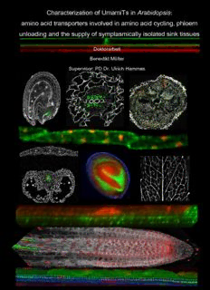
Characterization of UmamiTs in Arabidopsis: amino acid transporters involved in amino acid cycling, phloem unloading and the supply of symplasmically isolated sink tissues Doktorarbeit Benedikt Müller Supervisor: PD Dr. Ulrich Hammes Zusammenfassung Characterization of UmamiTs in Arabidopsis: amino acid transporters involved in amino acid cycling, phloem unloading and the supply of symplasmically isolated sink tissues Dissertation zur Erlangung des Doktorgrades der Naturwissenschaften (Dr. rer. nat.) der Fakultät für Biologie und Vorklinische Medizin Universität Regensburg Vorgelegt von Benedikt Müller aus Tiefenbach im April 2016 contents PhD Supervisor: PD Dr. Ulrich Hammes 1st Mentor: Prof. Dr. Thomas Dresselhaus 2nd Mentor: Prof. Dr. Ralph Hückelhoven 2 contents Das Promotionsgesuch wurde eingereicht am: Die Arbeit wurde angeleitet von: PD Dr. Ulrich Hammes Unterschrift: ________________ (Benedikt Müller) 3 contents contents 1. ZUSAMMENFASSUNG.................................................................................................... 10 2. SUMMARY ........................................................................................................................ 12 3. INTRODUCTION ............................................................................................................... 14 3.1. Human nutrition: a challenge for the future................................................................... 14 3.2. Seeds: an evolutionary hallmark for plants and human civilization ............................... 15 3.3. Transport processes in higher plants .............................................................................. 17 3.3.1. Transport between cells ..............................................................................................17 3.3.2. Transport between organs ...........................................................................................18 3.3.2.1. Long distance transport in the phloem..................................................................19 3.3.2.2. Long distance transport by the xylem...................................................................21 3.4. From nitrogen to amino acids…. a journey with breakpoints......................................... 22 3.5. Amino acid transporters in plants .................................................................................. 24 3.5.1. APC- transporter family .............................................................................................25 3.5.2. AAP- transporter family .............................................................................................25 3.5.3. Amino acid transporters from other gene families ........................................................25 3.6. Transport processes at symplasmically isolated sink tissues ........................................... 26 3.6.1. The seed: an endogenously induced symplasmically isolated sink tissue ........................27 3.6.2. The root-knot: an exogenously induced symplasmically isolated sink tissue...................30 3.7. The identification of UmamiTs ....................................................................................... 33 4. AIM OF THE PROJECT ................................................................................................... 36 5. RESULTS .......................................................................................................................... 37 5.1. Vascular anatomy in the seed ......................................................................................... 37 5.2. The seed: a symplasmically isolated domain................................................................... 40 4 contents 5.3. Distribution of secondary plasmodesmata during seed development .............................. 42 5.4. Hormonal regulation of the reproductive tissue: dynamics during seed development .... 46 5.4.1. Cytokinin response in the reproductive tissue...............................................................46 5.4.2. Auxin response in the reproductive tissue ....................................................................49 5.4.3. Auxin dynamics in the reproductive tissue ...................................................................54 5.4.4. PIN3 localization during seed development .................................................................59 5.4.5. D6 protein kinase localization during seed development ...............................................70 5.5. Expression of SCR during ovule and seed development .................................................. 72 5.6. B-type cyclin expression in ovule and seed development................................................. 74 5.6.1. CYCB1.2 ..................................................................................................................74 5.6.2. CYCB1.3 ..................................................................................................................77 5.7. UmamiTs are plasma membrane localized and expressed in various tissues ................... 78 5.7.1. Subcellular localization of UmamiTs ...........................................................................78 5.7.2. UmamiT expression in different tissues: focus on the RNA level and promoter activity .81 5.8. UmamiT promoter activity and protein localization in planta ......................................... 83 5.8.1. Leaves ......................................................................................................................83 5.8.2. Stem .........................................................................................................................87 5.8.3. Inflorescence .............................................................................................................89 5.8.4. Anthers .....................................................................................................................92 5.8.5. Replum, transmitting tract and funiculus .....................................................................95 5.8.6. SAM .........................................................................................................................98 5.8.7. Hypocotyl ............................................................................................................... 102 5.8.8. Germination ............................................................................................................ 104 5.8.9. Seed........................................................................................................................ 106 5.8.9.1. UmamiT14 localization in the seed .................................................................... 107 5.8.9.2. UmamiT11 localization in the seed .................................................................... 111 5.8.9.3. UmamiT29 localization in the seed .................................................................... 112 5.8.9.4. UmamiT28 localization in the seed .................................................................... 117 5.8.9.5. UmamiT37 localization in the seed .................................................................... 119 5.9. Characterization of the unloading zone ........................................................................ 120 5.9.1. UmamiT localization in functional transfer cells ........................................................ 120 5.9.2. Differentiation of UmamiT-positive cells is not regulated by APL .............................. 122 5.9.3. UmamiT postive cells are connected with companion cells ......................................... 124 5 contents 5.9.4. UmamiT-positive cells are connected with sieve elements .......................................... 126 5.9.5. Whole mount immunolocalization of the unloading zone: sieve elements are nucleated and physically connected to UmamiT-positive cells ................................................... 129 5.9.6. UmamiT-positive cells at the unloading zone show colocalization with PIN3............... 131 5.9.7. Auxin response in UmamiT14-positive cells during seed development ........................ 133 5.10. Root .......................................................................................................................... 135 5.10.1.1. Introduction of the marker lines in roots used in this thesis .................................. 135 5.10.1.2. Promoter activity of UmamiTs in the root........................................................... 139 5.10.1.3. Tissue specific localization of UmamiTs in the root ............................................ 145 5.10.1.4. Polar distribution of UmamiT14 in the root ........................................................ 150 5.10.1.5. UmamiTs and the protophloem paradoxon ......................................................... 152 5.10.1.6. Analysis of UmamiT-GFP overexpression in the root ......................................... 154 5.10.1.7. UmamiTs in the root: interplay with auxin ......................................................... 156 5.10.1.8. Immunolocalization of different marker lines in roots ......................................... 159 5.10.1.9. Characterization of UmamiT-positive cells in the root by immunolocalization...... 162 5.10.1.10. Validation of the sieve element identitiy of UmamiT positive cells by ...................... marker lines .................................................................................................... 167 1.1.1.3.1. UmamiT14 colocalized in cells with SE-ENOD promoter activity ....................... 168 1.1.1.3.2. UmamiT14 colocalized with APL in differentiating sieve elements ..................... 169 1.1.1.3.3. UmamiT14 colocalized with PD1 in sieve elements ............................................ 170 5.11. Analysis of the root-knot nematode feeding site by different marker lines: focus on giant cell associated tissue ....................................................................................................... 172 5.12. Expression of UmamiTs during root knot nematode infestation................................ 178 5.12.1. Characterization of UmamiT-positive cells in the root-knot by immunolocalization... 186 5.13. Characterization of knock-out plants ....................................................................... 194 5.13.1. Analysis of the level of free amino acids ................................................................... 197 5.13.2. Phenotypical characterization of roots in umamit mutants ........................................... 200 5.13.2.1. Root length in single knock-out background....................................................... 200 5.13.2.2. Cripple plants in the double knock-out background............................................. 201 5.13.2.3. Lugol staining of roots from umamit mutants ..................................................... 205 5.13.2.4. mPS-PI staining of roots from umamit mutants .................................................. 206 5.13.2.5. Whole-mount in situ RNA hybridization in the mutant background of UmamiTs 209 5.14. UmamiTs: filling the gaps......................................................................................... 213 6 contents 5.14.1. Localization of UmamiT41 in the seed and silique ..................................................... 215 5.14.2. Localization of UmamiT15 in the seed ...................................................................... 216 5.14.3. Localization of UmamiT2 in the seed ........................................................................ 217 5.14.4. Localization of UmamiT25 in the seed ...................................................................... 218 5.14.5. Localization of UmamiT21 in the seed and siliques .................................................... 219 5.14.6. Localization of UmamiT10 in the seed ...................................................................... 220 5.14.7. Localization of UmamiT34 in the seed ...................................................................... 221 5.14.8. Localization of UmamiT23 in the seed and embryo .................................................... 222 5.14.9. Localization of UmamiT1 in the seed and embryo...................................................... 224 6. DISCUSSION .................................................................................................................. 226 6.1. The seed: nutrients on a complex route. ....................................................................... 226 6.2. Hormonal dynamics during seed development ............................................................. 229 6.3. UmamiTs: the missing link in the amino acid supply of symplasmically isolated sink tissues and nitrogen cycling between xylem and phloem.......................................................... 232 6.3.1. Candidate UmamiTs are plasma membrane localized and broadly expressed in the vasculature of the above ground vegetative tissue....................................................... 232 6.3.2. UmamiTs are spatio-temporally distinct expressed in the seed and impact amino acid composition and yield .............................................................................................. 235 6.3.3. Integrating UmamiTs with hormonal dynamics and assimilate routes during seed development ............................................................................................................ 241 6.3.4. UmamiTs in the physiological gap of symplasmically isolated sink tissues .................. 243 6.3.5. UmamiTs during interaction with root-knot nematodes: supply of symplasmically isolated giant cells.................................................................................................... 245 6.3.6. UmamiTs in the root: a role in amino acid cycling ..................................................... 249 6.3.7. UmamiTs in the root: phloem-dependent impact on stem cells .................................... 252 6.4. UmamiTs as key transporters for amino acid cycling and the supply of symplasmically isolated sink tissues: a comprehensive summary .......................................................... 257 7. FUTURE ASPECTS........................................................................................................ 260 8. MATERIAL AND METHODS.......................................................................................... 262 8.1. Plants ........................................................................................................................... 262 8.1.1. Arabidopsis thaliana ................................................................................................ 262 7 contents 8.1.1.1. Growth on soil ................................................................................................. 262 8.1.1.2. Growth on plates .............................................................................................. 262 8.1.2. Lysopersicum esculentum (tomato)............................................................................ 263 8.1.2.1. Isolation of eggs from infected roots .................................................................. 263 8.1.2.2. Isolation of second-stage juveniles..................................................................... 263 8.1.2.3. Infection of Arabidopsis with Meloidogyne incognita ......................................... 264 8.2. GUS staining ................................................................................................................ 265 8.3. Sectioning of plant tissues............................................................................................. 265 8.4. Immunohistochemistry................................................................................................. 265 8.4.1. Embedding of plant tissue in methacrylate and sectioning........................................... 265 8.4.2. Immunolocalization on sections ................................................................................ 266 8.4.3. Whole mount immunolocalization............................................................................. 266 8.5. Propidium iodid staining of plant tissues ...................................................................... 266 8.6. Whole-mount in situ RNA hybridization ...................................................................... 267 8.7. mPS-PI staining............................................................................................................ 267 8.8. Seed measurement........................................................................................................ 267 8.9. Amino acid analytic...................................................................................................... 267 8.10. Statistical data processing......................................................................................... 268 8.11. Microscopy ............................................................................................................... 268 8.12. Molecular work ........................................................................................................ 268 9. LITERATURE.................................................................................................................. 269 9.1. References .................................................................................................................... 269 9.2. Publications .................................................................................................................. 278 10. APPENDIX .................................................................................................................. 279 10.1. Colocalization study ................................................................................................. 279 8 contents 10.2. Amino acid analytics................................................................................................. 280 11. DANKSAGUNG .......................................................................................................... 281 ERKLÄRUNG......................................................................................................................... 283 9
Description: