Characterization of Bioreducible Poly(B-amino Ester) Nanoparticles for siRNA Delivery PDF
Preview Characterization of Bioreducible Poly(B-amino Ester) Nanoparticles for siRNA Delivery
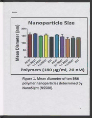
Characterization of Bioreducible Poly(B-amino Ester) Nanoparticles for siRNA Delivery by Bolivia Hurtado De Mendoza A Thesis Submitted in Partial Fulfillment of Requirements of the CSU Honors Program for Honors in the degree of Bachelors of Science in Biology College of Letters and Sciences Columbus State University Thesis —_——" ol (N, GAUL Date 05/06/2013 Committee Member * Se N > Date 05/06/2013 o ie Hows I/-A : UV; Committee Member * Xue a Hen tf — Date 05/06/2013 1% Director, Honors Proframt ac by teeny Date 05/06/2013 ABSTRACT Glioblastoma multiforme (GBM) is one of the most malignant brain tumors affecting adults. It is characterized by necrotic tissue and abnormal vasculature, making it highly resistant to current cancer treatments. A promising alternative to standard cancer therapeutics is the use of drug delivery systems such as polymeric nanoparticles (NP) that deliver silencing RNA (siRNA) exclusively to tumor cells for gene knockdown. Presently, nonviral delivery vectors, such as poly(B-amino ester) (PBAE) NP, are beneficial delivery systems because they are less immunogenic and easier to chemically modify than vectors delivered by viruses. Although current formulations of PBAEs allow cargo release via hydrolytic degradation, the ideal delivery vector should have an environmentally triggered siRNA release system to increase safety and transfection efficacy. We hypothesized that the addition of disulfides to the main backbone of PBAE NPs would reduce cytotoxicity and increase siRNA transfection efficacy. Glutathione, a reducing agent present in the cytosol, can reduce a disulfide bond into two thiols and thus increase NP degradability for safety and immediate siRNA release. In this study, we characterized the binding, physical, and siRNA delivery properties of reducible PBAE NP. Polymer-siRNA binding strength was analyzed by a gel retardation assay. NP size and zeta potential were measured using NanoSight and zetasizer instruments, respectively. siRNA transfection efficiency was assessed by the amount of green fluorescent protein detected in GBM cells using a fluorescence plate reader. This study revealed that the addition of disulfide groups to PBAEs increased their siRNA binding strength, lowered cytotoxicity, and increased siRNA transfection efficiency. The best polymer showed 76% GFP knockdown while maintaining high cell viability twenty days following transfection. This novel class of polymers can serve as improved nonviral delivery vectors for a wide array of RNA interference (RNA1) targets, including those associated with malignant tumors. INTRODUCTION Cancer is a leading cause of death worldwide and it is estimated that cancer mortality will increase to exceed 11 million individuals in 2030.'* Current treatments include invasive surgery, radiation therapy, and chemotherapy;>’ nevertheless these therapies sometimes exhibit high toxicity and can cause deleterious side effects to patients undergoing treatment.’ Chemotherapy is currently the most effective treatment for tumors in the metastatic stage.” It is a combinatorial drug therapy that targets rapidly proliferating cells by impeding certain steps in the DNA replication process.” However, a critical issue in chemotherapy is the lack of tumor selectivity and demise of healthy tissues during treatments.”’ Another challenge in chemotherapy is multidrug resistance (MDR), which decreases the efficacy of anti-cancer drugs for treatment. MDR occurs when cancer cells become resistant to a broad range of anti-cancer drugs after being exposed to one chemotherapeutic agent.’ A potential alternative to present cancer therapeutics is the use of nanoparticle-based drug delivery systems, where nanoparticles can specifically target and deliver drugs to cancer cells, while avoiding harm to normal cells. This targeting is facilitated by the enhanced permeability and retention effect (EPR), a phenomenon that allows nanoparticles to actively or passively pass through leaky tumor vasculature and aggregate at tumor sites.”’ Nanoparticles sized 50-200 nm in diameter have been known to take advantage of the EPR phenomenon.’ Nanoparticles for drug delivery are beneficial because of their scalability in production, increased intracellular stability, and adjustable tissue specificity. Currently, siRNA—based therapies present the following barriers: quick systemic clearance, low cellular uptake, and obliteration by nucleases.'” However, polymeric nanoparticles have been known to show advantageous qualities for siRNA delivery because they can be easily chemically modified and can provide protection for siRNA from serum nuclease degradation.’ Nanoparticles can be used to deliver silencing RNA (siRNA) exclusively to cancer cells to silence genes associated with tumorigenesis in a therapeutic process called RNA interference (RNAi). Once in the cytoplasm, siRNA can induce the degradation of its homologous messenger RNA target, therefore inhibiting the synthesis of a protein.’ Presently, viral delivery vectors have high transfection efficiency, because of their natural ability to efficiently insert genetic cargo into cells.” They can also easily condense nucleic acids, making their transportation to the cytoplasm easier.'' Nevertheless, they exhibit potential mutagenesis, high immunogenicity, small cargo capacity, and are difficult for chemical modification.’ Due to these immunological toxicities, no therapeutic treatments have been approved into the market.'” Alternatively, non-viral delivery vehicles display advantageous attributes, such as lower cytotoxicity, reduced immunogenicity, large cargo capacity, and feasibility in large scale production, but lack high transfection efficacy.’ Cationic polymers for siRNA delivery are most effective for gene therapy when the following factors are accomplished effectively. Polymer-siRNA binding must be strong to prevent dissociation or degradation of the polymer/siRNA complex. Binding is possible through the electrostatic attraction between cationically charged amine groups on a polymer and anionic siRNA. The cationic charge on a nanoparticle is also important for endocytotic cellular uptake because it should be cationic enough to become attracted to the negatively charged proteoglycans found on the phospholipid membrane surface. After endocytosis, the nanoparticle needs to successfully escape the endosome to avoid becoming degraded by the acidifying environment of a lysosome. A major cause of low transfection efficacy has been known to be the inefficient escape of the polymer/siRNA complex from the endosome.'® Therefore, the secondary and tertiary amines on the polymer structure serve as a buffering system that induces osmotic swelling and endosomal lysis, this process is known as the proton sponge effect. It allows the polymer-siRNA complex to make its way into the cytoplasm safely. Now to release the siRNA from the polymer/siRNA complex, the polymer’s acrylate bonds are cleaved by water molecules in the cytoplasm and the polymer degrades to reduce the possibility of cytotoxicity.’ Linear cationic polymers have the ability to bind and condense nucleic acids, such as siRNA into stable nanoparticles.'® Biodegradable cationic poly(B-amino esters) (PBAEs) nanoparticles are attractive non-viral delivery vectors because of several characteristics. They have an efficient buffering system that allows endosomal escape, an ability to bind siRNA into a stable nanoparticle/siRNA complex, increased transfection efficiency, reduced cytotoxicity, and improved biodegradability.” PBAEs are formed through a Michael addition of diacrylates to primary or bis-secondary amines.® Small modifications to a polymer’s chemical structure with varying end groups can cause large changes to its size and so they are chemically modified." We hypothesized that the addition of disulfides to the main backbone of PBAE NPs would reduce cytotoxicity and increase siRNA transfection efficacy. Glutathione found in high concentrations of about 1-8mM in the cell cytoplasm versus 5-50uM in blood serum can serve as a reducing agent to reduce a disulfide bond into two thiols." This can degrade the nanoparticle allowing the siRNA to dissociate and induce the obliteration of its homologous mRNA. We seek to discover the best siRNA/polymer formulation that achieves the highest siRNA transfection efficacy and lowest cytotoxicity in Glioblastoma 319 cells. This study aims to characterize the binding, physical, and siRNA delivery properties of bioreducible poly (B-amino ester) nanoparticles with disulfide linkages. MATERIALS AND METHODS Polymers Small aliquots of ten polymers at 100 mg/mL in dimethyl sulfoxide (DMSO) plus dessicant were stored at 4°C. Glioblastoma cell cultures Glioblastoma (GB) astrocyte cells were cultured in Dulbecco’s Modified Eagle Medium (DMEM 11995, Invitrogen, Carlsbad CA) with 10% Fetal Bovine Serum (FBS) and 1% anti-anti. Cells were kept in an incubator sustaining a humid 5% CO2 atmosphere at 37°C. Cells had their media removed and resupplied with fresh media every Monday, Wednesday, and Friday for nine weeks. Nanoparticle Sizing and Zeta Potential Analysis A NanoSight NS500 instrument (NanoSight Ltd. Wiltshire, UK) was used to size nanoparticles. A triplicate of each polymer sample was performed. Both polymer and scRNA concentrations were diluted in sodium acetate. Additionally, each scRNA/polymer solution was diluted 1:100 in phosphate buffered saline (PBS). A Zetasizer Nanoseries Nano-ZS90 was used to measure nanoparticle zeta potential. Both polymer and scRNA were diluted in sodium acetate buffer. Each diluted solution was mixed and left to incubate for 10 minutes for particle formation. Then 200 pl of particle/scRNA solution was diluted in 800 ul of PBS. After, a diluted particle solution was injected into a zeta cell and placed in the Zetasizer apparatus for analysis. Polymer-siRNA binding study: Gel Retardation Assay Polymer concentrations ranging from 0-600 w/w were diluted in different concentrations of sodium acetate buffer. Then a constant concentration of scrambled RNA (scRNA) was diluted in a constant concentration of sodium acetate. After, the diluted polymer solution was mixed with the diluted scRNA solution and left to incubate for ten minutes for particle formation. The loading buffer was 30% water diluted glycerol with no dye. This loading buffer was added to the polymer/scRNA particles in the first row of wells. To prevent any interference with polymer/scRNA binding, dyes were excluded. To observe the reducing action of glutathione on a particle disulfide bond, SmM glutathione was added to the polymer/scRNA particles in the second row of wells. Particle samples from both wells were loaded into a 1% agarose gel with ethidium bromide staining. Gel ran for 20 minutes at 100 V and was exposed to UV to see the bands. siRNA Transfection and Cell Viability Assay Cells were plated in 96-well tissue culture treated plates at a concentration of 15,000 cells per well. DMEM was added to all wells and plates were placed in the incubator at 37°C overnight. Lipofectamine 2000 (Invitrogen) was mixed with Optimem I (Invitrogen) following specific directions from the manufacturer’s protocol. Lipofectamine served as a positive control for the transfection experiment. Then siRNA and scRNA were both diluted in sodium acetate and pipetted into a 96-well round bottom plate. Polymers were also diluted in sodium acetate and aliquoted out into the same 96-well round bottom plate. Polymer solutions were then mixed with the scRNA solutions on one row and siRNA solutions on another row. Particle solutions incubated for ten minutes for particle formation and then were added to GB cells. After transfection, plates were placed in incubator for four hours, followed by a removal of media and a resupply of fresh DMEM. Cell viability was examined by the CellTiter 96 AQeous One MTS assay (Promega). Media was removed from cells and cell titer reagent was added to transfected cells and incubated for one hour. Cells with only media and no nanoparticle/siRNA/scRNA solution (control) were normalized. GFP knockdown in transfected cells was monitored for 20 days by a measuring absorbance of each well using a BioTek Synergy 2 plate reader. Statistical Treatment of Data GraphPad Prism 5 software was used for all statistical analysis. Results in line and bar graphs indicate +/- standard error of mean. Results Nanoparticle Size ) m : n ( r 3 e t e m 2 a i D 50 - n a 0- e M A AA A A A 2A dd aA CSHE Echt cht E ct Tct IcTht Tcht ICheEt SChet Ch He Me NS We Ms Polymers (180 g/m, 20 iM] ATR A Faure 1. Mean Alamiciep oftt en 2 BR6 polymer nanoparticles determined by NanoSight (NS500).
