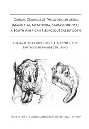
Caudal cranium of Thylacosmilus atrox (Mammalia, Metatheria, Sparassodonta), a South American predaceous sabertooth PDF
Preview Caudal cranium of Thylacosmilus atrox (Mammalia, Metatheria, Sparassodonta), a South American predaceous sabertooth
CAUDAL CRANIUM OF THYLACOSMILUS ATROX (MAMMALIA, METATHERIA, SPARASSODONTA), A SOUTH AMERICAN PREDACEOUS SABERTOOTH AN ALIA M. FORASIEPI, ROSS D. E. MACPHEE, AND SANTIAGO HERNANDEZ DEL PINO BULLETIN OF THE AMERICAN MUSEUM OF NATURAL HISTORY CAUDAL CRANIUM OF THYLACOSMILUS ATROX (MAMMALIA, METATHERIA, SPARASSODONTA), A SOUTH AMERICAN PREDACEOUS SABERTOOTH anai.Ta m. forasiepi IANIGLA, CCT-CONICET, Mendoza, Argentina ROSS D.E. MacPHEE Department of Mammalogy, American Museum of Natural History, New York SANTIAGO HERNANDEZ DEL PINO IANIGLA, CCT-CONICET, Mendoza, Argentina BULLETIN OF THE AMERICAN MUSEUM OF NATURAL HISTORY Number 433, 64 pp., 27 figures, 1 table Issued June 14, 2019 Copyright © American Museum of Natural History 2019 ISSN 0003-0090 CONTENTS Abstract.3 Introduction.3 Materials and Methods.7 Specimens.7 CT Scanning and Bone Histology.8 Institutional and Other Abbreviations.12 Comparative Osteology of the Caudal Cranium of Thylacosmilus and Other Sparassodonts .. .12 Tympanic Floor and Basicranial Composition in Thylacosmilus.12 Tympanic Floor Composition in the Comparative Set.19 Petrosal Morphology.26 Tympanic Roof Composition and Middle Ear Pneumatization in Thylacosmilus and Comparative Set.36 Features Related to Nerves and Blood Vessels in Thylacosmilus and Comparative Set.38 Discussion.48 Major Osteological Features.48 Major Vascular Features.49 Cerebrospinal Venous System in Thylacosmilus and Other Sparassodonts.49 Carotid Foramen Position in Marsupials and Placentals.52 Acknowledgements.55 References.55 Appendix 1: Glossary.62 2 ABSTRACT The caudal cranium of the South American sabertooth Thylacosmilus atrox (Thylacosmilidae, Sparassodonta, Metatheria) is described in detail, with emphasis on the constitution of the walls of the middle ear, cranial vasculature, and major nerve pathways. With the aid of micro-CT scanning of the holotype and paratype, we have established that five cranial elements (squamosal, alisphenoid, exoccipital, petrosal, and ectotympanic) and their various outgrowths participate in the tympanic floor and roof of this species. Thylacosmilus possessed a U-shaped ectotympanic that was evidently situated on the medial margin of the external acoustic meatus. The bulla itself is exclusively com¬ posed of the tympanic process of the exoccipital and rostral and caudal tympanic processes of the squamosal. Contrary to previous reports, neither the alisphenoid nor the petrosal participate in the actual tympanic floor, although they do contribute to the roof. In these regards Thylacosmilus is distinctly different from other borhyaenoids, in which the tympanic floor was largely membranous (e.g., Borhyaena) and lacked an enlarged ectotympanic (e.g., Paraborhyaena). In some respects Thy¬ lacosmilus is more similar to hathliacynids than to borhyaenoids, in that the former also possessed large caudal outgrowths of the squamosal and exoccipital that were clearly tympanic processes rather than simply attachment sites for muscles. However, hathliacynids also exhibited a large alisphenoid tympanic process, a floor component that is absent in Thylacosmilus. Habitual head posture was inferred on the basis of inner ear features. Large paratympanic spaces invade all of the elements participating in bounding the middle ear, another distinctive difference of Thylacosmilus compared to other sparassodonts. Arterial and venous vascular organization is relatively conservative in this species, although some vascular trackways could not have been securely identified without the avail¬ ability of CT scanning. The anatomical correlates of the internal carotid in relation to other basicra¬ nial structures, the absence of a functional arteria diploetica magna, and the network for venous return from the endocranium agree with conditions in other sparassodonts. INTRODUCTION sidered to have the most primitive morphology found in the group (e.g., Simpson, 1948; Mar¬ Sparassodonta is a wholly extinct radiation of shall, 1981; Goin et al., 1986; Goin and Candela, predaceous metatherians thought to be endemic 2004). Expressing this point in the form of a defi¬ to South America. Fossils of metatherians with nition by ancestry, Sparassodonta is the group hypercarnivorous dentitions have been found on that includes the common ancestor of Patene and other continents, but in general they are too frag¬ all its descendents (e.g., see Forasiepi, 2009; mentary to provide any certainty regarding their Engelman and Croft, 2014; Forasiepi et al., 2015; likely affinities (Beck, 2015), or are controversial Suarez et al, 2016; Muizon et al., 2018). for other reasons (Carneiro, 2018; see discus¬ Although there is broad agreement regarding sions in Muizon et al., 2018). Sparassodonta is the overall taxonomic composition of Sparasso¬ generally regarded as a monophyletic clade of donta, there are also persistent areas of contro¬ stem marsupials (e.g., Szalay, 1982; Marshall and versy. The earliest taxa for which sparassodont Kielan-Jaworowska, 1992; Rougier et al., 1998; affinities have been claimed include the Early Babot, 2005; Forasiepi, 2009; Engelman and Paleocene Tiupampan species Mayulestes ferox Croft, 2014; Forasiepi et al., 2015; Suarez et al., and Allqokirus australis (e.g., Muizon, 1994, 2016; Muizon et al., 2018). Nearly 60 different 1998; Muizon et al, 1997, 2018). In a recent phy¬ species of sparassodonts have been recognized, logenetic analysis presented by Muizon et al. the majority of Neogene age (fig. 1). Patene, a (2018), Mayulestes, Allqokirus, and Patene are taxon represented in Eocene deposits in Argen¬ shown as forming a monophyletic group at the tina, Brazil and Peru, has been traditionally con¬ base of Sparassodonta. However, at least in the 3 4 BULLETIN AMERICAN MUSEUM OF NATURAL HISTORY NO. 433 case of Mayulestes, alternative phylogenetic posi¬ Ma Epochs SALMAs Sparassodonta diversity 0— HOLOC. Bon.+ Luj.+Platan tions also have been proposed (Rougier et al., PLEIST. Ensenadan 1998; Goin, 2003; Babot, 2005; Forasiepi, 2009; Marplatan Qz Engelman and Croft, 2014; Forasiepi et al., 2015; Chapadmalalan ]LU -O Montehermosan Suarez et al, 2016; Wilson et al., 2016). Undisputed records of Sparassodonta, includ¬ Huayquerian ing ones for Patene simpsoni, begin in the Early IQ- Chasicoan Eocene (Itaboraian) and extend through to the Mayoan Pliocene (Chapadmalalan), when the last of them Laventan disappeared (Babot and Forasiepi, 2016; Prevosti M 15— Colloncuran and Forasiepi, 2018) (fig. 1). Forasiepi (2009) divided non-Tiupampan taxa into two principal Santacrucian XPjnturah) ' groups, Hathliacynidae and Borhyaenoidea, the 20- Colhuehuapian latter comprising Borhyaenidae, Thylacosmilidae, and (possibly) Proborhyaenidae (see Babot, 2005; Forasiepi et al, 2015; Babot and Forasiepi, 2016). 25 This arrangement agrees with earlier treatments, such as those of Marshall (1978,1981; Marshall et Deseadan al., 1990). However, Proborhyaenidae did not emerge as a natural group in all studies (Babot, 30- 2005; Forasiepi et al., 2015). Tinguirirican Most sparassodonts can be viewed as special¬ ized hypercarnivores (Wroe et al., 2004; Prevosti 35— et al., 2013; Zimicz, 2014, Lopez-Aguirre et al., Mustersan 2016; Croft et al, 2018), with only 10% of species 40 qualifying as omnivores or mesocarnivores uti¬ 2 Barrancan lizing the criteria of Prevosti and Forasiepi M (2018). Perhaps the most famous of all sparas¬ 45— Vacan sodonts is the so-called “marsupial sabertooth,” Thylacosmilus atrox (Riggs, 1933, 1934; figs. 2-5). <- Riochican 50 FIG. 1. Diversity of sparassodonts through time. Itaboraian Lines representing the diversity curve based on number of species (black blocks) as predicted by the fossil record (Lazarus taxa not included). Pos¬ 55- sible fossil record biases include hiatuses (time spans lacking known fossils), different taxon dura¬ tions, and averaging of biochronological units. 60- M Other biases which may mask the real diversity pattern include inadequate or imprecise chrono¬ Peligran logical ages for associations determined by radio- 65 Tiupampian metric dating, as well as differences in sampling effort across temporal units or geographical regions (Prevosti and Forasiepi, 2018). Dot cluster indicates the temporal span of Thylacosmilus atrox from the Late Miocene (Huayquerian) to Pliocene (Chapad¬ malalan). Scale: 1:1 species. 2019 FORASIEPI ET AL.: CAUDAL CRANIUM OF SPARASSODONTA 5 FIG. 2. Artistic reconstruction of Thylacosmilus atrox by Jorge Blanco. Its hypertrophied upper canines and correlated detailed descriptions of the paratype (FMNH cranial modifications have inevitably invited P14344; figs. 4, 9, 10). They identified several otic comparisons to Smilodon and other sabertooth features of Thylacosmilus that, if correctly inter¬ placentals —a classic example of convergent evo¬ preted, would not only be novel for sparassodonts lution (e.g., Riggs, 1933, 1934; Simpson, 1971; but unique among metatherians. These identifica¬ Marshall, 1976a, 1977a; Turnbull, 1978; Turnbull tions are given special attention in this paper. and Segall, 1984; Churcher, 1985). Thanks to the availability of pCT scanning, we But the dental apparatus is not the only region have been able to reevaluate Turnbull and Segalls of striking morphological specialization seen in (1984) descriptions, which by necessity had to be sabertooth skulls: the auditory regions of Smilo¬ largely based on surface anatomy. However, unex¬ don and Thylacosmilus are likewise remarkably pected difficulties in identifying certain features modified compared with those of their close rela¬ required examination of other fossil evidence for tives (Turnbull and Segall, 1984). In his initial morphological variation in the sparassodont cau¬ description of Thylacosmilus, Riggs (1934) pro¬ dal cranium. This was required in any case because vided a brief treatment of its cranial anatomy, the literature on metatherian cranial morphology based mainly on the holotype (FMNH P14531; is limited compared with that available for euthe- figs. 3, 6-8). Turnbull and Segall (1984) reevalu¬ rians. Although metatherians and eutherians are ated and extended Riggs’ interpretations, adding phylogenetic sisters, these two clades have fol- 6 BULLETIN AMERICAN MUSEUM OF NATURAL HISTORY NO. 433 A B FIG. 3. Thylacosmilus atrox, holotype, FMNH P14531 (Hualfin, Catamarca, Argentina; Corral Quemado For¬ mation; Chapadmalalan, mid-Pliocene). Skull in lateral (A) and dorsal (B) views. Abbreviations: cod, occipital condyle; earn, external acoustic meatus; FR, frontal; MX, maxilla; NA, nasal; nc, nuchal crest; PA, parietal; rt, rami temporales; s, sagittal crest; SQ, squamosal; tl, temporal line. 2019 FORASIEPI ET AL.: CAUDAL CRANIUM OF SPARASSODONTA 7 lowed different evolutionary (and morphological) ing such wide ranges in size, even in fossils of simi¬ trajectories for the past -130 Ma (O’Leary et al., lar ontogenetic age. In fact, even though less 2013). As a result, conditions in fossil metatheri- pronounced than in Thylacosmilus, substantial ans need to be interpreted on their own terms, morphological variability has been demostrated for and not merely viewed as special instances of what other sparassodonts (e.g., in Cladosictis patagonica, can be observed in eutherians. expressed as differences in mandibular size and shape; Echarri et al., 2017), just as it has in extant mammals generally (e.g., Lindenfors et al., 2007). MATERIALS AND METHODS It is important to note that Turnbull and Segall (1984) made modifications to the holotype and Specimens paratype in the course of their investigation. First, Thylacosmilus: The specimens principally the apparent outlines of several sutures were utilized for this study are the holotype (FMNH drawn with indelible ink on both specimens. P14531; figs. 3, 6-8), which is in good condition Although many of these tracings correspond fairly overall, and the paratype (FMNH P14344; figs. 4, well to actual boundaries between bone territories 9, 10), which has suffered considerable erosional as seen in the scans, in some cases they do not, or damage but retains a reasonably intact caudal the situation is ambiguous. For a given suture to cranium. Additional details on basicranial con¬ be named as such in this paper there must be struction were retrieved from T. atrox MMP secure evidence of it, either macroscopically or in 1443-M (figs. 5, 11). Other specimens reviewed the segmental data. This issue principally affects in this paper are listed in table 1. evaluation of bullar composition. Although both Goin and Pascual (1987) have shown that the specimens can be described as reasonably well numerous Late Miocene/Pliocene thylacosmilid preserved, bullar surfaces display hairline breaks species named in the 20th century cannot be mean¬ that cannot be easily distinguished from sutures. ingfully distinguished from one another and should In a second modification of the fossil material, therefore be regarded as synonyms of Thylacosmilus Turnbull and Segall (1984) removed most of the atrox, the earliest valid name apart from an unused right bulla of the paratype in order to better senior synonym (Achlysictis lelongi, now sup¬ explore middle ear anatomy. Fortunately, the pressed; see Goin and Pascual, 1987) coined by detached bulla was kept with the rest of the skull, Ameghino (1891). The amount of individual varia¬ permitting us to provide some additional inter- tion within T. atrox as currently defined is certainly pretrations of its morphology. substantial (e.g., Thylacosmilus FMNH P14531, Other Metatheria: The comparative set of P14344, and MMP 1443-M; figs. 3-5). Because of fossil and extant specimens utilized in this paper their notable difference in size, the holotype and is listed in table 1. Certain features whose rela¬ paratype were thought by Riggs (1934: 9) to be of tions or ontogeny are uncertain were traced in greatly different ontogenetic ages. He described the serially sectioned pouch young of Monodelphis paratype as “a young adult specimen,” basing his domestica (head length, 8.5 mm; postnatal day assessment on the fact that “the mandible is nearly 12; DUCEC), Perameles sp. (head length, 17.5 one fourth smaller in linear dimensions than that mm; ZIUT), and Macropus eugenii (head length, of the holotype.” However, we found that the denti¬ 29 mm; ZIUT). For additional information on tions were notably worn in both specimens, indi¬ these specimens, see Sanchez-Villagra and cating that any difference in their ontogenetic ages Forasiepi (2017). Other data were obtained from could not have been very considerable. It is possible the literature. that sexual dimorphism, perhaps in combination Anatomical nomenclature: The nomen¬ with or amplified by individual variation (Goin and clature used in the descriptions largely follows Pascual, 1987), played an important role in produc¬ MacPhee (1981) and Wible (2003); a few new BULLETIN AMERICAN MUSEUM OF NATURAL HISTORY NO. 433 4 cm FIG. 4. Thylacosmilus atrox, paratype, FMNH P14344 (Rio Santa Maria, Chiquimil, Catamarca, Argentina; Andalgala Formation; Huayquerian, Late Miocene). Skull in A, dorsal and B, ventral views. Abbreviations: AL, alisphenoid; BO, basioccipital; C, upper canine; cc, carotid canal; cod, occipital condyle; cps, caudal paratympanic space; ctpsq, caudal tympanic process of squamosal; earn, external acoustic meatus; EX, exoc- cipital; fo, foramen ovale; for, foramen rotundum; FR, frontal; hfr, rostral hypoglossal foramen; ips, inferior petrosal sinus; lps, lateral paratympanic space (= epitympanic sinus); MX, maxilla; nc, nuchal crest; PA, parietal; rt, rami temporales; s, sagittal crest; sjf, secondary jugular foramen; sof, sphenoorbital fissure; SQ, squamosal; tl, temporal line; tpex, tympanic process of exoccipital. Arrow indicates direction to or position of features hidden by other structures. terms are introduced and defined where they slice thickness of 0.625 mm at 120 kV and 200 first occur. For all other definitions of anatomical mA. This scan resulted in a final stack of 433 terms, see appendix 1. slices of 307 x 471 pxl. Thylacosmilus atrox (paratype, FMNH P14344). Scanned on a GE Phoenix vtomex s240 CT Scanning and Bone Histology pCT scanner in the laboratory of Zhe-Xi Luo Thylacosmilus atrox (holotype, FMNH (University of Chicago) with a slice thickness of PI4531). Originally scanned by Larry Witmer 0.07 mm at 210 kV, 190 mA. The scan resulted in (Ohio University) at O’Bleness Memorial Hos¬ a final stack of 2022 slices of 1493 x 1180 pxl. pital in Athens, Ohio, using a General Electric Sipalocyon gracilis (AMNH VP 9254). Scanned Light Speed Ultra MultiSlice CT scanner with a on a GE Phoenix vtomex s240 pCT scanner 2019 FORASIEPI ET AL.: CAUDAL CRANIUM OF SPARASSODONTA 9 FIG. 5. Thylacosmilus atrox MMP 1443-M (Chapadmalal, Buenos Aires, Argentina; Chapadmalal Formation; Chapadmalalan, mid-Pliocene). Skull in A, lateral and B, dorsal views. Abbreviations: cod, occipital condyle; earn, external acoustic meatus; FR, frontal; MX, maxilla; NA, nasal; nc, nuchal crest; PA, parietal; rt, ramus temporalis; s, sagittal crest; SQ, squamosal; tl, temporal line.
