Calciphylaxis - a challenging & solvable task for plastic surgery? A case report. PDF
Preview Calciphylaxis - a challenging & solvable task for plastic surgery? A case report.
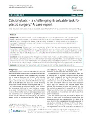
Tsolakidisetal.BMCDermatology2013,13:1 http://www.biomedcentral.com/1471-5945/13/1 CASE REPORT Open Access – Calciphylaxis a challenging & solvable task for plastic surgery? A case report Savas Tsolakidis*, Gerrit Grieb, Andrzej Piatkowski, Ziyad Alharbi, Erhan Demir, David Simons and Norbert Pallua Abstract Background: Calciphylaxis (calcific uremic arteriolopathy) is rareand its pathogenesis is notfully understood. Indeed, Calciphylaxis presents a challenge through the course of itsmanagement which involve different specialities but unfortunately this disease so farhas a poor prognosis. We herein present, inthis case report, a multidisciplinary approach involving plastic surgeons withspecial regards to reconstructive approach after debridement procedures. Case presentation: We present a 21 years old malewith a BMI of 38,2, who was transferred to our department from anotherhospital. Calciphylaxis has been diagnosed after receiving anticoagulation withphenprocoumon after a single event of pulmonary embolism. The INR on admission was 1,79. Hehad necrotic spots on both sides of the abdominal wall and on both thighs medially. During this time heunderwent several reconstructive procedures in our department. Conclusion: It can be suggested that this agonizing disease needs indeed a multidisciplinary approach involving Nephrologists, Dermatologists, Intensive Care Physiciansand Plastic Surgeons,taking into consideration that surgical correction can achieve further improvementina specialized centre. Notwithstanding, further cohortstudies should be approached clinically to insight thelight on this disease withspecial regard to theprognosis after this approach. Keywords: Calciphylaxis, Phenprocoumon, Natriumthiosulphate, Skin Grafting, Buried Chip skin grafting Background The differential diagnosis is challenging and includes Calciphylaxis (calcific uremic arteriolopathy) is a rare and vasculitis,cholesterol emboli anddiabetic ulcers. poorlyunderstooddiseasewhichdemonstratesachallenge Calciphylaxis can bestaged into two phases. Phase one for different specialities. Media calcifications of arterioles is characterized by pruritus, cutaneous laminar erythe- and fat tissue are distinctive and primarily affect the skin mas, which isunspecific and therefore can easily be mis- but in some cases involvement of nerve sheaths, visceral interpreted in the first place. Phase two shows painful organs and muscles have been described. Even if diag- ulceration and necrosis [2]. Preferably affected areas are nosed inanearly phase, the chances ofsuccess inhealing the lower extremities. There are some cases described areverylowandthemortalityisexceptionallyhigh[1]. where also the abdomen and visceral organs were It is characterized by extremely painful, partly necrotic affected. The mortality in phase one is about 30%, but skin ulcerations, which can be found almost on any part the mortality in phase two is extended up to 80% [3]. of the body. Hallmark of this disease is a fortified calcifi- These typical ulcerations and necrosis were predomin- cationofthearteriolesmainlyinthesubcutaneoustissue antly presented on the abdomen and both thighs of the and sometimes even calcification of peripheral nerve patientwe treated. sheathsandfatcellbodiescanbefound[1].Theprimary This rare disease was first described by Bryant and clinical picture is multifaceted and can delay diagnosis. White in 1898, where a condition of calcifications and cutaneous necrosis was circumscribed [4]. Selye et al. were the first, who highlightened the pathophysiology of calciphylaxis, labelled the disease in 1962 and charac- *Correspondence:s.tsolakidis@me.com terised it as a systemic hypersensitivity reaction. In ani- DepartmentofPlasticSurgeryandHandSurgery,BurnCenter,Medical Faculty,RWTHAachenUniversity,Pauwelsstrasse30,52074Aachen,Germany mal experiments, Selye et al. could show an induction of ©2013Tsolakidisetal.;licenseeBioMedCentralLtd.ThisisanOpenAccessarticledistributedunderthetermsoftheCreative CommonsAttributionLicense(http://creativecommons.org/licenses/by/2.0),whichpermitsunrestricteduse,distribution,and reproductioninanymedium,providedtheoriginalworkisproperlycited. Tsolakidisetal.BMCDermatology2013,13:1 Page2of4 http://www.biomedcentral.com/1471-5945/13/1 calcification of various organs via the combination of defined edges in the upper and lower thighs and the ab- sensitizing agents, like Dihydrotachysterol, Vitamin D2, domen, where some showed signs of necrosis. Further D3 and parathyroid hormone followed by exposure to a histological examinations showed typical signs of micro- challenger, as there are for example metallic salts (iron, thrombosis and calcification of the arterioles subcutane- aluminium)or trauma [5-7]. Ketteler et al. referred the ouslyandsubstantiated thediagnosis ofcalciphylaxis. condition can lead to an imbalance of two calcification The patient needed high doses of opiates over the inhibiting proteins; Matrix-Gla and Fetuin A, which are whole duration of inpatient treatment. Unfortunately the Vitamin Kdependant[8].Inanimalexperimentsitcould initial calcifications became necrotic and possible surgi- be shown that an essential lack of these proteins can caloptions wereevaluated. lead to calcification of arteries and even rupturing of the A combination of Natriumthiosulphate was adminis- aorta [9,10]. tered intravenously, which is a common therapy for Even the risk factors seem to be controversial. Predis- calciphylaxis and a specific surgical approach to this posing factors are an acute or chronic renal failure, complex disease was initiated when the patient arrived obesity, diabetes, hyperparathyroidism, female Cauca- at our department. The patient had necrosis on the sians and an elevated calcium phosphorus product. abdomen and both thighs as well as partially superin- Further presumably risk factors are phenprocoumon, fected wounds with oxacillin resistant staphylococcus vitamin K deficiency (phytonadione), lack of fetuin A aureus (Figure 1). A bone scintigraphy revealed no protein, malignancy, alcoholic liver disease, connective increased activity of calcium carbonate in thighs or the tissue disease, protein C and S deficiency [11]. Interest- abdominal area. ingly the majority of these patients had normal serum After the patient consented to a surgical treatment we calcium, normal serum phosphorus, normal calcium started our surgical approach with extensive debride- phosphorus product, normal serum parathyroid hor- ment of the affected areas on the abdominal wall and mone levels and normal serum creatinine levels. The both thighs (Figure 2). A consecutive VAC therapy for mortality rate in this study was 52% with sepsis being the lesions on the thighs was mandatory to establish a theleading cause [12]. clean environment. At this level, attempt to close the Although the risk remains to cause another compli- cated ulcer, a histological sample excision also including subcutaneous tissue is mandatory. The sample has to be stained with alizarin red or silver-nitrate to differentiate Calciphylaxis from different diseases[11]. Fine et al. suggested bone scintigraphy as one of the first steps to substantiate the diagnosis. In their study, only one of the investigated cases had negative bone scan [13] .The mortality in phase one was 33%. After de- velopmentofulcers themortality reached 80%[13,14]. In this case report, we highlight a multidisciplinary treatment of a patient, suffering from Calciphylaxis, which after10monthsledtoa successfulresult. Casepresentation We report on an obese 21 years old male with a Body Mass Index (BMI) of 38,2, who presented to our depart- ment with Calciphylaxis after phenprocoumon intake in consequence of pulmonary embolism. The target partial thrombin time (pTT) was between 50-60s. In addition the patient lost overtime 100 kg body mass after getting a gastric banding in 2007. Comorbidities were a dilative cardiomyopathy, insulin dependant diabetes, obesity, allergic asthma, hypothyreoidism and lack of anithrom- binefactor III(AT3). Calciphylaxis was already diagnosed histologically in the former hospital, where he was treated with Figure1Showsthepatient'swoundsonboththighsandthe Natriumthiosulphate intravenously for about 9 weeks. abdominalwallonadmissioninourdepartment. Initial symptoms were tender and painful lesions with Tsolakidisetal.BMCDermatology2013,13:1 Page3of4 http://www.biomedcentral.com/1471-5945/13/1 patient into rehabilitation, where his status was further improved by mobilization, physiotherapy and lymphatic drainagemassages. Conclusion Calciphylaxis still remain a challenge and need further investigation to understand this disease although differ- ent causes and numerous therapeutic strategies have beendescribed.Afurtherchallengeinthe clinicalsetting is that the therapy for Calciphylaxis is not clearly defined. Independently of the causing agent (factor), the present therapeutic strategy enfolds pain control, sur- gery, calcium binding agents, phosphorus reducer, corti- sone, andantibiotic therapy. Regarding our patient, we treated him with a combin- Figure2Showsexemplarythewoundsonthethighafter ation of surgical approach with intravenous application initialdebridementsinourdepartment. of binding agents which have led to a successful out- come.However,it shouldbestatedthatlocalflaptechni- ques showed no benefit but merely buried skin grafting wounds on the abdomen via advancement flaps were was finally crowned with success. Therefore an interdis- unsuccessful. ciplinary approach to this disease via supporting drug A main problem to deal with was not only a perman- therapy and surgical measures may be a promising ent level of extensive pain which was encountered way to treat patients suffering from Calciphylaxis. The through our pain specialists in concordance with the patient was then discharged into rehabilitation centre WHO – pain schedule with opiates but also the recur- without further wound problems in his soft tissues rent wound infections with complicated germs, which and until recently showed no signs of a relapse of Calci- were treated with different antibiotic patterns. Over time phylaxis(Figure 3). the Calciphylaxis therapy was adjusted and complemented Notwithstanding, despite the fact that this surgical withPamidronat,abiphosphonateagenttoreducedecom- approach may lead to a satisfactory result in our patient position of osseous structures and Renagel (Sevelamer), a further studieswitha larger patient cohortwill be needed phosphatebinder. todetermineitsefficiencyforthesepatients. Initial skin grafts did not show satisfactory results in the beginning of the surgical treatment; hence another Consent attempt with skin grafts in buried chips technique was Written informed consent wasobtained from thepatient preceded 6monthsafteradmission. for publication of this case presentation and any accom- From the plastic surgical point of view, the complexity panying images. of the wounds demanded a combination of consecutive debridements, jet lavages, repetitive VAC therapies and Buried chip skin grafting (both thighs and abdomen). In addition dressings were changed with an ointment pad andsilvercoatingdressing(AtraumanAg,PaulHartmann Ges.m.b.H,WienerNeudorf–Austria)as wellasantisep- ticsolution(Polyhexanid,Lohmann&RauscherGmbH& Co. KG, Neuwied – Germany) on a daily basis. The pa- tient could be sufficiently mobilized and had no further signs of infection. Daily sea salt baths and physiotherapy sessionsimproved the patient’s status until hewas ableto walkaloneontheward. Finally, pain medication could be reduced drastically and sedoanalgesia was not further necessary for dressing changes. The laboratory on discharge showed a single slight elevated phosphate of 1.88 (0.84-1.48 mmol/l). Cal- cium was in normal range (2.10-2.60 mmol/l). Creatinine Figure3Showstheclinicalsettingaftersuccessfulskin graftingprocedures.Thewoundswereclosed. was 1.5 (0.7-1.2). We could successfully discharge the Tsolakidisetal.BMCDermatology2013,13:1 Page4of4 http://www.biomedcentral.com/1471-5945/13/1 Competinginterests Theauthorshavedeclaredthatnoconflictofinterestexists. Authors’contributions STdraftedthemanuscriptandparticipatedintheanalysis.GG,AP,ZAand DSconceivedthedesignandparticipatedinthetreatmentandcontributed intherevisionprocess.EDtreatedthePatientintheintensivecareunit.NP participatedintheoperationsaswellasrevisedandeditedthemanuscript. Allauthorsreadandapprovedthefinalmanuscript. Acknowledgement TheauthorswouldliketothankMr.WernerKriegelforthegreatworkin photoselectionfortheselectedpatient. Received:2August2012Accepted:7January2013 Published:14January2013 References 1. KettelerM:Casereport.NEnglJMed2001,345:1119–24. 2. FineA,FlemingS,LeslieW:Calciphylaxispresentingwithcalfpainand plaquesinfourcontinuousambulatoryperitonealdialysispatientsand inonepredialysispatient.AmJKidneyDis1995,25(3):498–502. 3. FineA,ZachariasJ:Calciphylaxisisusuallynonulcerating:Riskfactors, outcomeandtherapy.KidneyInt2002,61:2210. 4. BryantJH,WhiteWH:Acaseofcalcificationofthearteriesand obliterativeendarteritisassociatedwithhydronephrosisinachildaged sixmonths.Guy`sHospitalReports1898,55:17. 5. SelyeH:Calciphylaxis.Chicago-USA:UniversityofChicagoPress;1962. 6. SelyeH,GentileG,PioreschiP:Cutaneousmoltinducedbycalciphylaxis intherat.Science1961,134:1876–1877. 7. SelyeH,GabbianiG,StrebelR:Sensitizationtocalciphylaxisby endogenousparathyroidhormone.Endocrinology1962,71:554–558. 8. KettelerM,SchlieperG,FloegeJ:Calcificationandcardiovascularhealth: newinsightsintoanoldphenomenon.Hypertension2006,47:1027–34. 9. MurshedM,SchinkeT,McKeeMD,KarsentyG:Extracellularmatrix mineralisationisregulatedlocally;differentrolesoftwoGla-containing proteins.JCellBiol2004,165:625–30. 10. LuoG,DucyP,McKeeMD,PineroGJ,LoyerE,BehringerRR,KarsentyG: Spontaneouscalcificationofarteriesandcartilageinmincelacking matrixGlaprotein.Nature1997,386:78–81. 11. KettelerM,BiggarPH,BrandenburgVM,SchlieperG,WestenfeldR: Epidemiology,PathophysiologyandTherapyofCalciphylaxis.Deutsches Ärzteblatt2007,104(50):3481. 12. SagarUN,MylesW,SternsRH,HixJK:CalciphylaxisfromNonuremic Causes:ASystematicReview.ClinJAmSocNephrol2008,3(4):1139–1143. 13. FineA,ZachariasJ:Calciphylaxisisusuallynonulcerating:Riskfactors, outcomeandtherapy.KidnIntern2002,61:2210–2217. 14. PodymowT,WherrettC,BurnsKD:Hyperbaricoxygeninthetreatmentof calciphylaxis:acaseseries.NephrolDialTransplant2001,16:2176–2180. doi:10.1186/1471-5945-13-1 Citethisarticleas:Tsolakidisetal.:Calciphylaxis–achallenging& solvabletaskforplasticsurgery?Acasereport.BMCDermatology2013 13:1. Submit your next manuscript to BioMed Central and take full advantage of: • Convenient online submission • Thorough peer review • No space constraints or color figure charges • Immediate publication on acceptance • Inclusion in PubMed, CAS, Scopus and Google Scholar • Research which is freely available for redistribution Submit your manuscript at www.biomedcentral.com/submit
