Table Of Content~
SpringerWienNewYork
Geza Zboray- Zsolt Kovacs- Gy6rgy Kriska .
Kinga Molnar- Zsolt Palfia
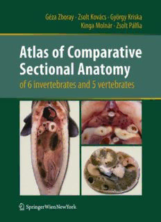
Atlas of Comparative
Sectional Anatomy
of 6 invertebrates and 5 vertebrates
SpringerWienNewYork
Dr.GezaZboray.Dr.Kinga Molnar-ZsoltPallia
DepartmentofAnatomy,CellandDevelopmentalBiology,FacultyofScience
Eotvost.orandUniversity,Budapest,Hungary
Dr.ZsoltKovacs
DepartmentofZoology, FacultyofNaturalandTechnicalSciences,SavariaCampus
UniversityofWestHungary,Szombathely,Hungary
Dr.GyorqyKriska
SectionforMethodologyinBiologyTeaching,FacultyofScience
Eotvost.orandUniversity,Budapest,Hungary
Thisworkissubjectto copyright.
All rights arereserved,whetherthe whole or partofthe material
isconcerned,specificallythose oftranslation,reprinting,re-useof
illustrations, broadcasting, reproduction by photocopying ma
chinesorsimilarmeans,andstorageindatabanks.
ProductLiability:Thepublishercangivenoguaranteeforallthein
formationcontainedinthisbook.Thisdoesalsorefertoinformation
aboutdrugdosageandapplicationthereof.Ineveryindividualcase
the respective user must check its accuracy by consulting other
pharmaceuticalliterature.
The use of registered names,trademarks, etc. in this publication
does not imply, even in the absenceofaspecific statement,that
suchnamesareexemptfrom the relevantprotectivelawsandregu
lationsandthereforefreeforgeneral use.
©2010Springer-VerlaglWien
Printed inAustria
SpringerWienNewYorkispart of
SpringerScience+BusinessMedia
springer.at
Typesetting:Camerareadybytheauthors
Printing:HolzhausenDruck&Medien GmbH,1140Vienna,Austria
Printed onacid-free andchlorine-freebleached paper
SPIN:12704408
With411figurespartlycoloured
Library ofCongressControl Number: 2010929491
ISBN978-3-211-99762-8 SpringerWienNewYork
OPENING RECOMMENDATION
A universal goal of science is "to explain the complex visible by some simple invisible" as
Jean Perrin, Nobel laureate physicist, put it. In other words, we try to find the rule that gov
erns what we see and what we experience. We have an insatiable desire to explain the world
around us, perhaps because it is advantageous to reduce uncertainties in our future. A pre
requisite to understanding our world is recognising, identifying and classifying living organ
isms that we use, we fear or we simply marvel at. At first sight the only common thing that
connects an earthworm, a cockroach and a rat is that they all move, apparently voluntarily,
and from this we can safely assume that they are animals. This is a pleasing simple rule be
cause it is also visible. When we look inside these animals, at first sight there are few, if any,
common features to explain their forms. Only when we learn to differentiate and recognise
organs may we find the simple rules that they are all equipped to exchange materials with the
external world and are equipped and programmed to generate offspring.
In the last thirty years, spectacular progress has been made in developmental biology re
vealing how these forms and organ systems come about. The same sets of genes control the
formation of body parts with similar functions but different designs from vertebrates to in
sects and other animals. By expressing or activating these master genes it is possible to grow
an eye on the leg of an insect, or to grow muscle or liver cells in a dish from undifferentiated
stem cells. For example, the gene Pax-6 governs eye formation in all bilaterian animals stud
ied. The set of Hox genes, coding for eight homeotic proteins contain a conserved DNA bind
ing homeobox domain, was discovered in the fruit fly as responsible for the orderly sequence
of body parts from head to the tip of the abdomen. Homologous clusters of Hox genes gov
ern the formation of the orderly segmental development of the vertebrate body including our
brain together with its insatiable desire to explain the world. Still, the student looking for or
der and homologies in different animals needs to find out what is where.
The authors of this atlas help us to recognise these simple rules of composition in all the
commonly studied animals, by taking us on a wonderful journey through their bodies. One
species of six clades of invertebrates (the large roundworm of pigs, Ascaris suum; the com
mon earthworm, Lumbricus terrestris; the swan mussel, Anodonta cygnea; the edible snail,
Helix pomatia; the North American crayfish, Orconectes limosus, and the giant cocroach,
Blaberusgiganteus) and one species of fivevertebrate classes (the common carp, Cyprinus car
pio; the edible frog, Rana esculenta; the red-eared terrapin, Trachemys scripta elegans; the
chicken, Gallus domesticus, and a laboratory strain of the black rat, Rattus rattus) are shown
in photographs of unique whole body section series. The animals were fixed in order to pre
serve the positions of the organs, then sectioned by various methods to display the relation
ships of different body parts in several planes. Beautifully presented full size graphics, on fac
ing pages to each photograph, explain what we see in the photographs. The atlas comes with
digital illustrations providing stereoscopic views of the specimens. This atlas is of immense
5
help to those who want to learn or teach how the common body parts such as the muscula
ture, digestive system, the circulation, the gills and lungs and the reproductive organs are lo
cated in response to the changes in body shape. Of course, the atlas is only a guide and not a
replacement for dissecting the animals and identifying the organs in situ.
That is exactly what I did a long time ago without the aid of an atlas, as a first year biolo
gy undergraduate at Lorand Eotvos University, under the guidance of my teacher, the first au
thor Dr Geza Zboray in Budapest where these specimens were prepared recently. As we mar
velled the "endless forms most beautiful" as Sean B. Carroll expressed it in his book of the
same title, we learned not only to recognise body parts and the relationships of systems from
the earthworm to the rat, but also the rigour of observation and accurate recording. This at
las provides a great example how simple preparations can teach us so much about the animal
and its representation. Today, as a neuroscientist and microscopist, remembering my roots, it
is a special honour for me to warmly recommend this book to all those interested in the liv
ing world.
Peter Somogyi, FRS, FMedSci 15. February 2010
Professor of Neurobiology
The University of Oxford
6
ACKNOWLEDGEMENTS
We have got valuable supporting comments and advice from our highly regarded col
leagues: Dr. Eva Fekete, professor, University of Szeged, Faculty of Science, Szeged, Hungary;
Dr. Katalin Halasy, professor, Szent Istvan University, Faculty of Veterinary Medicine,
Budapest, Hungary; Dr. GaborHo1l6si, associate professor, University of Debrecen, Faculty of
Science, Debrecen, Hungary; Dr. Erzsebet Hornung, associate professor, Szent Istvan
University, Faculty of Veterinary Medicine, Budapest, Hungary; Dr. Janos Kovacs, professor
emeritus, Eotvos Lorand University, Faculty of Science, Budapest, Hungary.
We are grateful for their true helpfulness, and for their confidence in our efforts and work.
We gratefully remember the late Dr.Janos Vajda professor (Semmelweis University, Faculty
of Medicine, Budapest, Hungary), who gave valuable pieces of advice when we started this
work.
For their useful recommendations and technical assistance we are grateful to Dr. Gabor
Juhasz (Eotvos University, Faculty of Science, Research Group of Proteomics, Budapest,
Hungary), and to Tamas Torok (University of West Hungary, Savaria Campus, Faculty of
Natural and Technical Sciences, Szombathely, Hungary).
We are grateful to Dr. Mikl6s Sass professor (Eotvos Lorand University Faculty of Science,
Budapest, Hungary) for supporting our efforts, and reading the manuscript.
We thank Mrs. Zsolt Pdlfia (Sarolta Sipos) (Eotvos Lorand University Faculty of Science,
Budapest, Hungary) for the editorial work and related corrections of the text.
The text was translated into English by Dr. Orsolya Szab6-Salfay.
We thank Dr. Attila L. Kovacs associate professor (Eotvos University, Faculty of Science,
Budapest, Hungary) for carefully reading and improving the manuscript.
We are grateful for Dr. Peter Somogyi, Fellow of the Royal Society, Professor of Neurobio
logy, MRC Unit Director (UK) for his preface.
The authors also wish to extend special thanks to Dr. Claudia Panuschka and Mag. Karim
Ernst Karman, editors of the Springer Press who provided their expretise to assist and pro
mote our work.
We are also grateful to our family and loved ones for their patience and support.
The Authors In February, 2010.
7
THE AUTHORS
Dr. Geza Zboray
assistant professor
Eotvos Lorand University, Faculty of Science
Department of Anatomy, Cell- and Developmental Biology
Budapest, Hungary
Dr. Zsolt Kovacs PhD
associate professor
University of West Hungary, Savaria Campus
Faculty of Natural and Technical Sciences
Department of Zoology, Laboratory of Neurobiology
Szombathely, Hungary
Dr. Gyorgy Kriska PhD
assistant professor
Eotvos Lorand University, Faculty of Science
Section for Methodology in Biology Teaching
Budapest, Hungary
Dr. Kinga Molnar PhD
assistant professor
Eotvos Lorand University, Faculty of Science
Department of Anatomy, Cell- and Developmental Biology
Budapest, Hungary
Zsolt Palfia MSc
biol.-eng., lecturer
Eotvos Lorand University, Faculty of Science
Department of Anatomy, Cell- and Developmental Biology
Budapest, Hungary
8
CONTENTS
Introduction to the atlas 11
The structure of the atlas 11
Materials and methods 12
Anatomical abbreviations 15
INVERTEBRATES (Invertebrata)
The roundworm of pigs (Ascaris suum) 17
The earthworm (Lumbricus terrestris) 25
The swan mussel (Anodonta cygnea) 41
The roman snail (Helix pomatia) 53
The spiny-cheek crayfish (Orconectes limosus) 73
The giant cockroach (Blaberus giganteus) 95
VERTEBRATES (Vertebrata)
The carp (Cyprinus carpio) 115
The edible frog (Rana esculenta) 141
The red eared slider (Iracbemys scripta elegans) 169
The domestic fowl (Gallus gallus domesticus) 193
The laboratory rat (Rattus rattus) 227
Index 273
9
Description:This atlas contains 189 coloured images taken from transversal, horizontal and sagittal sections of eleven organisms widely used in university teaching. Six invertebrate and five vertebrate species – from the nematode worm (Ascaris suum) to mammals (Rattus norvegicus) – are shown in detailed ima

