AN ANTIGENIC ANALYSIS OF PLANT AND SOIL CORYNEBACTERIA, AND THEIR RELATIONSHIP TO ANIMAL STRAINS OF THIS GROUP PDF
Preview AN ANTIGENIC ANALYSIS OF PLANT AND SOIL CORYNEBACTERIA, AND THEIR RELATIONSHIP TO ANIMAL STRAINS OF THIS GROUP
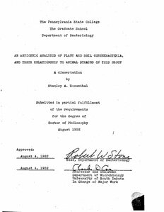
The Pennsylvania State College The Graduate School Department of Bacteriology AN ANTIGENIC ANALYSIS OP PLANT AND SOIL CORYNEBACTERIA, AND THEIR RELATIONSHIP TO ANIMAL STRAINS OP THIS GROUP A dissertation by Stanley A. Rosenthal Submitted in partial fulfillment of the requirements for the degree of Doctor of Philosophy August 1952 Approved: August 4, 1952 - , , ol/ August 4, 1952 Professor and Chairman Department of Microbiology University of South Dakota In Charge of Major Work TABLE OF CONTENTS Page I. INTRODUCTION AND REVIEW OP THE LITERATURE . . . . 1 | II. METHODS . . . . . ........................... . 7 I 1 A. Cultures.................................. 7 j B. Preparation of Antigen Suspensions ........ 7 1. Plant and soil attains............... 7 2. Animal strains. ................... 7 C. Preparation of Antisera............... 11 D. Adsorption of Antisera . .......... 12 ' E. Procedures for Tube Agglutination............ 12 1. Plant and soil strains.................. 12 2. Animal strains......................... 12 P. Procedures for a Serological Survey of the Corynebacteria....................... 12 III. RESULTS AND DISCUSSION........................... 14 A. Intra-species Studies........ ............ 14 1. £. michiganense .................. 14 2. £. poinsettiae........................35 3. C. insldiosum................. 35 4. £. flaccumfaoiens . . . 39 5. £. sepedonicum............... 39 B. Inter-species Studies....................... 41 C. Antigenic inter-relationships between the Plant and -Animal Corynebacteria..............65 37504;; Page IV. GENERAL DISCUSSION. . . . . . ' . 75 V. SUMMARY . . . . ................. 79 VI. BIBLIOGRAPHY................ ;. . . . . . . . . . 81 VII. ACKNOWLEDGEMENTS. . . . . . . . . . . . . . . . . 85 I. INTRODUCTION AND REVIEW OP THE LITERATURE Corynebacterium diphtheriae has received considerably more attention by bacteriologists than other corynebacteria because of its great medical importance. Early interest was primarily focused on the soluble toxin of this organism, and it was not until the therapeutic and prophylactic properties of antitoxic serum had become firmly established that the or ganism itself became the subject of serological investigation. The earlier work in this field has been summarized by Morton (1940) and McLeod (1943), who concluded that the 3 physio logical types are antigenically distinct, with each sero logical group composed of a number of sub-groups. Hewitt (1947) found a total of 53 sero-types within the physiological varieties, with the mltis type being the most heterogeneous. Hoyle (1942), using an alcohol-soluble antigen in the complement-fixation test, concluded that mltis, gravis and intermedlus types contained the same group antigen shared by C. hofmannl, in addition to an antigen specific for C. diphtheriae. By means of the slide agglutination test, Perris (1950b) was able to group 794 C. diphtheriae strains into 14 sero-types, with only 3 strains remaining unclassified. Both Oeding (1950) and Lautrop (1950) have demonstrated the presence of thermolabile and thermostable antigens in C. diphtheriae. The group-specific "0" antigens were heat-stable and were present in all 3 physiological varieties. Lautrop (1950) found the same "0" antigen present in over 300 strains of £. diphtheriae, with most strains containing an additional "0* antigen. The heat-labile antigens were apparently type- specific. Substances responsible for serological specificity in C. diphtheriae have been isolated. Wong and T'ung (1938) first reported that they had isolated a polysaccharide which was shared by all 3 physiological varieties; however, they (Wong and T*ung, 1939b) later demonstrated 2 group-specific polysaccharides. Their report is in accord with the findings of Oeding (1950) that the heat-stable group antigen was re sistant to treatment with trypsin and absolute alcohol. Morton (1940) suggested that Wong and T*ung (1938), in their first paper, used strains which were of the same serological group, although they differed culturally. Shortly thereafter, they (Wong and T»ung, 1939c; 1940) isolated an alkali-soluble protein from diphtheria organisms. Five serological types of C. diphtheriae each yielded an alkali-soluble protein which precipitated only with homologous antiserum. These data strongly indicate that an alkali-soluble protein is the sub stance responsible for type-specificity in C. diphtheriae. Freeman and Minzel (1952) have recently suggested that C. diphtheriae strains be grouped on the basis of glucose and starch fermentation in heart infusion broth and then typed serologically, using type-specific antisera. The interesting and Important problem of the sero logical differentiation of C. diphtheriae has been hindered by the auto-agglutination of a large number of strains. This property has undoubtedly contributed to the large number of conflicting reports which have appeared in the literature. Various methods have been used to obtain stable suspensions from auto-flocculating strains. Robinson and Peeney (1936) and Hewitt (1947) used NaOH solutions to obtain stable anti gen suspensions, while Murray (1935) was successful with 1.3$ NaHCOa ai*d mechanical separation of clumped cells. In the slide agglutination test, Ferris (1950a,b) used 5$ saline, although titers were slightly lower than those obtained when physiological saline was employed. Ewing (1933) stabilized auto-agglutinating strains by heating and shaking, and Eagle- ton and Baxter (1923) used glycerin and heat. Minzel and Freeman (1950) incorporated Tween 80 into broth media when growing diphtherial antigens. After testing 200 strains of C. diphtheriae and various diphtheroids, they concluded that the use of Tween 80 broth gave suspensions which were more stable than those prepared by other methods. Relatively little work has been reported on the sero logical relationships between £. diphtheriae and other members of the genus. Bailey (1925), using tube agglutination pro cedures, found no significant cross reactions among diphtheria organisms, C. hofmanni and C. xerose. On the other hand, Wong and T'ung (1939a) found that a polysaccharide isolated from C. xerose reacted equally well with all antisera prepared against the various cultural types of C. diphtheriae and that C. xerose antiserum precipitated polysaccharides prepared from C. diphtheriae. Antiserum prepared against C. hofmanni did not react with the above polysaccharides, nor did heterologous antisera precipitate C. hofmanni polysaccharide. These data support the report of Bull and McKee (1924), who found that C. hofmanni did not bind complement in the presence of extracts of virulent and avirulent diphtheria organisms. Of the 3 strains of C. hofmanni used in this latter study, 2 shared antigenic components, while 1 strain appeared to be entirely unrelated to the others. On the other hand, Hoyle (1942) found that a lipoid antigen was shared by C. diphtheriae and G. hofmanni, but that C. hofmanni also contained a specific antigen. Lautrop (1950) reported that the "O" antigen of £. diphtheriae is also present in £. hofmanni and C. ovis. The serological relationships among strains of C. equi have been published and confirmed (Bruner, et al, 1939; Karl- son, £t al, 1940; Bruner and Edwards, 1941; Woodroofe, 1950). These organisms are highly type-specific, and species-specific antigens can not be demonstrated by agglutination tests. Woodroofe (1950) divided 16 strains of C. equi into 3 main groups, with 3 strains agglutinating only in homologous anti- serum. Bruner and Edwards (1941) found that 29 of 34 strains fell into 4 distinct groups which were further subdivided into types. The remaining 5 strains were not related antigenically to the main groups or to each other. The species-specific antigen of C. equi was demon strated by removing the type-specif1c antigen with HC1 at 100 C. Heating the cells with acid at 60 C did not remove the type-specific antigen (Bruner, et al, 1939). All strains examined contained the species-specific antigen, which was A demonstrated by eomplement-fixation tests on the extracted cell residues; but not all antisera contained the species- specific antibody (Bruner, et al, 1939). Merchant (1935) has investigated the serological relationships among corynebacteria causing animal diseases. He found that C. pyogenes contained a species-specific anti gen; antisera prepared against 2 strains agglutinated all 5 of his C. pyogenes cultures. Only 1 C. pyogenes strain ag glutinated in 2 of 4 C. renale antisera and in 1 C. pseudo- tuberculosis antiserum. C. pseudotuberculosis apparently contained a species-specific antigen, but species-specific antibody could not be demonstrated in all C. pseudotuberculosis antisera. Three C. renale antisera agglutinated 4 to 6 of 12 C. pseudotuberculosis strains. Most of Merchants C. renale cultures contained a species-specific antigen. Marked cross reactions were observed when G. renale antigens were reacted with C. pseudotuberculosis antisera. Four strains classified as £• resale on the basis of fermentation reactions, morphology cultural characteristics and source of isolation were found to have a closer serological relationship to C. pseudotuber culosis than to C. renale. Merchant also found serological cross reactions among 1 strain of C. diphtheriae, 2 of C. renale, and 1 of C. pseudotuberculosis. In addition, Lautrop (1950) reported on the antigenic relationship between C. diphtheriae and C. ovis (C. pseudotuberculosis). Feenstra, e_t al (1945) have shown the presence of a species-specific antigen in C. renale, confirming the earlier work of Merchant (1935), but only 1 of 5 C. renale antisera agglutinated all strains. Although some work has been done on the animal and human corynebacteria, a siirvey of available literature has failed to reveal any serological studies on the plant and soil diphtheroids. Conn and Dimmick (1947) have objected to the inclusion of the plant and soil diphtheroids in the genus Corynebacterium (Jensen, 1934; Starr and Pirone, 1942). On the basis of morphological and physiological studies, they felt that the plant diphtheroids now classified in the genus Corynebacterium show sufficient differences from the type species to suggest that they do not belong in this genus. Their opinions, nevertheless, were not based on antigenic studies. They also felt that not all of the plant coryne bacteria belong in the same genus. Therefore, the purpose of this study is to investigate the antigenic structure of the plant and soil corynebacteria and the possible occurrence and distribution of their antigens within the animal coryne bacteria, in an attempt to clarify the relationships among members of this ill-defined group of micro-organisms as well as their status with respect to the genus Corynebacterium. II. METHODS A. Cultures Many of the cultures were obtained through the kind ness of several individuals throughout the country, and others were purchased from the American Type Culture Collection. The species designations of the cultures used in this study are the same as those given to the author, except where those names were in conflict with the designations given in Bergey's Manual of Determinative Bacteriology (Breed, et al, 1948). A list of the cultures and their sources is given in table 1. B. Preparation of Antigen Suspensions 1. Plant and Soil Strains These antigens were grown on Eugonagar (Baltimore Biological Laboratory) in Pyrex Blake bottles for 1 to 3 days at room temperature, collected in small amounts of formalinized saline1, filtered through sterile cotton and washed once with formalinized saline. The cells were resuspended in small amounts of formalinized saline, and these heavy suspensions were kept in the refrigerator as stock antigens. When tube antigens were needed, the stock suspensions were diluted with formalinized saline to a dilution which allowed 85$ light transmittance using a Coleman Universal Spectrophotometer at 650 A. 2. Animal Strains When auto-agglutinating antigens were studied, a modification of the method of Minzel and Freeman (1950) was 10.5$ formalin and 0.85$ NaCl in distilled water.
