AN ANATOMICAL, HISTOLOGICAL, AND EMBRYOLOGICAL STUDY OF THE RETROCEREBRALCOMPLEX IN THE HONEY BEE (APIS MELLIFERA L.) PDF
Preview AN ANATOMICAL, HISTOLOGICAL, AND EMBRYOLOGICAL STUDY OF THE RETROCEREBRALCOMPLEX IN THE HONEY BEE (APIS MELLIFERA L.)
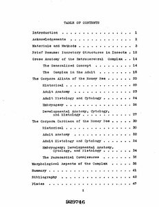
TABLE OF CONTENTS Introduction............ 1 Acknowledgements.......... 2 Materials and Methods................ 3 Brief Resume: Incretory Structures in Insects • 12 Gross Anatomy of the Retrocerebral Complex . . 14 The Generalized Concept ................. 14 The Complex in the A d u l t ............. 15 The Corpora Allata of the Honey Bee • • • • • • 20 Historical.............................. 20 Adult A n atomy.......... .23 Adult Histology and Cytology ..........24 Embryogeny.............................. 26 Developmental Anatomy, Cytology, and Histology......................27 The Corpora Cardiaca of the Honey Bee • • • • • 30 Historical.............................. 30 Adult A na tomy..........................32 Adult Histology and Cytology........... 34 Embryogeny; Developmental Anatomy, Cytology, and Histology........... 34 The Paracardial Commissures ............ 36 Morphological Aspects of the Complex • 36 Summary...................................... 41 Bibliography ............................ 42 P l a t e s .................. , . . . 4 7 i 929746 AN ANATOMICAL, HISTOLOGICAL, AND EMBRYOLOGICAL STUDY OF THE RETROCEREBRAL COMPLEX IN THE HONEY BEE (APIS MELLIFSRA L.) DISSERTATION Presented in Partial Fulfillment of the Requirements for the Degree Doctor of Philosophy in the Graduate School of The Ohio State University by BLAKE BJ. HAN AN, B.A., M.Sc. The Ohio State University 1952 Approved by* INTRODUCTION Personal experiences in various phases of applied beekeeping came at a time when fundamental research was being conducted on insect parahormones and great medical advances were being made through the appli cation of vertebrate hormones to humans. The applied aspects of endocrinology to the field of apiculture appeared to be a fertile field for research. Cer tain questions were particularly intriguing. To what extent were incretory structures present in the honey bee? Could such secretions as they produce be used in the artificial regulation of processes and behavior such as ovulation, secretion of food and venom, and swarming? An analysis of the proper approach to the field of research involved fundamental studies on incretory structures of the honey bee, their physi ological role, and the applied aspects of endocrinol ogy in the field of apiculture. Arduous search through the literature revealed little reliable infor mation on the anatomy of such structures in the honey bee as are known to produce parahormones in other insects. Thus a study of the structures would have to be undertaken before an understanding could be achieved as to the possible role of parahormones in the honey bee. The so-called retrocerebral complex of the honey bee was chosen initially for investigation and the entire research program confined to its structures and relationships. It is hoped that the results of this investiga tion will lead into research on the physiological role of these structures and thence, possibly, into the applied aspects of endocrinological api culture. ACiQTO'WXEDGEMENTS Sincere gratitude is extended to all who have had a part in the completion of this work. Prom among such individuals who have provided insight, assistance, cooperation, inspiration, and materials, the writer wishes to express his special apprecia tion to the following: Professors Winston E. Dunham, Charles A. Reese, Willard C. Iflyser; and John A. Knierim, a graduate assistant; as well as to Ernest and Ronald Powler. 2 MATERIALS AND METHODS COLLECTING Honey beeB in which adult structures were studied were secured from several sources* Old workers were collected from flowers, and young workers were obtained from brood nests of bee colonies* Drone bees, regard less of age, were collected from the Ohio State Uni versity apiary and the writer’s apiary. Virgin queens were reared by using a modified Doolittle culture method and also by procuring queen cells from swarming colonies and incubating them at 92° * 1°P. Spent queens were obtained from beekeepers as a result of their requeening programs* Larvae and pupae of known ages were collected in colonies in several apiarieB, FIXATION Adult material collected early in the study was fixed in a 10^ formalin cold-blooded Ringer* s fixative. ThiB was not too satisfactory because staining tech niques used later were found to be more effective after Zenker’s fixation. Even with double fixation, mater ial fixed originally in formalin was not nearly of the cytological quality as that fixed originally in Zen ker’s. The advantage of formalin fixation lay in its rather non-critical fixation time limit. Con- sequently, the formalin technique was employed when material was collected by others and fixation time could not "be controlled. After fixation in either solution, all tissues were stored in 70% alcohol. Tissues fixed in Zenker’s fluid were washed in iodized 70% alcohol before storage. Larvae and pupae obtained by the author were fixed for 12 to 18 hours in Zenker* s solution, then washed in iodized 70% ethyl alcohol, and finally stored in 10% alcohol. Pupae furnished by Dr. W. C. Myser of Ohio State University had been labelled as to age and had been fixed for 18 to 24 hours in Peterson* s fluid (K.A.A.D.) which has the following formula: Kerosene, commercial grade 1 part Glacial acetic acid 2 parts Ethyl alcohol, 95% 10 parts Dioxane 1 part Following fixation in this fluid, the material had been rinsed quickly in water and stored in 10% alcohol. It was discovered that Peterson* s fluid was not a suitable fixative for critical histological or cytological study of honey bee tissues. It has many good qualities as a larval fixative. Cells thus fixed take nuclear stainB well, but do not produce as clear-cut cellular characteristics under magni fications greater than approximately 100X as do either Bouin*s or Zenker’s solutions. TECHNIQUE; ADULT TISSUES Attempts were made to section the entire head capsule and contents. This procedure was unsuccess ful because of the heavy aclerotization; therefore the following method was used to remove the retro cerebral complex from the head. While the anesthe tized insect was held in the fingers, the occipital region of the head was clipped off. The mouth parts, ventral head region, and then the lateral aspects of the head were similarly removed. This left in place adjacent to the neck, the greater portion of the brain and esophagus, the retrocerebral complex, and a more or less square portion of the frons and antennal region that still hung to these parts anter iorly. The insect was pinned in a small dissecting dish in 70% alcohol and, with the aid of a dissecting microscope, the frontal wall of the head capsule was then detached and the remaining soft tissues includ ing the complex were clipped free of the thorax at the neck. 5 After the complex was removed from the head capsule, it was dehydrated through an alcohol series. The tissues were kept for 10 to 15 minutes first in 8 5 then 95^, and finally in 100/& ethyl alcohol. After dehydration, the tissues were cleared in xylene for 5 to 10 minutes. Cedarwood oil was used initially hut in the case of these particular adult tissues, xylene was found to he more rapid and at the same time cause no great amount of overhardening or shrinkage. Tissues thus dehydrated and cleared were trans ferred directly to the melted imbedding medium, 52° - 55°F. Fisher’s Tissuemat. Infiltration time on adult tissue was never less than two hours and more frequently overnight. The problem of orienting small pieces of tissues was difficult. This was eventually solved by putting the tissue, together with the Tissuemat, into a warmed 3 mm slide well. At the moment the Tissuemat had cooled sufficiently to hold the material in the desired position,a drop of cold water was pulled acroBB the surface of the Tissuemat with a pipette to harden the surface rapidly. The whole slide was then im mersed in cold water to reduce crystal size. This procedure may not be new but it has several things in its favor when imbedding small material. It reduces the chances of poor orientation and thus cuts down the number of replicas needed to obtain one satis factory series. The imbedded material is also very easily labelled and stored in the slide well. After imbedding, the block was cut from the well and trimmed for sectioning. This was facilitated by the lower side of the block being already flat on removal from the slide well. Sections of adult tis sues were finally standardized at 6 microns in thickness. Sections under 6 microns did not sec tion or flatten well, whereas those above 8 microns were too thick for critical cytological studies. A number of nuclear stains were tested on these tissues. The stains that yielded the best results were iron hematoxylin, methylene blue, and phospho- tungstic acid hemotoxylin. The latter stain was finally used exclusively. Methylene blue produced beautifully stained neural tissue, but staining times were so critical and the washing effects of alcohol were so acute, that only a small percentage of the finished slides were of high quality. Most of the hematoxylins (Ehrlich*s, Delafield's, etc.) produced too diffuse an effect for critical cyto logical use. Both iron and phosphotungstic acid hematoxylin (Jones, 1950) yielded very clear, clean- line staining. The latter stain was chosen primarily "because it produced a greater degree of differential staining, especially in muscular, glandular, and neural tissues. Cytoplasmic counter stains of eosin, and orange G. were tried but discontinued since they added little, if any, value to the differential staining effect mentioned above. All sections were mounted serially in Piccolyte, TECHNIQUE« POST-EMBRYOLOGICAL TISSUES Basically, the slide preparation technique was patterned after Dr. Myser* s methods in his study on the post-embryogeny of the honey bee. The following brief outline will describe the handling of larval and pupal material. From storage, after employing either K.A.A.D. or Zenker* s fixation, pupal heads and necks were clipped from the thorax and dehydrated upward through an alcohol series. From absolute alcohol the speci mens were passed through gradually increasing con centrations of cedarwood oil. Later xylene was substituted for the slower cedarwood oil technique. This was satisfactory, however, only when the tissues had been dehydrated completely (4 hours or more in two washes of abso-
