Amanita ochroterrea and Amanita brunneiphylla (Basidiomycota), one species or two? PDF
Preview Amanita ochroterrea and Amanita brunneiphylla (Basidiomycota), one species or two?
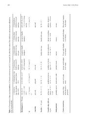
NE.uMyt.s Diaa 2v1is(o4n):, 1A7m7a–n1i8ta4 o(2c0h1ro1t)errea and Amanita brunneiphylla (Basidiomycota) 177 Amanita ochroterrea and Amanita brunneiphylla (Basidiomycota), one species or two? Elaine M. Davison Department of Environment and Agriculture, Curtin University, GPO Box U1987, Perth, Western Australia 6845 Western Australian Herbarium, Department of Environment and Conservation, Locked Bag 104, Bentley Delivery Centre, Western Australia 6983 Email: [email protected] Abstract Davison, E.M. Amanita ochroterrea and Amanita brunneiphylla (Basidiomycota), one species or two? Nuytsia 21(4): 177–184 (2011). Amanita ochroterrea Gentilli ex Bas and A. brunneiphylla O.K.Miller are robust, macroscopically similar mushrooms described from the south-west of Western Australia. According to the protologue of A. brunneiphylla, the main difference between them is the presence (in A. ochroterrea) or absence (in A. brunneiphylla) of clamp connections. However, in the current study abundant clamp connections have been observed in the holotype and paratypes of A. brunneiphylla. As other microscopic characters are indistinguishable, A. brunneiphylla is synonymised with A. ochroterrea, and an expanded description presented. Introduction Amanita species are large, conspicuous mushrooms with a worldwide distribution. They are readily recognized to the generic level in the field, but the majority are difficult to separate into species solely on their appearance. Microscopic characters are usually needed before collections can be confidently identified. The most commonly used microscopic characters are spore size and shape, their response to Melzer’s iodine, the presence of clamp connections in the basidiome especially at the base of basidia, the structure of the subhymenium and underlying lamella trama, the pileipellis, and the structure of the universal veil on the pileus. All of these characters are used to separate species and construct diagnostic keys (Bas 1969; Reid 1979; Tulloss 1994; Wood 1997). In 1953 Gentilli described several collections of Amanita spp. from Kings Park, Perth, Western Australia which included two large specimens with an earthy buff cap, buff lamellae and pale buff spore print that he called A. preissii (Fr.) Sacc. forma ochroterrea Gentilli (Gentilli 1953). This forma was not validly published since it was not accompanied by a Latin description or diagnosis. Gentilli sent these specimens to Bas who considered them to be so distinctive that he described them as a new species, A. ochroterrea Gentilli ex Bas (Bas 1969; MB308574). Following a visit to the south-west of Western Australia in 1989, Miller described A. brunneiphylla O.K.Miller (MB358165), a robust species with a dull white cap, light brown lamellae and a pale yellow spore print. He recognised that this was similar to A. ochroterrea, but separated the two species on the presence (in A. ochroterrea) or absence (in A. brunneiphylla) of clamp connections, together with small differences in spore size and colour of the lamellae (Miller 1991). 178 Nuytsia Vol. 21 (4) (2011) This paper reports a re-examination of the holotype and paratypes of A. brunneiphylla held at the Western Australian Herbarium (PERTH). These are compared with the published description of A. ochroterrea. Additional collections named as A. ochroterrea in PERTH, and macroscopically similar specimens held in private collections in Western Australia have also been examined. As a result, A. brunneiphylla is synonymised with A. ochroterrea and an expanded description is provided. Materials and methods Methodology follows that of Tulloss (2000, 2008). Colours are from the Royal Botanic Garden, Edinburgh (1969) and colour codes in the form of ‘p3A2’ are from Kornerup and Wanscher (1978). Dried material was rehydrated in 10 % NHOH or 3 % KOH and stained with 1 % Congo Red. Biometric 4 variables for spores follow Tulloss and Lindgren (2005), i.e. ‘L = the average spore length computed for one specimen examined and the range of such averages, L´ = the average spore length computed for all spores measured, W = the average spore width computed for one specimen examined and the range of such averages, W´ = the average spore length computed for all spores measured, Q = the ratio of length/breadth for a single spores and the observed range of the ratio of length/breadth for all spores measured, Q = the average value of Q computed for one specimen examined and the range of such averages, Q´ = average value of Q computed for all spores measured’. Comparison of Amanita ochroterrea and A. brunneiphylla The macroscopic descriptions of A. ochroterrea and A. brunneiphylla are similar; however, the protologue of A. brunneiphylla states that the flesh is ‘firm, dull white tinted grey at the base’ (Miller 1991). An image (E567) of a paratype (PERTH 007565259, O.K. Miller OKM 23747, as reproduced in Figure 1A, shows that the flesh is tinted brown, however, this may have resulted from drying. Neither Gentilli (1953) nor Bas (1969) comment on the colour of the context. The microscopic characters of the holotype and paratypes of A. brunneiphylla do not differ from the type description of A. ochroterrea (Table 1). Basidiospore dimensions, amyloid reaction, size of basidia, and shape of lamella edge cells in all mature collections are similar to those of the type description of A. ochroterrea. Clamp connections are present and abundant in all of these collections; thus the original description of A. brunneiphylla is neither supported by the holotype nor the paratypes. These observations did not detect any significant differences between these taxa. As a result, A. brunneiphylla is synonymised with A. ochroterrea. An expanded description of A. ochroterrea is provided. Expanded description of Amanita ochroterrea Amanita ochroterrea Gentilli ex Bas, Persoonia 5: 505–506, figures 278–281 (1969). Amanita preissii (Fr.) Sacc. f. ochroterrea Gentilli, W. Austral. Naturalist 4: 30, figure 3 (1953), (nom. inval., Art. 36.1). Type: Perth, Kings Park, Western Australia, June 1953, J. Gentilli s.n. (holo: L). (MB308574). E.M. Davison, Amanita ochroterrea and Amanita brunneiphylla (Basidiomycota) 179 Amanita brunneiphylla O.K.Miller, Canad. J. of Bot. 69: 2694 (1991). Type: Murdoch University campus, Western Australia, 7 May 1989, O.K. & H.H. Miller, E.M. & P.J.N. Davison OKM 23621 (holo: PERTH 07587473, image E 511). (MB358165). Basidiome small to very large (Figures 1A, B). Pileus 35–170 mm diam, up to 15 mm thick, hemispheric when young, becoming more or less plane with a depressed centre with age, cream, pale buff to pale olivaceous buff (p1A2–p2B4–p4B3), margin of the pileus appendiculate, non-sulcate, no surface staining or bruising reaction. Universal veil on pileus (Figure 1C) adnate, forming a soft thin crust over the whole pileus, sometimes with small floccose warts in the centre, sometimes with thick felty angular flattened warts in the centre, cream, pale grey olivaceous, pale vinaceous buff, pale buff to pale olivaceous buff (p1A2–p3B5–p4B3). Lamellae close, free to narrowly adnate, 5–15 mm broad, ventricose, buff, olivaceous buff, hazel (p1B2–p5C4–6), drying buff to snuff brown (p5C4–F7), often with two tiers of plentiful lamellulae, the shorter truncate, the longer attenuate, lamella margin lighter in colour and slightly fimbriate. Stipe length (bottom of pileus context to top of bulb) 60–115 mm; width at mid-stipe 14–37 mm, more or less equal, solid, pale olivaceous buff, pale buff (p2B4–p4B3), furfuraceous or covered in mealy scales below the partial veil. Partial veil superior, descendent, soft, fugacious, initially greenish cream darkening to pale olivaceous buff (p1A2–B2). Bulb 55–72 × 28–44 mm, initially ovoid, narrowing with age, olivaceous buff, encrusted with sand. Remains of universal veil at the base of the stipe soft ridges and scales, in some specimens forming girdles; loose patches often remaining in the soil. Flesh cream, straw, to pale olivaceous buff (p1A2–p2B5) in both pileus and stipe, sometimes darkening on exposure to air. Smell mild and earthy when young, stronger when older. Spore deposit cream (p3A2–3) to buff. Figure 1. Amanita ochroterrea. A – showing the brownish colour of freshly exposed flesh (paratype of PERTH 07565259, O.K. Miller OKM 23747); B – Amanita ochroterrea (PERTH 08334897, B-S 168); C – surface of pileus (PERTH 08059632, E.M. & P.J.N. Davison EMD 6-2008). Photographs by N.L. Bougher (A), L.I. Little (B) and E.M. Davison (C) 180 Nuytsia Vol. 21 (4) (2011) atype collections A. brunneiphylla PERTH 07565259 paratype (8.5–)9.5–11(–11.5) × 4.5–6 (15/1) from lamella 1.7–2.3, mean 2.0 amyloid 30–50 × 9–11 globose, clavate to cylindric, <20 wide ramose Present and abundant in all tissues ylla with observations from holotype and par A. brunneiphylla A. brunneiphylla PERTH 07587562 PERTH 07564465 paratypeparatype no spores, basidia no spores, basidia immatureimmature basidia immaturebasidia immature clavate to spherical, clavate to pyriform, 15–40 × 10–1520-28 × 14-20 ramoseramose Present and abundant present and abundant in all tissuesin all tissues ea and A. brunneiph A. brunneiphylla PERTH 07587473 holotype (8–)9–12 × 4.5–5 (20/1) from lamella 2.0–2.7, mean 2.2 amyloid 38–45 × 9–10 pyriform to clavate, up to 25 × 12 inflated ramose present and abundant in all tissues r r s of Amanita ochrote A. brunneiphylla type description (Miller 1991) (8–)9–10.8 × 4.1–5 1.8–2.3, mean 2.1 amyloid 34–38 × 7–10 pyriform to clavate, 18–26 × 9–15 isodiametric cells none seen in any tissue n e type descriptio A. ochroterrea type description (Bas 1969) (10–)11–13 (–13.5) × 5–6.5 (20/1) 1.9–2.4, mean 2.1 amyloid 50–55 × 11–12 globose to clavate, 15–35 × 15–20 probably ramose present at base of basidia and in pileipellis h Table 1. Comparison of tof A. brunneiphylla. Basidiospores L × W (µm) Q Amyloidy Basidia L × W (µm) Lamella edge cells (size in µm) Subhymenium Clamp connections E.M. Davison, Amanita ochroterrea and Amanita brunneiphylla (Basidiomycota) 181 Basidiospores (Figure 2A) [161/9/8] (8–) 9.5–13(–15) × (4–)4.5–6 (–6.5) µm, L = 10.1–11.7 µm; L′ = 11.0 µm; W = 4.7–5.6 µm; W′ = 5.2 µm; Q (1.67–) 1.82–2.44(–3.00), Q = 2.00–2.23; Q′ 2.13) hyaline, colourless, thin-walled, smooth, amyloid, elongate to cylindrical, infrequently bacilliform, adaxially flattened, with apiculus sublateral, truncate, about 1 × 1 µm, with granular contents. Pileipellis difficult to delimit, merging into both universal veil and pileus context, not or slightly gelatinized at the centre, gelatinization of the hyphal walls in some specimens near the pileus margin; hyphae 2–10 µm wide, thin walled, hyaline, orientation mainly radial with some interweaving. Pileus context tissue yellow in NHOH, hyphae 6–30 µm wide, thin walled, hyaline, dominant, mainly with 4 radial orientation; inflated cells up to 60 × 250 µm, thin walled, colourless. Lamella trama bilateral; width of central stratum 40–60 µm, hyphae 5–20 µm wide, no inflated cells seen; subhymenial base 25–60 µm wide, hyphae 4–20 µm wide, dominant orientation is initially about 30° from the vertical with the hyhae bending round in a smooth curve to the subhymenium, inflated cells up to 20 × 70 µm, infrequent; subhymenium (Figure 2B) 20–40 µm wide, ramose to inflated ramose, thin-walled; lamella margin cells (Figure 2C) globose, pyriform to clavate up to 10–15 × 25–40 µm. Basidia [132/7/7] (30–)35–60(–65) × (7.5–)9–11(–12.5) µm, thin-walled, yellow contents in NHOH, about 90 % four- 4 spored about 10 % two-spored, sterigmata up to 5 µm long by 2 µm wide at the base; basal clamps present. Universal veil on the pileus (Figure 2E) comprising abundant mainly ellipsoidal to spherical, rarely pyriform to clavate, venticose to fusiform cells up to 70 × 150 µm, but most smaller, inflated cells terminal or in short chains; filamentous hyphae 5–10 µm wide, frequently branched, irregularly disposed, hyphae more abundant in the proximal part of the universal veil, while the inflated cells are more abundant in the distal part of the universal veil; some gelatinization of the walls of both filamentous and inflated hyphae evident in older specimens. Universal veil on the stipe base disordered tissue of terminal spherical and clavate cells up to 30 × 150 µm and filamentous hyphae 3–12 µm wide. Stipe context acrophysalidic, with acrophysalides up to 30 × 300 µm dominant, filamentous hyphae 3–13 µm wide with mainly axial orientation. Partial veil (Figure 2D) of dominant spherical to clavate or pyriform inflated cells 15–20 × 50 µm, but occasionally up to 30 × 300 µm, and infrequent, mainly radial, filamentous hyphae 2–8 µm wide. Vascular hyphae present but infrequent in all tissues, 2–15 µm wide, occasionally branched, thin walled, with yellow glassy contents in NHOH, frequently 4 serpentine, no knots or concentrations of vascular hyphae noted in any tissue. Clamp connections present and frequent in all tissues. Collections examined. WESTERN AUSTRALIA: Murdoch University campus, 25 Apr. 1990, E.M. & P.J.N. Davison EMD 12-1990 (PERTH 08059640); Melville, 12 May 2008, E.M. & P.J.N Davison EMD 6-2008 (PERTH 08059632); near Southern Cross, 23 June 1974, B. Dell & K. Elson s.n. (PERTH 00768804, UWA 1862); Swan View, 20 May 2005, K. Griffiths B-S 168 (PERTH 08334897); Murdoch University campus, 7 May 1989, O.K. & H.H. Miller, E.M. & P.J.N. Davison, OKM 23621 (PERTH 07587473, E 511 holotype of A. brunneiphylla); Kings Park, 21 May 1989, O.K. & H.H. Miller OKM 23660 (PERTH 07587562, E 529 paratype of A. brunneiphylla); Moore River, 23 May 1989, O.K. & H.H. Miller OKM 23720 (PERTH 07564465, E 560 paratype of A. brunneiphylla); Regans Ford, 30 May 1989, O.K. & H.H. Miller OKM 23747 (PERTH 07565259, E 567 paratype of A. brunneiphylla); Kings Park, 26 May 1982, anon. s.n. (PERTH 07607512, E 239). Rejected collections. Corrigin shire, J. Catchpole, I.C. Tommerup & N.L. Bougher s.n., 8 July 1999 (PERTH 07658508, E 6235); Grass Patch, T.C. Daniell s.n., 18 Aug. 1984 (PERTH 00918326, UWA 2966) (green form); Gleneagle, R.N. Hilton s.n., 18 June 1975 (PERTH 00771961, UWA 2001); Walpole Nornalup National Park, K. Syme 29:87, 24 May 1987 (PERTH 07575270, UWA 3510). 182 Nuytsia Vol. 21 (4) (2011) Figure 2. Amanita ochroterrea. A – spores from spore print; B – squash of young basidia and subhymenium, clamp connections indicated by arrows; C – lamella edge cells; D – cells from the partial veil, clamp connection indicated by an arrow; E – vertical section through the universal veil on the pileus, the proximal part is at the bottom. A, C, D, E (PERTH 08059632, E.M. & P.J.N. Davison EMD 6-2008); B (PERTH 07587562 O.K. Miller OKM 23660, paratype of A. brunneiphylla). Scale bars A = 10 µm, B–E = 20 µm. E.M. Davison, Amanita ochroterrea and Amanita brunneiphylla (Basidiomycota) 183 These collections have been rejected for the following reasons: PERTH 07658508 has a saccate volva and probably resides in Section Amidella; PERTH 00771961 and PERTH 07575270 have been rejected because they have low Q: 1.64 and 1.72 respectively; PERTH 00918326 has a much wider central stratum and wider lamellae. Distribution and habitat. Solitary or gregarious, in sandy soil in dry sclerophyll woodland and sand plain, often associated with Eucalyptus marginata Sm. and Corymbia calophylla (Lindl.) K.D.Hill & L.A.S.Johnson. Amanita ochroterrea is a distinctive species that is widely distributed in the south- west of Western Australia from the Moore River (31°00′ S, 115°30′ E) to Southern Cross (31°13′ S, 119°18′ E) although it does not appear to be common. It has not been recorded in South Australia (Grgurinovic 1997) or eastern Australia (Wood 1997). Fruiting period. April to August. Diagnostic features. Robust basidiomes with buff pileus, brown gills, yellowish spore print and clamp connections throughout. Suggested common name. Brown-gilled amanita. Notes. Bas (1969) placed A. ochroterrea in Amanita (subsection Solitariae Bas) Stirps Grossa in part because of the irregularly disposed remnants of the universal veil on the pileus. He commented, however, that these remnants were difficult to analyse. The collections examined here support Bas’ placement of A. ochroterrea in Stirps Grossa because the basidia have clamp connections, Q is less than 2.2, the universal veil on the pileus forms a subfelted layer and is composed of irregularly disposed inflated cells intermixed with hyphae. Acknowledgements The Western Australian Herbarium (PERTH) is thanked for the loan of collections of A. ochroterrea and A. brunneiphylla. H.H. Miller kindly provided additional information about the O.K. Miller collections from Western Australia. N.L. Bougher is thanked for an image of PERTH 007565259. K. Griffiths is thanked for the gift of collection B-S 168 and L.I. Little is thanked for an image of this collection. I thank R.E. Tulloss, N.L. Bougher and K.R. Thiele for constructive discussions and feedback on the manuscript. References Bas, C. (1969). Morphology and subdivision of Amanita and a monograph of its section Lepidella. Persoonia 5: 285–579. Gentilli, J. (1953). Amanitas from King’s Park, Perth. The Western Australian Naturalist 4: 25–34, 59–63. Grgurinovic, C.A. (1997). Larger fungi of South Australia. (The Botanic Gardens of Adelaide and State Herbarium: Adelaide.) Kornerup, A. & Wanscher, J.H. (1978). Methuen handbook of colour. (Methuen: London.) Miller, O.K. (1991). New species of Amanita from Western Australia. Canadian Journal of Botany 69: 2692–2703. Reid, D.A. (1979). A monograph of the Australian species of Amanita Pers. ex Hook. (Fungi). Australian Journal of Botany Supplementary Series No. 8: 1–97. 184 Nuytsia Vol. 21 (4) (2011) Royal Botanic Garden, Edinburgh. (1969). Flora of British fungi: colour identification chart.(Her Majesty’s Stationery Office: Edinburgh.) Tulloss, R.E. (1994). Type studies in Amanita Section Vaginatae I: some taxa described in this Century (studies 1–23) with notes on description of spores and refractive hyphae in Amanita. Mycotaxon 52: 305–396. Tulloss, R.E. (2000). Note sulla metodologia per lo studio del genere Amanita (Agaricales). Bollettino del Gruppo Micologico G. Bresadola Trento 43(2): 41–58 Tulloss R.E. (2008). Notes on methodology for study of Amanita (Agaricales). In: Tulloss, R.E & Yang, Z.L. Studies in the genus Amanita Pers. (Agaricales, Fungi) http://www.amanitaceae.org/content/uploaded/pdf/methodsb.pdf. [accessed 12 May 2008]. [English original of Tulloss (2000)] Tulloss, R.E. & Lindgren, J.E. (2005). Amanita aprica – a new toxic species from western North America. Mycotaxon 91: 193–205 Wood, A.E. (1997). Studies in the genus Amanita (Agaricales) in Australia. Australian Systematic Botany 10: 723–854.
