A synopsis of Trichocladium species, based on the literature PDF
Preview A synopsis of Trichocladium species, based on the literature
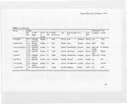
Fungal Diversity 2 (March 1999) A synopsis of Trichocladium species, based on the literature Teik-Khiang Goh* and Kevin D. Hyde Fungal Diversity Research Project, Department of Ecology and Biodiversity, The University of Hong Kong, Pokfulam Road, Hong Kong; *email: [email protected] Goh, T.K. and Hyde, K.D. (1999). A synopsis of Trichocladium species, based on the literature. Fungal Diversity 2: 101-118. The genus Trichocladium is reviewed, based on the literature, with a synopsis of 18 accepted species. A key to the species and a composite diagram of the conidial morphology of accepted species are provided. Short comments are given to the 22 Trichocladium names which are considered unacceptable or doubtful inthe genus. Key words: freshwater fungi, Hyphomycetes, mitosporic fungi, soil fungi, systematics, taxonomy. Introduction The genus Trichoc/adium Harz (1871), based on the lectotype species T asperum, includes dematiaceous species of hyphomycetes producing solitary, thick-walled, more or less pyriform to clavate phragmoconidia, from micronematous or semi-macronematous, mononematous conidiophores (Hughes, 1952; Ellis, 1971). Species of Trichoc/adium are frequently isolated from soil (Dixon, 1968; Domsch and Gams, 1972) or encountered in aquatic environments (Crane and Shearer, 1978; Kohlmeyer and Volkmann Kohlmeyer, 1995; Hyde and Goh, 1998, 1999; Hyde et al., 1999). In our surveys of fungal diversity on submerged wood (Goh and Hyde, 1996; Hyde and Goh, 1998, 1999; Hyde, Goh and Steinke, 1998; Hyde et al., 1999) we frequently encountered Trichoc/adium species. This has prompted us to carry out the present synoptic work on the genus. It is evident that the genus Trichoc/adium is becoming heterogeneous and species identification has become difficult. The genus has included species producing effuse colonies on natural substrata (e.g. Hughes, 1969) as well as species forming sporodochioid conidiomata (e.g., Crane and Shearer, 1978). The genus has also included species with small or large conidia, e.g., T minimum and T macrosporum, respectively (de Hoog and Grinbergs, 1975; 101 Kirk, 1981). A few species have been reported to have conidia with germ pores (Hughes, 1969), and some have conidia which may be verrucose (Nath, 1978), deeply lobed (Sutton, 1975), or formed in chains (Batista and Upadhyay, 1965). Previous synoptic work on Trichocladium species was carried out by Hughes (1952, 1958) who briefly discussed the status of historical species published during the past century. A synopsis of six Trichocladium species was presented in tabulated form (Dixon, 1968). Taxonomic keys to some Trichocladium species were given in Pidoplichko and Kirilenko (1972), Moustafa and Ezz-El din (1990), and Kohlmeyer and Volkmann-Kohlmeyer (1995). Several species have been recently added to the genus (Tubaki, Tan and Ogawa, 1993; Matsushima, 1996; Hyde and Goh, 1999; Hyde et al., 1999). There are presently 40 binomial names in Trichocladium, and some have been previously transferred to other genera (de Hoog and Grinbergs, 1975; Palm and Stewart, 1982; Kirk, 1983; Zucconi and Lunghini, 1997). In the present paper, we present a bibliographic reflection on the genus and consider eighteen species to be acceptable. Eight species have been transferred to other hyphomycete genera; six names are listed as facultative synonyms of accepted Trichocladium species or of other hyphomycetes; and eight names are considered unacceptable in Trichocladium, or their identity remains doubtful because of insufficient information. The characters of the 18 acceptable species are presented for comparison in Table 1. A key to these species is also given and their conidia are illustrated (Figs. 1-18) to facilitate identification. Taxonomy Acceptable species ofTrichocladium Key to acceptable species of Trichocladium 1. Conidia laterally attached on conidiogenous hyphae, 2-celled, proximal cell subglobose, distal cell triangular and pointed at the apex . ............................................................................................... T variosporum 1. Conidia not laterally attached on conidiogenous hyphae, morphology not as above 2 2. Conidia rough-walled, ornamented or lobed 3 2. Conidia smooth-walled 5 3. Conidia tuberculate or lobed, lacking germ pores .T lobatum 3. Conidia reticulate to coarsely roughened, with germ pores 4 102 Fungal Diversity 2 (March 1999) 4. Conidia coarsely roughened, mostly I-septate, with a single, apical germ pore T asperum 4. Conidia reticulate, 2-3-septate, with several germ pores T ismailiense 5. Occurring on dead palm material.. 6 5. Isolated from soil or occurring on wood, Juncus, or from marine/ freshwater habitats 7 6. Conidia ellipsoidal to obovoid, 1-4-septate, very variable in dimension, 16 42 x 11-23 !lm, distal cell hemispherical; occurring on dead leaf sheaths of Rhopalostylis T novae-zelandiae 6. Conidia pyriform, 1-2-septate, 15-20 x 10-15 !lm, distal cell subglobose and the largest; occurring on Nypa T nypae 7. Conidia mostly 1-2-septate, rarely 4-5-celled, with a terminal germ pore in the distal cell 8 7. Conidia lacking germ pores 9 8. Conidia 8-12 !lm wide, 1-3-celled, ellipsoidal or clavate, distal cell hemispherical with a rounded apex T canadense 8. Conidia 6-7.5 !lm wide, predominantly 3-celled, occasionally 4-celled, pyriform or clavate, distal cell ellipsoidal with apointed apex. T pyriforme 9. Conidia mostly with 3 or more cells 10 9. Conidia never more than 3-celled 14 10. Conidia distinctly constricted at the septa and appearing monilioid, dista1 cell usually larger and subg10bose; occurring on submerged wood 11 10. Conidia not constricted or slightly constricted at septa, ellipsoidal or clavate, distal cell usually hemispherical; isolated from soil or occurring on wood in terrestrial habitat 13 11. Colonies sporodochioid; conidia obovoid to pyriform, with distal cell distinctly darker than the rest of the cells T achrasporum 11. Colonies effuse; conidia clavate or monilioid, with cells more or less the same degree of pigmentation 12 12. Conidia straight, dark brown, distal cell up to 20 !lm diam ....T constrictum 12. Conidia curved, light brown, distal cell up to 14(rarely 17 !lm) diam . ..................................................................................................... T lignicola 103 13. Vegetative hyphae verruculose; conidia 12-24 x 8-14 /lm, not constricted at septa, distal cell hemisphericaL T taiwanense 13. Vegetative hyphae smooth; conidia 20-40 x 11-16 /lm, slightly constricted at septa, distal cell hemispherical or sometimes subglobose T opaqum 14. Conidia small, 8-12 x 4-5 /lm, ellipsoidal, always 2-celled, distal cell brown and larger, proximal cell subhyaline T minimum 14. Conidia larger, width greater than 6 /lm and usually over 10 /lm long ..... 15 15. Conidia mostly less than 20 /lm long, moderately to strongly constricted at the septa; occurring in marine or brackish habitats 16 15. Conidia 20-33 /lm long, not constricted or slightly constricted at the septa; occurring on wood in terrestrial or freshwater habitats 17 16. Conidia curved, distal cell fuscous, elongate ellipsoidal, subobtuse at the apex, 7-10 /lm wide; occurring on dead culms ofJuncus T medullare 16. Conidia straight, distal cell reddish-brown, subglobose to ellipsoidal, rounded at the apex, 15-20 /lm wide; occurring on submerged wood . ........................................................................................... T alopallonellum 17. Conidia 9-15 /lm wide, distal cell oblong, pale to medium olivaceous brown; occurring on wood submerged in freshwater. .T englandense 17. Conidia 12-18 wide, distal cell ovate to subglobose, dark brown to black and opaque; occurring on wood in terrestrial habitats T nipponicum Figs. 1-18. Conidiogenous hyphae and conidia of Trichoc/adium spp., drawn approximately at the same magnification for comparison. All bars = ]0 Jlm. 1. T achrasporum, redrawn with reference to Kohlmeyer and Volkmann-Kohlmeyer (I995). 2. T alopallonellum, redrawn with reference to Ellis (197]). 3. T asperum, redrawn with reference to Ellis (1971). 4. T englandense, redrawn with reference to Hyde and Goh (1999). 5. T cons/ric/um, redrawn with reference to Schmidt (1974). 6. T canadense, redrawn with reference to Hughes (1959) and Ellis (1971). 7. T ismailiense, redrawn with reference to Moustafa and Ezz-El-din (1990). 8. T lignincola, redrawn with reference to Schmidt (1974).9. T loba/um, redrawn with reference to Sutton (1975). 10. T medullare, redrawn with reference to Kohlmeyer and Volkmann Kohlmeyer (1995). 11. T minimum, redrawn with reference to de Hoog and Grinbergs (1975). 12. T nipponicum, redrawn with reference to Matsushima (1996). 13. T novae-zelandiae, redrawn with reference to Hughes (1969). 14. T nypae, redrawn with reference to Hyde e/ al. (1999). IS. T opacum, redrawn with reference to Ellis (1971). 16. T pyriforme, redrawn with reference to Ellis (1971). 17. T /aiwanense, redrawn with reference to Matsushima (1983). 18. T variosporum, redrawn with reference to Zachariah e/ al. (1981). 104 Fungal Diversity 2 (March 1999) ft'" "••:•.;.•,'•••••• .'.'~(,"<,--. "'.-:":,:~.. ----= -~ 12 ,~, . ~~ , -. V· • :.":;...•'. )'...... ._',..,1~:.~.'."5 :~.r( .•,;: .. ~~....... ,..,';~" ".:,....."... . ..• _18 105 Table 1msncmss.tppacmttocmphposwsdosrssosssoSrr3(2N2c3nyyoyo11hphlnuuuuuyeynevoo2rodae--yoo--dlrdrn--ahamsobbbbbarreenn5-3i56vnieli33noniietppHng)defl.mmmmmrfocggffrr(aiaNNpapapalaaas3odioraoleredoolia-uoorlataobbbbbbbiiocrrrpeeeeel,r7irrersssrisssst..rrrcorclbteeeombsmsotessssssrrrrriofsmmmmmmmmm)veaiacisssegggggipeeeeeeieAAiuuodetldoeeels-nnnnnnneeeeeooohoooodagigsndssmrsmmnnnrfttdddddtttttttoooeoooololthoehytttiirorottttttteeupgdugoihhhhhhh22f222dCrr111cnt11hinhncssiissa64s505sccseas720d72tcccdtowCMUwEEGGNttgsttutslwtt--tuurl---rrrr---aaahe--arrenmr23ra433ocsseugaaslsaaa211oKoee.rr22aaladalaavdoiuutupel96vvii702bnryiiirr686ooii58doielllrggAggebnolbbmmggbhgeelilaapaddilodhhophheepggddhhgeadyhrrrsrrrmtciax,xxxxxxxtaaxxttdnsttrooooooeurllttlhasttpnnroooioegeuuuuuusibeecscocscyyaoocccbbirbornnnnnnosgxadopslllilllvbvoooi2pg(lddddddaaacblaaa2uihlsia2oasssl(oevvvaeeeeeevvvgtu-leeel-el-ouvra3rddddddaaaaaa,h,b3r)merfsatttttti3tg)eeeseeeectael,,,,,aoc,lbcoepseted Trichoc/adium spewcioeosd. wwwmwooooaooonodddnde JSuunbcsutrsatum Type TTTTTT aeamliosnslmeobpgdpaealuaarti(u8u8lln71l1/lilml-m-d-0ae5mo121ne-r-n24)012ense5e0slelum s98111i--z92012e---50211276 straight T achrasporuTTTmcclioagnnnsaitndrceicontlsuaem ocuvlaml Species 106 Fungal Diversity 2 (March 1999) Table sc1poceclposowspwassosNi2clyb.hlloohoqyvngaralae-ooroaoiiaiilurev(sh5vlillplomoHipvptcl.rfaataapscofaaarssddaoeoeNTtotoJEICBtailbbutbbrrooiler,rconeissssssssaerbclassrlnussaimdhr.fmmmmmmmmtmcpdoesvdeeiuieeiirsittaweaoaigfnne,Zolnnnoaooooooooialenfmnnnnsssndhnttptttieaiooooooooed-lletotlooteiniiauottttttttyipggnnnhhhhhhhhsl2d28Cet1111athheeesssa5sae0-cs5222dntto2tttl1wtUlr-ou-rrr----drn)a4ohhtoehc32saaa2222irara.tiulelK0veeveviii3ii0044edllrpsrpgggalagbmmmaailexooluippoohhhnhgttdauuxxxxxbxeiieiseiitttgtlsssnnnnoooxupppgpttpepeoddcibbleehhhedolllayybeetodd233llraeee2rurddriripmse-(-vrrr,pp-iisey3-5iiiffs3aessccc4oortooaaai)errfiilllmm,oddr,,aaamsbbbrrrtsssoooreeeouuunnnssnnnntttttgrrdddaaeeeiidddgghhtt 22-5 sheaths pSaulbmstratum Type TT onpypaac(466ue18111f---m0-.2115l971-m---.141125)5873 sIze straight T minimum TTTT pnnvyaoiprrvipiafoooesnr-pmziToceeurlaumtanmidwiaaenens1e6-42 x ellipsoid, Species 107 1. Trichoc/adium achrasporum (Meyers and Moore) Dixon ex Shearer and Crane, Mycologia 63: 244 (1971). (Fig. 1) Culcitalna achraspora Meyers and Moore, American Journal of Botany 47: 349 (1960). == Trichocladium achraspora Dixon, Transactions of the British Mycological Society 51: == 163 (1968), nom. nudo This marine fungus, first described as Culcitalna achraspora (Meyers and Moore, 1960), produces conidia in sporodochioid form on wood. Species of Trichocladium producing conidia in the form of compact sporodochia have been transferred to Bactrodesmium (Palm and Stewart, 1982; Zucconi and Lunghini, 1997). Kohlmeyer and Kohlmeyer (1979), however, considered that the degree of conidiophore aggregation in C. achraspora could not be considered as a sporodochium, and they accepted its relocation in Trichocladium. The placement of this species in Trichocladium has been also recognised by Hughes (1969), Shearer and Crane (1971), and Roldan and Honrubia (1989). Halosphaeria mediosetigera Cribb and 1. Cribb, a marine ascomycete, has been reported to be the teleomorph of T achrasporum (Shearer and Crane, 1977). 2. Trichocladium alopallonellum (Meyers and Moore) Kohlm. and Volkm.- Kohlm., Mycotaxon 53: 352 (1995). (Fig. 2) Humicola alopallonella Meyers and Moore, American Journal of Botany 47: 346 (1960). == Trichocladium alopallonellum is a marine species (Meyers and Moore, 1960; Kohlmeyer and Volkmann-Kohlmeyer, 1995), with conidia that are mostly 2-septate and pyriform, with a fuscous, subglobose distal cell. The inclusion of this fungus in the genus Humicola Traaen has been considered inappropriate because the conidia of Humicola species are all one-celled (Ellis, 1971; DeBertoldi, Lepidi and Nuti, 1972). 3. Trichocladium asperum Harz, Bulletin de la Societe Imperiale de Naturalistes de Moscou 44: 125 (1871). (Fig. 3) This species was selected by Hughes (1952) to be the lectotype of Trichocladium because Harz (1871) did not designate a type species for the genus. This species has been well described and illustrated (Hughes, 1952; Matsushima, 1985). It is unique in having coarsely warted, two-celled conidia with a single germ pore in each cell. There has been some taxonomic confusion involving T asperum and Dicoccum asperum (Corda) Sacc. (syn. Sporidesmium asperum Corda). For discussion and treatment of this taxonomic confusion, see Hughes (1952) and Hughes and Pirozynski (1972). 108 Fungal Diversity 2 (March 1999) 4. Trichocladium canadense S. Hughes, Canadian Journal of Botany 37: 857 (1959). (Fig. 6) Conidia in this species are 1-3-septate, but are mostly I-septate, the distal cell always bearing a terminal germ pore (Hughes, 1959). Germ pores have also been noted in conidia of T. asperum, T. ismailiense, and T.pyriforme (Dixon, 1968; Hughes, 1969; Moustafa and Ezz-El-din, 1990). A synanamorph producing phialospores in some isolates of T. canadense has been noted by Hughes (1959), the only Trichocladium species reported to have a synanamorph. 5. Trichocladium constrictum I. Schmidt, Mycotaxon 24: 419 (1985).(Fig. 5) = Trichocladium angelicum Roldan and Honrubia, Mycotaxon 35: 353 (1989). This species was described from submerged wood from the Baltic Sea. The name was published without designation of the holotype (Schmidt, 1974), but was validated later (Schmidt, 1985). The conidia are unique in having 2-5 cells that are strongly constricted at the septa, and resemble chains of balls (Schmidt, 1974). Trichocladium angelicum, which was also described from submerged wood and has the same conidial morphology and dimensions (Roldan and Honrubia, 1989), is placed here as a synonym of T constrictum. Some conidia of T achrasporum resemble those of T constrictum in having constricted cells, but they are shorter and darker (Meyers and Moore, 1960; Ellis, 1976). 6. Trichocladium englandense Goh and K.D.Hyde, Mycological Research 103: (In press). (Fig. 4) This is a species reported from submerged wood (Hyde and Goh, 1999). It is distinct in having conidia with an oblong to cylindrical, pale brown distal cell. 7. Trichocladium ismailiense A.F. Moustafa and E.K. Ezz-El-din, Nova Hedwigia 50: 255 (1990). (Fig. 7) This species was isolated from saline soil in Egypt (Moustafa and Ezz-EI din, 1990). The presence of several germ pores in the distal cell of the conidia, and the pigmentation of the aerial mycelium may suggest some superficial resemblance to species of Gilmaniella Barron. The mode of conidiogenesis in Gilmaniella, however, differs from that in Trichocladium (i.e., mono- or polyblastic versus holothallic, respectively, sensu Cole and Samson, 1979). 109 8. Trichocladium lignincola I. Schmidt, Mycotaxon 24: 420 (1985). (Fig. 8) This species was described from wood submerged in a river in Germany. The name was first published without designation of the holotype (Schmidt, 1974), but was later validated (Schmidt, 1985). It is similar to T constrictum in having multicellular conidia which are strongly constricted at the septa (Schmidt, 1974). However, the conidia are curved and are lighter in colour than those of T constrictum. 9. Trichocladium lobatum B. Sutton, Antonie van Leeuwenhoek 41: 181 (1975). (Fig. 9) This species is unique in having spherical conidia which are ornamented with flabelliform, spathulate or petaloid lobes (Sutton, 1975). 10. Trichocladium medullare Kohlm. and Volkm.-Kohlm., Mycotaxon 53: 349 (1995). (Fig. 10) This is a marine species reported from dead, standing culms of Juncus roemerianus from saltmarshes (Kohlmeyer and Volkmann-Kohlmeyer, 1995). The conidia are comparable to those of T alopallonellum, another marine species. However, in T medullare, the dista1 cell of the conidium is elongate ellipsoidal and fuscous, whereas it is subglobose to ellipsoidal and reddish brown in T alopallonellum. 11. Trichocladium minimum de Hoog and Grinbergs, Transactions of the British Mycological Society 64: 341 (1975). (Fig. 11) This species was isolated from soil in Chile. It can be recognised by its small conidia, consisting of a brown distal cell and a subhyaline proximal cell (de Hoog and Grinbergs, 1975). 12. TricllOcladium nipponicum K. Matsush. and Matsush., Matsushima Mycological Memoirs 9: 40 (1996). (Fig. 12) This is a typical Trichocladium with clavate conidia borne on repent hyphae (Matsushima, 1996). The conidia have a dark brown to black, opaque distal cell, and thus superficially resemble those of Melanocephala. Melanocephala species, however, have distinct conidiophores which proliferate percurrently, and conidia with a distinct basal frill when detached. 13. Trichocladium novae-zelandiae S. Hughes, New Zealand Journal of Botany 7: 153 (1969). (Fig. 13) 110
