A new species of the genus Cerviniopsis from Sagami Bay, Japan and reinstatement of the genus Neocervinia, with a report on the male of Neocervinia itoi Lee & Yoo, 1998 (Copepoda: Harpacticoida: Aegisthidae) PDF
Preview A new species of the genus Cerviniopsis from Sagami Bay, Japan and reinstatement of the genus Neocervinia, with a report on the male of Neocervinia itoi Lee & Yoo, 1998 (Copepoda: Harpacticoida: Aegisthidae)
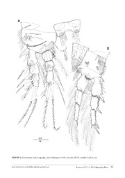
Zootaxa 3575: 27–48 (2012) ISSN 1175-5326 (print edition) www.mapress.com/zootaxa/ ZOOTAXA Article Copyright © 2012 · Magnolia Press ISSN1175-5334(online edition) urn:lsid:zoobank.org:pub:CCE1F328-3A3E-48E8-8B99-8AA0A1C18A23 A new species of the genus Cerviniopsis from Sagami Bay, Japan and reinstatement of the genus Neocervinia, with a report on the male of Neocervinia itoi Lee & Yoo, 1998 (Copepoda: Harpacticoida: Aegisthidae) EUN-OK PARK1, MOTOHIRO SHIMANAGA2, SUK HYUN YOON3& WONCHOEL LEE1, 4 1Department of Life Science, College of Natural Sciences, Hanyang University, Seoul 133-791, Korea 2Aitsu Marine Station, Center for marine environmental studies, Kumamoto University, Matsushima, Amakusa, Kumamoto 861-6102, Japan 3Fisheries & Ocean Information Division, National Fisheries Research & Development Institute, Haean-ro 152-1, Gijang-eup, Gijang-gun, Busan 619-705, Korea 4Corresponding author, E-mail: [email protected] Abstract A new aegisthid copepod, Cerviniopsis reductasp. nov. is described from the deep sea in Sagami Bay, Japan. The new species has superficial resemblance to C. minutiseta Ito, 1983 in the armature formula of swimming legs. However they differ from each other in the shape of setae of the swimming legs, the distal margin of operculum, length of caudal rami, and the location of setae on P5 exopod. Also, the male of Neocervinia itoi Lee & Yoo, 1998 is described on the basis of samples collected from around the type locality in Sagami Bay, Japan. Sexual dimorphism of N. itoi male can be observed in the fused rostrum, atrophied mouthparts, P5, and P6. The sixth leg is symmetrical and both gonopores are presumably active, based on the presence of two spermatophores internally in the genital segment. This paper reports for the first time on the sexually dimorphic characters in the genus Neocervinia Huys, Mobjerg & Kristensen, 1997, reinstating its generic status with the newly revealed male characters. Key words: Cold Seep, Taxonomy, Deep-Sea Copepoda, Cerviniopseinae, Cerviniinae Introduction Harpacticoid copepods are known to be a dominant group in the hyperbenthos at the Hatsushima cold-seep site in Sagami Bay (Toda et al. 1994). So far three species of deep-sea dwelling harpacticoid copepods were described taxonomically based on the samples from Sagami Bay: Neocervinia itoi Lee & Yoo, 1998, Normanella bifida Lee & Huys, 1999 and Nudivorax todai Lee & Huys, 2000 (see Lee & Yoo 1998; Lee & Huys 1999, 2000). Benthic copepods are dominant in bathyal Sagami Bay and have been studied in several aspects, including sex ratio, reproductive activity (Shimanaga & Shirayama 2003), and temporal patterns in diversity and species composition (Shimanaga et al. 2004). Especially deep-sea cerviniids have been a focus of several studies, which included their distributional characteristics, sex ratio and gut content (Shimanaga et al. 2008, 2009). As a result of an ongoing study on the harpacticoid community in Sagami Bay (Shimanaga et al. 2009), a new Cerviniopsis Sars, 1903 species was discovered. In addition, the male of Neocervinia itoi was also collected from the area for the first time. The genus Neocervinia was erected (Huys et al. 1997) originally to accommodate N. tenuicauda (Brotskaya, 1963) and N. unisetosa (Montagna, 1981), with N. itoi as the third known species of the genus (Lee and Yoo 1998). Seifried (2003) synonymized Neocervinia Huys, Møbjerg & Kristensen, 1997 and Pseudocervinia Brodskaya, 1963 with Cervinia Norman in Brady, 1878 (Aegisthidae), but the generic status of the former is discussed and reinstated herein based on the newly observed characters. Accepted by T. Karanovic: 6 Nov. 2012; published: 7 Dec. 2012 27 Material and methods Copepods were collected from Sagami Bay, Japan from 2000 to 2005 using a multiple corer. Details of the sampling techniques used were provided in Shimanaga et al. (2008). Specimens were fixed in 5% buffered seawater formalin and subsequently preserved in 70% ethanol. Copepods were dissected in lactic acid and mounted on slides in polyvinyl lactophenol mounting medium. All drawings have been prepared using a camera lucida on Olympus BX50 differential contrast interference microscope. The terminology for body and appendages follows Huys et al. (1996). Abbreviations used in the text are: P1–P6, first to sixth thoracopod; exp(enp)-1(2,3) to denote the proximal (middle, distal) segment of the exopod (endopod), ae, aesthetasc. Type specimens are deposited in the Marine Biodiversity Institute of Korea (MABIK). Scale bars in the figures are indicated in μm. Systematics Family Aegisthidae Giesbrecht, 1892 Subfamily Cerviniopseinae Brodskaya, 1963 GenusCerviniopsis Sars, 1903 Cerviniopsis reducta sp. nov. (Figures 1–8) Type locality. Sagami Bay, Japan (35° 04.4' N 139° 32.3' E). The water depth ranged from 750 to 770m. Specimens examined. Holotype: male (CR00179981) dissected on seven slides. Paratypes: one female (CR0000179982) dissected on eight, and one male (CR00179983) on eight slides, respectively. All specimens are from the type locality and collected by Dr. M. Shimanaga on May 20 2002. Description. Male. Total body length 967 μm (measured from tip of rostrum to posterior margin of caudal rami). Maximum width 211μm measured at posterior margin of cephalothorax. Body surface armed with sensilla and minute denticles (Figs. 1A, C–D). Prosome (Fig. 1A) 5-segmented, comprising cephalosome and 4 free pedigerous somites. P1 bearing somite fused to cephalosome, but with surface suture line showing original segmentation. Cephalothorax smooth (Fig. 1A), with few sensilla and smooth posterior margin. Short tripod shaped wrinkle present in middle of cephalosome. Succeeding two prosomites with thin comb-like hyaline frills forming smooth posterior margin. Rostrum well developed, elongated and triangular-shaped with pointed anterior apex, clearly fused to cephalosome (Fig. 1A–B). Dorsal surface smooth with 1 pair of sensilla near middle of lateral margin of rostrum dorsally and 1 tube pore on ventral surface. Urosome (Figs. 1A, C–D) 6-segmented, comprised of P5-bearing somite, 4 free abdominal and anal somites. Urosomites 3–5 denticulated dorsoventrally with well developed hyaline frill. P5 -bearing somites with smooth dorsoventaral surface and few spinules along lateral posterior angles. Anal somite (Fig. 1D) with well-developed operculum with spinulate posterior margin and flanked by pair of secretary pores. Caudal rami (Fig. 1A) ten times longer than wide; seta I shortest, located at proximal half; seta II 2.5 times longer than seta I, and located laterally; seta III as long as seta II; caudal setae IV pinnate, slightly longer than caudal ramus; caudal seta V pinnate, twice longer than IV; seta VI shorter than seta III and located on distal inner corner; seta VII bare, located near distal end of caudal ramus, as long as seta II, and triarticulated. Antennule (Fig. 2A) 7-segmented, segment 1 largest with several rows of spinules on surface. Segment 2 with few spinules on anterior surface. Aesthetascs on segments 2, 3, 4, and 7. Armature formula: 1-[1 pinnate], 2-[11 pinnate + 1 ae], 3-[3 bare + 3 pinnate + 1 ae], 4-[2 bare + 7 pinnate + (1+ae)], 5-[1 pinnate], 6-[2 pinnate], 7-[5 bare + 1 strong pinnate + 1 pinnate + 3 rigid spines + 1 acrothek]. Apical acrothek consisting of well-developed aesthetasc fused basally to 1 slender naked seta and 1 strong pinnate bent spine. 28 · Zootaxa 3575 © 2012 Magnolia Press PARK ET AL. FIGURE 1. Cerviniopsis reducta sp. nov., male (holotype): (A) habitus, dorsal; (B) rostrum; (C) urosome, ventral; (D) anal somite, dorsal; (E) P6, ventral; (F) P5, ventral. All scales in μm. NEW CERVINIID COPEPODS FROM SAGAMI BAY Zootaxa 3575 © 2012 Magnolia Press · 29 FIGURE 2.Cerviniopsis reducta sp. nov., male (holotype): (A) antennule; (B) antenna; (C) labrum. Both scales in μm. 30 · Zootaxa 3575 © 2012 Magnolia Press PARK ET AL. Antenna (Fig. 2B) 3-segmented, comprising coxa, allobasis, and free 1-segmented endopod. Coxa with several rows of spinules. Allobasis with 2 plumose abexopodal setae. Exopod 4-segmented with seta formula 2.1.1.020, respectively; all setae pinnate; apical outermost seta strongest. Free endopodal segment with long spinules along surface, and with 1 naked seta and 2 pinnate spines laterally and 1 geniculate, and 5 pinnate setae and 1 strong pinnate spine apically. Labrum (Fig. 2C) well developed; ventral margin ornamented with blunt median tooth, several sub-lateral row of blunt teeth, and short lateral spinules. Mandible (Fig. 3A) with large coxa bearing well-developed gnathobase; cutting edge with 6 major blunt teeth overlapping each other; accessory seta plumose. Mandibular palp well developed. Basis set on peduncle, wider than long with 4 pinnate setae; 3 setae set on small peduncle, respectively; several rows of long spinules along anterior surface. Endopod 1-segmented with 3 plumose lateral, 2 naked and 5 pinnate apical setae; row of spinules along outer lateral margin. Exopod 4-segmented with seta formula 2.1.1.2; segment 1 largest, and long spinules on anterior surface. Maxillule (Fig. 3B) with rows of spinules along anterior suface and outer lateral margins as figured. Praecoxa with well developed arthrite bearing 2 plumose anterior surface setae and 9 apical setae and spines. Coxa with epipodite represented by 1 seta; endite cylindrical with 1 naked, and 5 pinnate setae. Basis and endopod completely fused forming maxillulary allobasis with 5 pinnate and 6 naked slender setae. Exopod also incorporated into maxillulary allobasis and represented by 3 plumose setae. Maxilla (Fig. 3C–D), syncoxa with row of spinules on outer lateral margin and cylindrical 4 endites (2 praecoxal, 2 coxal); enditic seta formula 5.3.3.3; proximal praecoxal endite largest with 2 pinnate and 3 naked setae; all setae on distal praecoxal and coxal endites naked. Allobasis produced into long curved claw; accessory armature consisting of 1 geniculate spine and 1 slender seta distally, 1 slender seta and 1 strong pinnate spine proximally near articulation with endopod. Endopod 3-segmented; enp-1 with 1 geniculate spiniform, and 1 slender setae; enp-2 with 2 geniculate spiniform setae; enp-3 with 1 geniculate spiniform and 2 naked slender setae. Maxilliped (Fig. 3E) comprising syncoxa, basis, and 2-segmented endopod; rows of spinules present anteriorly and posteriorly as figured. Syncoxa elongated with 5 strong pinnate spines and 1 pinnate seta. Basis with 1 pinnate seta and 1 strong pinnate spine. Endopod 2-segmented; proximal segment with 2 plumose setae; distal segment with 2 strong pinnate apical spines, and 2 lateral setae (proximal one plumose, distal one naked). Swimming legs 1–4 (Figs. 4A, B; 5A, B) biramous, P1–P4 with 3-segmented exopod and 3-segmented endopod, and each ramus ornamented with setules and spinules along inner and outer margins as illustrated. Intercoxal sclerites well developed; ornamented with row of spinules in middle of anterior and posterior surface. P1 (Fig. 4A), praecoxa with row of spinules on anterior surfaces. Coxa wider than long and trapezoidal, ornamented with row of spinules on anterior and posterior surface and along outer margin. Basis with 1 outer plumose seta and 1 inner strong uni-pinnate spine and ornamented with row of long spinules on anterior surface extending posteriorly. Endopod subequal to exopod in length; enp-3 longest. Exp-1 largest; median outer spine of exp-3 distinctly shorter than others. P2 (Fig. 4B), praecoxa, small with row of spinules along anterior distal margin. Coxa with row of spinules on anterior and posterior surface, and along outer margin. Basis with 1 short plumose outer seta and spinules on inner lateral margin and row of spinules along distal margin near articulation with exopod; ornamented with row of long spinules along inner lateral margin and posterior surface with small patch of spinules on anterior and posterior surface. Endopod not extending to distal end of exopod; row of fine spinules along outer margin of each segment; enp-1 with elongated outer distal end forming sharp triangular tip; enp-3 slightly longer than two preceding segments. Each exopodal segments with row of spinules along outer margins; exp-1 largest, and exp-2 shortest; inner apical seta of exp-3 reduced, naked and small. P3 (Fig. 5A), praecoxa, small with row of spinules along anterior median distal margin. Coxa with row of spinules on anterior and posterior surface and along outer margin. Basis with 1 plumose outer seta and long spinules on inner lateral margin and row of spinules along distal margin near articulation with endopod. Endopod exceeding to distal end of exopod; row of spinules along outer and inner margin of each segment; enp-1 and enp-2 with elongated outer distal end forming sharp triangular tip; enp-3 slightly longer than two preceding segments; inner apical seta on enp-3 reduced, naked, and small. Each exopodal segments with row of spinules and setules along outer and inner margins; exp-1 largest and exp-2 shortest; inner apical seta of exp-3 reduced, naked and small. NEW CERVINIID COPEPODS FROM SAGAMI BAY Zootaxa 3575 © 2012 Magnolia Press · 31 FIGURE 3.Cerviniopsis reducta sp. nov., male (holotype): (A) mandible; (B) maxillule; (C) maxilla; (D) maxillary allobasis; (E) maxilliped. Scale in μm. 32 · Zootaxa 3575 © 2012 Magnolia Press PARK ET AL. FIGURE 4.Cerviniopsis reducta sp. nov., male (holotype): (A) P1, anterior; (B) P2, anterior. Scale in μm. NEW CERVINIID COPEPODS FROM SAGAMI BAY Zootaxa 3575 © 2012 Magnolia Press · 33 P4 (Fig. 5B), praecoxa, naked. Coxa with row of spinules on anterior and posterior surface, and along outer margin. Basis with 1 plumose outer seta and long spinules on inner lateral margin and row of spinules along distal margin near articulation with endopod. Endopod exceeding to distal end of exopod; row of spinules along outer and inner margin of each segment; enp-1 and enp-2 with elongated outer distal end forming sharp triangular tip; enp-1 shortest; enp-3 longer than enp-2; inner apical seta on enp-3 reduced, naked and small. Each exopodal segments with row of spinules and setules along outer and inner margins; exp-1 shortest, and exp-3 longest; inner apical seta of exp-3 reduced, naked and small. Armature formula as follows: Exopod Endopod P1 1.1.023 1.1.121 P2 1.1.222 1.2.221 P3 1.1.222 1.2.221 P4 1.1.122 1.2.120 P5 (Fig. 1F), baseoendopod forming short, outer setophore bearing plumose basal seta. Each endopodal lobe fused each other forming 1 narrow plate, without any ornamentation. Exopod long, narrow plate shaped, and 6.5 times longer than wide; whole surface covered with setules; with 1 strong pinnate apical, 1 short pinnate inner and 1 short naked outer lateral setae; outer lateral seta inserted at distal 1/3; 1 short spinous process present between apical and inner setae. Sixth pair of legs (Fig. 1E) not fused medially, symmetrical. Each P6 bilobate with outer lobe bearing elements consisting of 2 plumose setae; outer one shortest. Anterior surface naked and inner distal margin somewhat swollen without any ornamentation. Female. Total body length of examined samples 1,219 μm, much larger than in male (measured from tip of rostrum o posterior margin of caudal rami). Largest width presumably at posterior margin of cephalic shield (not measurable due to dorsoventrally depressed body). General body appearance same as in male. Rostrum (Fig. 6C) well developed, elongated and triangular-shaped with pointed anterior apex, clearly fused to cephalosome. Dorsal surface smooth with 1 pair of sensillae near middle of lateral margin of rostrum dorsally and 1 tube pore on ventral surface as in male. Urosome (Fig. 6B) 5-segmented, comprised of P5-bearing somite, genital double, and 3 free abdominal somites. All urosomite with pattern of surface ornamentation consisting of small spinules dorsoventrally. Hyaline frills of urosomites denticulate. Genital double somite (Figs. 6B; 8C) with transverse surface ridge dorsally and laterally forming spinous processes laterally indicating original segmentation; completely fused ventrally. Genital field positioned anteriorly, just above middle line between original segmentation (Fig. 6B); copulatory pore minute; gonopores fused medially forming single genital slit covered on both sides by well developed opercula derived from sixth legs; P6 elongate, with row of spinules along anterior and posterior margins, and 1 bare and 1 pinnate setae; outer seta longest and pinnate (Fig. 8C). Caudal rami (Fig. 6B) 14 times longer than wide, longer than those in male and with 7 caudal setae as in male. Antennule (Fig. 6A) 7-segmented, segment 1 largest with several rows of spinules on surface. Segment 2 with few spinules on anterior surface. Aesthetascs on segments 3, and 7. Armature formula: 1-[1 pinnate], 2-[8 pinnate + 1], 3-[3 pinnate + 2 geniculate + 6 +(1+ ae)], 4-[3 pinnate], 5-[1 + 1 pinnate], 6-[1 + pinnate], 7-[1 strong pinnate + 2 + 3 rigid spines + 1 acrothek]. Apical acrothek consisting of well-developed aesthetasc fused basally to 1 slender pinnate seta and 1 strong pinnate bent spine. Antenna, mandible, maxllule, maxilla, maxilliped, and P1 same as those in male. Swimming legs 2–4 (Figs. 7A–B, 8A) biramous, P2–P4 with 3-segmented exopod and 3-segmented endopod, and each ramus ornamented with setules and spinules along inner and outer margins as illustrated. Intercoxal sclerites well developed; ornamented with row of spinules in middle of anterior and posterior surface. Armature formula same as in male. P2 (Fig. 7A) endopod not extending to distal end of exopod; inner apical seta of exp-3 not reduced, long and pinnate. P3 (Fig. 7B) endopod not extending to distal end of exopod; inner apical seta on enp-3 not reduced, long, and pinnate. Inner apical seta of exp-3 not reduced, longer than outer apical one and pinnate. 34 · Zootaxa 3575 © 2012 Magnolia Press PARK ET AL. FIGURE 5.Cerviniopsis reducta sp. nov., male (holotype): (A) P3, anterior; (B) P4, anterior. Scale in μm. NEW CERVINIID COPEPODS FROM SAGAMI BAY Zootaxa 3575 © 2012 Magnolia Press · 35 FIGURE 6.Cerviniopsis reducta sp. nov., female (paratype): (A) antennule; (B) urosome, ventral; (C) rostrum. Both scales in μm. 36 · Zootaxa 3575 © 2012 Magnolia Press PARK ET AL.
