A New Species of Megacricetodon (Cricetidae, Rodentia, Mammalia) from the Middle Miocene of Northern Junggar Basin, China PDF
Preview A New Species of Megacricetodon (Cricetidae, Rodentia, Mammalia) from the Middle Miocene of Northern Junggar Basin, China
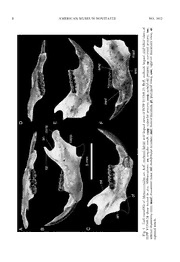
PUBLISHED BY THE AMERICAN MUSEUM OF NATURAL HISTORY CENTRAL PARK WEST AT 79TH STREET, NEW YORK, NY 10024 Number 3602, 23 pp., 13 figures, 2 tables April 9, 2008 A new species of Megacricetodon (Cricetidae, Rodentia, Mammalia) from the Middle Miocene of northern Junggar Basin, China SHUNDONG BI,1 JIN MENG,2 AND WENYU WU3 ABSTRACT Dental,mandibular,andpostcranialspecimensofMegacricetodonyein.sp.,aredescribed.The new specimens, including the complete dentition, mandible, and partial forelimb and hindlimb, represent the most complete materials known for the genus, provide valuable information concerningtheinterspecificvariationofthegenus,andleadtothereassessmentofthesuprageneric positionof Megacricetodon. Megacricetodon yei is characterized by having medium-size, clearly split anterocone of M1, presence of the labial spur of the anterolophule and the posterior spur of the paracone in some M1s,mediumtolongmesolophinM1-2,frequentoccurrencesofdoubleprotolophules,transverse orposteriorlydirectedmetalophuleofM2,andsingleanteroconidofthem1.Megacricetodonyeiis more closely related to Megacricetodon (5 Aktaumys) dzhungaricus than to any other species of Megacricetodon,butismorederivedthanthelatter.Basedonthenewinformation,thevalidityof the genusAktaumysis discussed. ThepostcranialfeaturesofMegacricetodonyeishowclearadaptationsforterrestrialhabits,but as in many ground-dwelling rodents living in burrows, it could also climb or dig. The associated fauna has been correlated to Tongxin fauna from the adjacent part of China and the Belometchetskya fauna of north Caucasus, equivalent to early Middle Miocene age, or MN 6 correlative.ThestageofevolutionofMegacricetodonyeiisconsistentwiththefaunalcorrelation. 1Department of Biology, Indiana University of Pennsylvania, Indiana, PA 15705. Research Associate, Section of VertebratePaleontology,CarnegieMuseumofNaturalHistory([email protected]). 2DivisionofPaleontology,AmericanMuseumofNaturalHistory([email protected]). 3InstituteofVertebratePaleontologyandPaleoanthropology,ChineseAcademyofSciences,Beijing100044,China. CopyrightEAmericanMuseumofNaturalHistory2008 ISSN0003-0082 2 AMERICAN MUSEUMNOVITATES NO. 3602 INTRODUCTION the Halamagai Formation between the Tieersihabahe section and Chibaerwoyi sec- Cricetid rodents of the genus Megacri- tion in 1998 and 2000. The bones were cetodon are among the most abundant mam- excavated mainly from a mudstone lens malian species in the Old World Cenozoic (about 2 m2) imbedded in grayish medium- fauna. Fahlbusch (1964) first recognized grained sandstone. Dozens of maxillae and Megacricetodon as a subgenus of the genus mandibles with complete dentitions, 25 isolat- Democricetodon, which he established at the ed teeth, and about 27 postcranial elements time, but later researchers have elevated the were recovered. Although no articulated subgenus to the genus level (Mein and elements were found, the ratio of the speci- Freudenthal, 1971a). The genus Megacri- mens, morphology, and size suggest that they cetodon is species-rich. To date, more than belong to the same species. Numerous speci- 25 fossil species have been recognized from mens of a distinctively larger taxon, the Miocene of Europe and Asia. Despite Cricetodon n. sp. were also recovered from their great diversity and widespread distribu- the same spot and have been described by tions, all the species of Megacricetodon are Bi (2005). Comparisons are specifically represented primarily by isolated teeth, often made to Cricetodon from Tieersihabahe collected by screen washing. Very few cranial since it represents the most complete skele- and postcranial bones are known. In addi- tal materials of extinct cricetids known to tion to numerous specimens of dentitions, date. here we describe the best preserved postcra- nial skeletal materials known for this genus from the northern Junggar basin, Xinjiang, MATERIALS AND METHODS China. The Junggar materials shed new light on the interspecific variation and anatomy of Terminology for dental morphology is Megacricetodon and help to clarify several after Mein and Freudenthal (1971a). taxonomic issues of the genus. Because their terminology has been well The northern Junggar basin of China has known, we prefer not to include a diagram yielded many fossiliferous localities, of which to illustrate the tooth structure. Positional Tieersihabahe-Chibaerwoyi Cliff (TCC) is an abbreviations of teeth follow the common extremely rich locality. Fossil mammals in alphanumeric convention of using uppercase this area were first discovered in 1982 by a versus lowercase letters to identify maxillary field team from the Institute of Vertebrate or mandibular teeth, respectively, and num- Paleontology and Paleoanthropology (IVPP), bers to indicate their placement in the tooth Chinese Academy of Sciences. Subsequently, row (for example, M1 and m1). The cranial fossil mammals, mainly large mammals, were and postcranial skeletal terminologies, wher- described, including carnivores (Qi, 1989), ever appropriate, follow those by Howell artiodactyls(Ye,1989),proboscideans (Chen, (1926), Rinker (1954), Cooper and Schiller 1988), lagomorphs (Tong, 1989), and rodents (1975), Carleton and Musser (1989), (Wu, 1988). Carleton and Olson (1999), and Gebo and Our field investigations, run from 1995 Rose (1993). through the present, have greatly improved Teeth and mandibles were imaged and the documentation of Tertiary strata in this measured using a Nikon SMZ 8 microscope area.Basedonthesenewdiscoveries,Yeetal. set at 20 3 magnifications, and measure- (2001a, 2001b, 2003) redefined the stratigra- ments were recorded to the nearest 0.01 mm. phicalunitsofTCC,inascendingorder,as:(1) The SEM photographs of some teeth were the Eocene-Oligocene Ulunguhe Formation, taken from uncoated specimens using a (2) the Late Oligocene Tieersihabahe Forma- Hitachi scanning electron microscope. All tion, (3) the Early Miocene Suosuoquan postcranial measurements were taken to Formation, (4) the Middle Miocene Hala- 0.05 mm using digital calipers. IVPP is the magai Formation, and (5) the late Middle abbreviation of the Institute of Verte- Miocene Kekemaideng Formation. The mate- brate Paleontology and Paleoanthropology, rials described here were collected from Beijing. 2008 BIETAL.: NEW SPECIESOFMEGACRICETODON 3 SYSTEMATIC PALEONTOLOGY imal end; V15350.5–6, 2 distal portions of left humeri; V15350.7, proximal portion of a right ORDERRODENTIABOWIDICH,1821 ulna; V15350.8–10, 3 proximal portions of left SUBORDERMYOMORPHABRANDT,1855 ulnae; V15350.11–12, 2 distal left ulnar ends; SUPERFAMILYMUROIDEAILLIGER,1811 V15350.13, a complete right femur except the FAMILYCRICETIDAEROCHEBRUNE,1883 brokendistalend;V15350.14–17,4fragmentary SUBFAMILYCRICETINAESTEHLIN&SCHAUB, rightfemurs; V15350.18–19, 2fragmentary left 1951 femurs. V15350.20–21, 2 complete right tibiae GENUSMEGACRICETODONFAHLBUSCH,1964 exceptthebrokenproximalend;V15350.22–23, 2 complete left tibiae except the broken pro- Megacricetodon yei, n. sp. ximal end; V15350.24–26, 3 fragmentary left tibiae;V15350.27,arightcalcaneus. HOLOTYPE: IVPP V15349.1, a fragmentary right maxilla with M1–M3 (fig. 3) LOCALITY AND AGE: Site XJ 98018 (46u 40.1289 N, 88u30.8469 E) at the Tieersihabahe REFERRED MATERIAL: IVPP V15349.2, a localityinthenorthernJunggarBasinofChina. fragmentary right maxilla with M1–M3; ThefirstsandbedoftheHalamagaiFormation, V15349.3, a right M1; V15349.4, a right M2; earlyMiddleMiocene. V15349.5-6,2rightM3;V15349.7,aleftmaxilla with M1–M3; V15349.8, a left maxilla with ETYMOLOGY: The species name, yei, is in honor of our colleague, professor Jie Ye, for M1–M3; V15349.9, a left maxilla with the his contribution to the study of Cenozoic zygomatic plate and M1; V15349.10–11, 2 left stratigraphy and mammals in northern M2;V15349.12–13,2leftM2;V15349.14–15,2 Xinjiang and discovery of the locality. left M3; V15349.16–19, 4 right upper incisors; V15349.20–22,3leftupperincisors;V15349.23, REPOSITORY: The specimens are housed in an almost complete left mandible except the the collections of the Institute of Vertebrate broken angular process; V15349.24, a left PaleontologyandPaleoanthropology,Chinese fragmentary mandible with the incisor and Academy of Sciences, Beijing. m1–3; V15349.25, a left fragmentary mandible DIAGNOSIS: A Megacricetodon species of with m1–m3; V15349.26, a left fragmentary mediumsize,M1withclearlysplitanterocone, mandible with the incisor and m1–m3; presence of the labial spur of the anterolo- V15349.27, a left fragmentary mandible with phule and the posterior spur of the paracone m1–m3; V15349.28, a left fragmentary mandi- in some M1s, medium to long mesoloph in blewiththeincisor;V15349.29,aleftfragmen- M1–2, frequent occurrences of the double tary mandible with the incisor and m1–m2; protolophules in M2 of the collection, M2 V15349.30, a left fragmentary mandible with metalophuletransverseorposteriorlydirected, the incisor, m1, and m3; V15349.31, a left m1 anteroconid simple, mesolophid of medi- fragmentary mandible with m2; V15349.32, a um length in m1–2, and presence of the left fragmentary mandible with m1–m2; entepicondylar foramen in the humerus. V15349.33-34, 2 left m1; V15349.35, a left m2; DIFFERENTIAL DIAGNOSIS: Differs from V15349.36–37,2isolatedm3;V15349.38,aright Megacricetodon (5 Aktaumys) dzhungaricus in fragmentarymandiblewiththeincisorandm1– having slightly smaller size; better bifurcated m3; V15349.39, a right fragmentary mandible M1 anterocone; shorter mesoloph in M1 and with the incisor and m2; V15349.40, a right M2;feweroccurrencesofthelabialspurofthe fragmentarymandiblewiththeincisorandm1– anterolophule in M1; fewer occurrences of m3; V15349.41, a right fragmentary mandible double protolophule and metalophule in M2. with m2–m3; V15349.42, a right fragmen- Differs from any other known species of tary mandible with m1–m2; V15349.43, a Megacricetodoninhavingthefollowingcombi- right fragmentary mandible with m2–m3; nation of features: well-bifurcated M1 antero- V15349.44,a right fragmentary mandible with cone; presence of a labial spur of the ante- m1–m2;V15349.45–46,2rightm3;V15350.1,a rolophule in many M1s, double protolophules baculum; V15350.2–3, 2 almost complete right inmostM2s,thepresenceoftheposteriorspur humeri except the broken proximal end; ofparacone,andlongmesolophinM1andM2, V15350.4, a left humerus except broken prox- andsinglem1anteroconid. 4 AMERICAN MUSEUMNOVITATES NO. 3602 Fig.1. SEMimagesofaleftfragmentarymaxillawiththeM1andtwoisolatedM1sofMegacricetodon yein.sp.showingvariations.A,ventralviewofthemaxilla(IVPPV15349.9);B,occlusalviewofarightM1 (V15349.3);C,occlusalviewofaleftM1(V15349.10).Arrowspointtolabialspuroftheanterolophulein both B and C. Abbreviations: inf, incisive foramen; sm, the oval patch for the superficial masseter; zp, zygomaticplate. DESCRIPTION (fig. 1, 3, 4). M1 is the largest upper cheek tooth with three roots, a major lingual one MAXILLA: Ofthecranialelements,onlythe and two minor buccal ones; the root support- left fragmentary maxilla is preserved in IVPP ing the anterocone extends slightly anteriorly. V15349.9(fig. 1).Thezygomaticplateisbroad The occlusal outline is longer than wide and and has a well- defined fossa on the ventral surfacefortheoriginoftheanteriorpartofthe muscle masseter lateralis profundus. The ante- riormarginoftheplateispartiallybroken,but the oval patch for the superficial masseter is preserved; itisprominent and located immedi- ately posteroventral to ventral end of the anterior margin. The posterior margin of the plate extends anterior to M1. The posterior portion of the incisiveforamen ispreserved,of which the posterior edge levels with the protocone of M1, as in the Cricetodon, but differing from those in most living cricetines (Cricetus and Mesocricetus) in which the incisiveforamenterminateanteriortoM1. UPPER TEETH: Seven upper incisors are preserved.Itsanteriorsurfaceisgentlyconvex and smooth. The enamel wraps slightly around onto both lateral and medial surface, but extends fartheron the lateral surface than onthemedialone(fig. 2).Thecrosssectionof the incisor is oval. Eight complete M1s are in the collection Fig.2. UpperrightincisorofMegacricetodonyei and five of them are preserved in the maxilla (IVPPV15349.16).A,lateralview;B,lingualview. 2008 BIETAL.: NEW SPECIESOFMEGACRICETODON 5 Fig.3. Occlusal(A),lingual(B),andlabial(C)viewsofuppercheekteethofMegacricetodonyei(IVPP V15349.1;holotype). slightly widens posteriorly. The anterocone is specimens.Theposteriorspuroftheparacone divided by a deep, longitudinal groove into ispresent,connectingtothemesolophinthree labialandlingualconules,ofwhichtheformer specimens. The mesoloph isof medium length is larger. There is a low ledge on its anterior in one specimen but is long and reaches the face in five of eight specimens. The anterolo- labial border of the occlusal surface in seven phule is connected to the lingual conule in six specimens.Theentomesolophisstrong,reach- of eight specimens or extends to a point ing the lingual border of the occlusal surface between the two conules in two M1s. The only in one specimen (fig. 4A). The metalo- low labial spur of the anterolophule is long phule is directed posteriorly, joining the and transverse in two specimens, of medium posteroloph immediately behind the hypo- length and joins the base of the paracone in cone. The posteroloph is long, extending to two specimens, and absent in the remaining the posterolabial part of the metacone. A 6 AMERICAN MUSEUMNOVITATES NO. 3602 Fig.4. UppercheekteethofMegacricetodonyei(IVPPV15349.2,V15349.7,V15349.1).Occlusalviewof upper cheek teeth showing variations owing to age and wear among individuals. Image B was flipped horizontallyto facilitate comparison. cingulum is usually developed and inflated to subquadrate (fig. 3, 4). The lingual and labial form a small cuspate between major cusps. branches oftheanterolopharewelldeveloped The sinus is transverse. and are of about equal lengths. In three M2s, Seven M2s are in the collection. The M2 is the protolophule is doubled; in the remaining also triple-rooted and its occlusal outline is four,onlytheanterior protolophuleispresent 2008 BIETAL.: NEW SPECIESOFMEGACRICETODON 7 and projects anterolingually. The single meta- M. similis, and extant cricetines (Cricetus and lophule extends anterolingually in one M2, Mesocricetus),thecoronoidprocessisfalciform transversely in two M2s, and posterolingually and very pronounced, with its tip higher than joining the posterior arm of the hypocone in thecondyloidprocess.Incontrast,thecoronoid fourM2s(fig. 4).Themesolophislonginfive processisreducedanditstipisslightlybelowor specimens and is of medium length in the aboutthesamelevelwiththecondyloidprocess remainingtwo.IntwoM2s,theposteriorspur in Cricetodon. The sigmoid notch is deep, ofthe paracone is present and connects to the distinctive from the shallow notch in mesoloph. The sinus is transverse. Cricetodon. The capsular process of the lower Eight M3s are in the collection. The M3 is incisoralveolusisconspicuous,lyingbelowthe much smaller than M1–2 and is also triple- baseofthecoronoidprocess.Indorsalview,the rooted. The labial anteroloph is well devel- condyloid process is longitudinally long in a oped in all M3s, butthe lingualone ispresent teardrop shape with its anterior tip bending only in two specimens. The protocone and slightlymedially.Theangularprocessismissing paracone are prominent, but the posterior from all the specimens. But judged from the cusps are reduced. The metalophule extends breakage, the angular notch is broad and anterolingually to connect the longitudinal somewhat oval. The pterygoid fossa for the crest. An axioloph originating from the insertionofthemedialpterygoidmuscleisdeep. longitudinal crest is directed anteriorly and LOWERTEETH: Nine lower incisors are pre- connects to the protolophule in two M3s served in mandibles. The lower incisor is (fig. 3). The hypocone and metacone are delicate compared to the upper incisor. It nearly merged into the posteroloph, which extends posteriorly under the cheek teeth and encircles the posterior sinus. terminates posteriorly at the level below the MANDIBLE: IVPP V15349.23 is a nearly base of the coronoid process. The tip of the complete mandible except for the broken lowerincisor isslightly lower thanthe occlusal angular process (fig. 5A, B, C). The lower surfaceofthecheekteeth(fig. 5F).Theenamel incisor is displaced from the alveolus and extends only to the labial side, covering about protrudesmoreanterodorsallythanitsoriginal one third of the labial surface. The incisor is position. The original position of the incisor is oval in cross section, but the anterior and preserved in V15349.24 (fig. 5D, E, F). The medial sides are flat. The anterior surface is dentary is thin; its ventral rim is convex under smooth and has no longitudinal ornaments. m1 and is concave under the last two molars. Thewearfacetoftheincisortipislong(fig. 7C). The anterior slope of the diastema is gently Fourteen m1s are present (fig. 6). The lower curved, whereas its posterior slope, anterior to molars are all double-rooted. The m1 ante- m1,issteep.Thelengthofdiastemais3.1 mm, roconidisa high,conicalcuspand situatedon less than that of the lower tooth row. On the the longitudinal axis of the tooth. Both the lateral surface of the mandible, the masseteric labialandlingualanterolophidsarewelldevel- crestsare weakerthanthose of Cricetodon; the opedandreachthebaseoftheprotoconidand superiorandinferiorcrestsconvergeanteriorly metaconid, respectively. The anterolophulid as a V shape, with its apex terminating at the extendsfromthe labial side of the anteroconid leveloftheanteriorrootofm1.Anovalmental and is connected to the anterior arm of the foramen is at the position anteroventral to the protoconid.Themetalophulidand hypolophu- m1; it opens anterolaterally and is best seen in lid extend slightly anterolabially. The mesolo- lateral view. On the medial surface of the phid is absent in seven, of medium length in mandible, the large mandibular foramen lies four, and long and reaches the lingual edge of anterovental to the base of the condyloid the tooth in three specimens. The ectomesolo- process. The ascending ramus is lateral and phidisabsentexceptforaweakonepresentin oblique tothe toothrow, similar tothatofM. one specimen. The sinusid is transverse. The similis (the only other Megacricetodon species longposterolophidclosestheposterosinusid. preservingthemedialsideofthemandible)and Sixteen m2s are in thecollection. The labial extant cricetines (Cricetus and Mesocricetus), anterolophidislongandconnectedtothebase butdifferingfromthatofCricetodon.InM.yei, of the protoconid. The lingual anterolophid is 8 AMERICAN MUSEUMNOVITATES NO. 3602 ofc,n, ms alviewsocess;iccrest; labidpreteri andonoimass ual,coror ngp,eri licup usal,cess;mc,s occlproa;s D–F,yloidfoss dd 3;noi 2og PPV15349.ess;conp,cn;pf,ptery Vocme sofIarprfora viewpsulntal alcame ngupp,nf, licam ndh;n; abial,aarnotcforame usal,langulbular ccln,ndi oaa C,s:m acricetodonyei.A–situ.Abbreviationassetericfossa;mf, gnm ei Morsf, ofcisma dibleowinrest; nhc ma4sric Left349.2asseteotch. 5.V15rmdn Fig.PPeriomoi IVinfsig 2008 BIETAL.: NEW SPECIESOFMEGACRICETODON 9 Fig.6. OcclusalviewsofaleftlowerincisorandcheekteethofMegacricetodonyei(A:IVPPV15349.23; B: V15349.25; C: V15349.24). Occlusal view of lower cheek teeth show variations owing to age and wear amongindividuals. short or absent in 14, except a strong one surface, particularly the enamel ridges, bears present in one specimen. The metalophulid numerouspitsandsomestriations.Thereisno and the hypolophulid are directed slightly shearing facet on the lingual and posterior anterolabially. The mesolophid is of medium sides of the tooth, suggesting that grinding length in nine, and short or absent in six was its primary function during mastication. specimens. The ectomesolophidis absent. The The orientation of the striations indicates a posterolophid is strong and descends to the primarily anteroposterior movement of the base of the entoconid. The wear of the lower lower jaw during mastication, with a minor m2 from a relatively old individual (IVPP component of lateral shift. V15349.24) is illustrated in figure 7A and B. Fourteenm3sareinthecollection.Thelabial The SEM image shows that the occlusal anterolophid is long and descends to join the 10 AMERICAN MUSEUMNOVITATES NO. 3602 Fig.7. Occlusalviewofm2andi1ofMegacricetodon(IVPPV15349.24).TheboxinAisenlargedinB. The arrowin Bindicates the orientation of the wearstriations. base of the protoconid, but the lingual one is dorsally. The tip, to which the cartilaginous short or absent. The mesolophid is of medium tissue adheres, is broken. lengthinthree,butabsentin10specimens.The HUMERUS: The humerus is represented by transverse hypolophulid reaches the longitudi- three immature specimens and two fragmen- nalcrestanteriortothehypoconid.Theposter- tary distal ends. The proximal end was olophid is connected to the entoconid and detached from all the immature specimens at encloses the posterosinusid. The entoconid is theproximalepiphysealline,sothattheshape reduced.Alingualcingulidalmostconnectsthe and extent of the head, greater tubercle, and entoconidandthemetaconid. lesser tubercle cannot be determined. The BACULUM: The baculum has a relatively remaining length of the humerus measures long shaft with an anchorlike base (IVPP 10.9 mm from the proximal epiphyseal line to V15350.1;fig. 8).Oneachsideofthebaseisa the distal articular surface, and 2.7 mm wide distally directed spike to which the corpora between the lateral and medial edges at the cavernosum attaches. The shaft is wider than distal end (IVPP V 15350.2). deep and expands slightly distally. In lateral The humeral shaft is straight, with a wide view, the two ends of the baculum bend distal end, large medial epicondyle, and prom-
